ISSN 1806-3713 © 2015 Sociedade Brasileira de Pneumologia e Tisiologia
http://dx.doi.org/10.1590/S1806-37562015000000235
Reversed halo sign
Edson Marchiori1,2, Gláucia Zanetti2,3, Bruno Hochhegger4,5
1. Universidade Federal Fluminense, Niterói (RJ) Brasil. 2. Universidade Federal do Rio de Janeiro, Rio de Janeiro (RJ) Brasil. 3. Faculdade de Medicina de Petrópolis, Petrópolis (RJ) Brasil. 4. Santa Casa de Misericórdia de Porto Alegre, Porto Alegre (RS) Brasil.
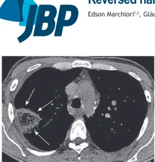
5. Universidade Federal de Ciências da Saúde de Porto Alegre, Porto Alegre (RS) Brasil. A 35-year-old man presented to the emergency room with chest pain accompanied by dyspnea. He reported having sustained a lower limb fracture and having been immobilized for 30 days.
A CT scan showed the reversed halo sign (RHS) with a reticular pattern, and the inal diagnosis was pulmonary infarction. The RHS found on HRCT of the chest is deined as a rounded area of ground-glass attenuation surrounded by a ring of consolidation. This sign was initially descri-bed as a sign speciic for organizing pneumonia (OP). Later studies identiied the RHS in a wide spectrum of infectious and noninfectious diseases. In Brazil, the most common infectious causes of the RHS are tuberculosis, paracoccidioidomycosis, and invasive fungal diseases (invasive pulmonary aspergillosis and mucormycosis). Among the noninfectious causes, OP, both idiopathic and secondary, is the most common. Other important causes are pulmonary infarction and sarcoidosis.
Although the RHS is considered a nonspeciic sign, a careful analysis of its morphological characteristics can narrow the differential diagnosis, helping the attending
physician to make a deinitive diagnosis. Two imaging patterns should be taken into account in order to make the diagnosis more speciic: the presence of nodules on the wall of or within the halo (nodular RHS); and a reticular pattern within the halo (reticular RHS).
The nodular RHS is generally found in active granulo-matous diseases, especially tuberculosis and sarcoidosis. It is also seen in some cases of paracoccidioidomycosis. Histopathological analysis of such cases has revealed that the formation of nodules is due to the presence of granulomas. With regard to the reticular RHS, the immunological status of the patient is the most important piece of clinical information needed in order to make the differential diagnosis. In immunocompromised patients, the primary diagnostic hypothesis is that of invasive fungal diseases. In immunocompetent patients, the reticular RHS corresponds, as a rule, to pulmonary infarction, usually secondary to thromboembolic disease. Suspicion of infarction from thromboembolic disease requires immediate conirmation by determination of D-dimers and CT angiography.
It should be borne in mind that the reticular and nodular patterns are not found in OP, which is the most common cause of the RHS. These considerations are important because the treatment for infectious conditions is completely different from that used for noninfectious conditions. Corticosteroid use, which is the treatment of choice for OP, can have harmful effects in patients with invasive fungal disease or active tuberculosis. Although the inal diagnosis should be based on the clinical mani-festations, the characteristics of the RHS can be quite useful in making the differential diagnosis. In some cases, lung biopsy may be necessary for the inal diagnosis.
RECOMMENDED READING
1. Marchiori E, Zanetti G, Hochhegger B, Irion KL, Carvalho AC, Godoy MC. Reversed halo sign on computed tomography: state-of-the-art review. Lung. 2012;190(4):389-94. http://dx.doi.org/10.1007/s00408-012-9392-x
J Bras Pneumol. 2015;41(6):564-564
564
Figure 1. CT scan (mediastinal window settings) showing the reversed halo sign in the right lung (white arrows). Note the reticular pattern within the halo.