Clinical status and detection of
periodontopathogens and
Streptococcus
mutans
in children with high levels of
supragingival biofilm
Abstract: Knowledge about the presence of some important oral patho-gens is an important step in better identifying children at risk for peri-odontal and/or caries diseases in later life. The purpose of this study was to detect the presence of Streptococcus mutans (Sm), Aggregatibacter ac-tinomycetemcomitans (Aa), Campylobacter rectus (Cr), Porphyromonas gingivalis (Pg), Prevotella intermedia (Pi), and Tannerella forsythia (Tf) in gingival bioilm samples from 196 children, and to assess whether any of these pathogens are more associated with gingival inlammation ex-tension and the Decayed/Missing/Filled teeth (DMFT/dmft) index. The subjects presented plaque index greater than 80% and were divided in 3 groups according to the bleeding index (BI): I) Low bleeding (≤ 30%), II) Medium bleeding (31 – 59%) and III) High bleeding (≥ 60%). The pres-ence of each pathogen was determined by PCR. The prevalpres-ence of Sm was 71.9% and the mean dmft/DMFT was 6.68. The prevalence in low, medium and high bleeding groups was 43.5%, 34.5% and 46.7% for Aa; 43.5%, 37.9%, and 36.7% for Cr; 99.1%, 100%, and 96.7% for Pg; 56.5%, 56.9%, and 66.7% for Pi; and 58.3%, 60.3%, and 56.7% for Tf, respectively. Pg (99.0%) was the most prevalent periodontal pathogen de-tected followed byTf (58.7%), Pi (58.2%), Aa (41.3%) and Cr (40.8%). Our study indicated that in this high plaque index population studied, a high prevalence of Sm and high mean DMFT were observed. In addi-tion, the presence of Pi was associated with the presence of inlammation (P < 0.05) whereas Cr was associated with periodontal health (P < 0.05).
Descriptors: Periodontal diseases; Dental caries; Dental plaque; Diagnosis.
Sheila Cavalca Cortelli(a) José Roberto Cortelli(a) Davi Romero Aquino(b) Marinella Holzhausen(a) Gilson Cesar Nobre Franco(c) Fernando de Oliveira Costa(d) Daniel Fine(e)
(a) PhD, Professor; (b)PhD student – Department
of Periodontology, University of Taubaté, SP, Brazil.
(c) PhD, Professor, Department of Oral Biology,
University of Taubaté, SP, Brazil.
(d) PhD, Professor, Department of
Periodontology, Federal University of Minas Gerais, MG, Brazil.
(e) PhD, Professor, Department of Oral Biology,
New Jersey Dental School, USA.
Corresponding author:
Sheila Cavalca Cortelli
Secretaria de Pós-Graduação em Odontologia - UNITAU
Rua Expedicionário Ernesto Pereira, 110, Centro - Taubaté - SP - Brazil
CEP: 12020-330
E-mail: [email protected]
Introduction
Periodontal diseases are infections in which spe-ciic bacteria play an important role. The continu-ous dental plaque accumulation may result in an imbalance between pathogenic species and the host defense mechanisms, which may lead to gingival in-lammation.1 In fact, poor oral hygiene status has
frequently been associated with a high prevalence or severity of periodontal disease.2-4
Current knowledge clearly shows that the mi-crobiota associated with healthy periodontal sites is quite different from that of inlamed sites. Increased frequency of detection of Aggregatibacter actino-mycetemcomitans (Aa), Porphyromonas gingivalis (Pg), Tannerela forsythia (Tf), Treponema denticola (Td), Prevotella intermedia (Pi), Fusobacterium nucleatum (Fn), Campylobacter rectus (Cr), and Eikenella corrodens (Ec) has been found in peri-odontal lesions.5
On the other hand, the bacterium Streptococcus mutans (Sm) is generally accepted to be the princi-pal etiological agent of dental caries due to its high cariogenic potential.6 Accordingly, higher levels of S. mutans have been associated with a higher risk for dental caries.7 Thus, microbiological diagnosis
of some of these speciic pathogens should improve our ability to identify individuals or sites at risk for developing oral diseases.
Therefore, it is reasonable to believe that knowl-edge about the colonization process that takes place early in life is an important step in better identifying which children need more effective oral treatments in order to minimize the risk of periodontal and/or caries diseases in later life. In view of the contro-versial data available on the early infection by oral pathogens, the aim of the present study was to detect the presence of selected oral pathogens, using PCR, in gingival marginal bioilm samples from Brazilian children with high levels of supragingival bioilm, and to assess whether any of these pathogens are more associated with gingival inlammation exten-sion and DMFT index.
Materials and Methods
Subject population
This study was conducted in accordance with
guidelines of the 196/96 resolution of the National Health Council (NHC) and was approved by the University of Taubaté Ethics Committee (protocol # 0076/01). The children’s parents or guardians received complete information regarding the objec-tives and procedures of the study and provided writ-ten informed consent.
One hundred and ninety six school children with mixed dentition (92 male and 104 female), ranging in age from 6 to 12 years (8.6 ± 1.4 years), from a public school in Taubaté (SP, Brazil) were enrolled in this cross-sectional study. The children in need of dental care were referred to the Dental Clinics of the University of Taubaté for appropriate treatment.
The children presented good general health and had not received treatment with antibiotics within the past 6 months prior to the study. In addition, the children selected for this experiment should present a high plaque index (minimum value of 80% for the Plaque Assessment Scoring System – PASS8).
Patients presenting systemic diseases, immuno-deiciency and/or use of orthodontic or prosthetic devices were excluded from this study.
Clinical examination and microbial sampling
The clinical exam and microbial samples collec-tion were performed in the children by two calibrat-ed examiners. Intra- and inter-examiner agreement was high (minimum kappa value was 0.83).
Clinical examination comprised the determi-nation of dental bioilm by PASS.8 A total of 208
children were initially examined in this study. Ac-cording to the cut-off point, plaque index > 80%, 12 children were excluded; therefore, 196 children (92 male and 104 female) completed the study. The bleeding index (BI)9 was also evaluated. Dental
sta-tus was assessed by the number of decayed, missing, illed teeth (dmft/DMFT), based on World Health Organization criteria (WHO).10 For BI and dmft/
DMFT, both dental arches were examined in their entirety. In accordance with BI, the children were divided into 3 groups: I) Low bleeding (≤ 30%), II) Medium bleeding (31% – 59%) and III) High bleed-ing (≥ 60%).
obtained from the mesial aspect of the same 5 PASS index teeth, i.e, irst molars and right upper incisor. In absence of irst molars or right upper incisor, sec-ond molars or left superior incisor were sampled. Be-fore the collection procedure, cotton rolls were ap-plied to prevent contamination of the sampling area with other oral luids. The supragingival bioilm was gently removed with sterile cotton pellets and gingival marginal bioilm samples were collected using sterile paper points (Tanari #30, Tanariman Industrial Ltda., Manacapuru, AM, Brazil) inserted to the depth of the gingival sulci during 60 seconds. This procedure was done for each of the ive sites previously selected and the paper points of each sub-ject were placed in a same microtube with 1 ml of reduced Ringer’s solution (0.9 g sodium chloride, 0.042 g potassium chloride, 0.025 g calcium chlo-ride, 100 ml distilled water) on ice. The samples were stored at –80°C until the analyses.
Microbial analysis by Polymerase Chain Reaction - PCR
The bacterial cells in the microtube were dis-persed using a VortexTM and centrifuged for 3 min
at 12,000 rpm. The genomic DNA was extracted from the bacterial pellet using a DNA puriication Kit (InstaGeneTM puriication matrix, BioRad
Labo-ratories, Hercules, CA, USA) according to the man-ufacturer’s instructions.
The polymerase chain reaction (PCR) method was performed according to the protocol described by Cortelli et al.11 (2008), using speciic primers for Streptococcus mutans12,Aggregatibacter actinomy-cetemcomitans,13 Porphyromonas gingivalis,13 Pre-votella intermedia,13 Campylobacter rectus13 and Tannerella forsythia.13
Statistical analysis
Analysis of variance (ANOVA) was used for
comparisons of the number of children in the differ-ent groups (Low, Medium and High bleeding). Stu-dent’s t post hoc test was applied when appropriate. The prevalence of periodontopathogens among the groups was analyzed by the Chi-squared test (X2).
The Kruskal-Wallis test was used to compare S. mu-tans and DMFT index.
Finally, Pearson’s test was used for the assessment of possible associations between S. mutans and the other periodontal pathogens (Aa, Cr, Pg, Pi and Tf). Statistical signiicance was established at 5%.
Results
Based on the Bleeding index (BI), the children were divided into three groups (Table 1). And, among children with high levels of supragingival bioilm we found a higher number of children with low bleeding scores (p < 0.05).
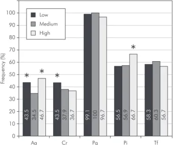
The microbial analysis by PCR indicated that 1 child (0.5%) was negative for all periodontal bac-teria studied; 41 children (20.9%) were positive for one species, 42 children (21.2%) were positive for two species, 36 children (18.7%) were positive for three species, 29 children (14.8%) were positive for four species and 47 children (24.0%) were positive for all bacteria studied. The most prevalent peri-odontopathogen was P. gingivalis(99.0%), followed by T. forsythia (58.7%), P. intermedia (58.2%), A. actinomycetemcomitans (41.3%) and C. rectus (40.8%). Finally, the overall prevalence of S. mutans was 71.9%.
The analysis of the prevalence of each bacterium among the different groups indicated that no statis-tically signiicant difference (p> 0.05) was observed in the detection frequencies of S. mutans (71.3%, 77.6% and 63.3% for Low, Medium and High bleed-ing, respectively), P. gingivalis (99.1%, 100% and 96.7% for Low, Medium and High bleeding, respec-tively) and T. forsythia (58.3%, 60.3% and 56.7%
Low Bleeding (≤ 30%) Medium Bleeding (31 – 59%) High Bleeding (≥ 60%) N 108 children* 58 children 30 children Gender 56 male / 52 female 24 male / 34 female 12 male / 18 female
Age 8.7 ± 1.4 years 8.6 ± 1.2 years 8.5 ± 1.5 years
* p < 0.05 by ANOVA, Student’s t post hoc test. Table 1 - Demographic
for Low, Medium and High bleeding, respectively). We also showed that a higher prevalence of P. intermedia was associated with gingival inlamma-tion extension, since its detecinlamma-tion frequency in the High Bleeding group (66.7%) was higher than in the Low (56.5%) and Medium (57.6%) bleeding groups. C. rectus was associated with periodontal health, because its detection frequency in the Low bleed-ing group (43.5%) was higher than in the Medium (37.9%) and High bleeding groups (36.7%). No sta-tistical difference was observed between Low and Medium bleeding (p > 0.05).
The detection frequencies for all bacteria studied are shown in Graph 1. A. actinomycetemcomitans was more prevalent in the Low (43.5%) and High bleeding (46.7%) groups when compared to the Me-dium bleeding group (34.5%).
The bacterial frequencies in the low, medium and high bleeding groups were also evaluated accord-ing to the gender and age of the participants. There were no signiicant differences (p < 0.05) regarding the bacterial frequencies among these groups.
In order to determine the association between S. mutansand the periodontal bacteria studied (A. actinomycetemcomitans, C. rectus, P. gingivalis, P. intermedia and T. forsythia), Pearson’s correla-tion coeficient (r) was calculated, and the results are summarized in Table 2. S. mutans did not show signiicant association with any periodontal bacte-ria. Among the periodontal pathogens evaluated, P. gingivalis showed the greater correlation coeficient with the presence of S. mutans, although with an
extremely reduced value (r = 0.1626), which makes this inding irrelevant.
A high percentage of children showed positive samples for S. mutans (71.9%) and the population presented a mean dmft/DMFT value of 6.68. When the dmft/DMTF index was determined for each studied group (6.96, 6.30 and 6.78, respectively for low, medium and high bleeding), no signiicant difference (p < 0.05) was found. In addition, when the mean frequency of S. mutans was determined for each studied group (0.71, 0.77 and 0.63,
re-Table 2 - Pearson’s correlation coefficient (r) and p value for S. mutans and the other oral bacteria analyzed (Aa – A. actinomy-cetemcomitans, Cr – C. rectus, Pg – P. gingivalis, Pi – P. intermedia and Tf – T. forsythia).
Aa Cr Pg Pi Tf
S. mutans r = 0.0168 r = 0.0104 r = 0.1626 r = 0.0918 r = – 0.0629
p = 0.8151 p = 0.8854 p = 0.0234 p = 0.2013 p = 0.3814
Table 3 - DMFT and medium frequency of S. mutans according to the studied group: Low, Medium and High bleeding.
Low Bleeding (Mean ± sd) Medium Bleeding (Mean ± sd) High Bleeding (Mean ± sd) DMFT 6.96 ± 3.78 6.30 ± 3.89 6.78 ± 4.27
S. mutans 0.71 ± 0.44 0.77 ± 0.41 0.63 ± 0.39
sd: Standard deviation.
0 40 50 60 70 80 90
30
20
10
100 Low
Medium
High
*
*
* *
Aa Cr Pg Pi Tf
4
3
.5
3
4
.5
4
6
.7
4
3
.5
3
7
.9
3
6
.7
5
6
.5
5
6
.9
6
6
.7
5
8
.3
6
0
.3
5
6
.7
9
9
.1
1
0
0
9
6
.7
Frequ
ency
(%)
Graph 1 - Detection frequencies (%) of each bacterium (Aa – A. actinomycetemcomitans, Cr – C. rectus, Pg – P. gingi-valis, Pi – P. intermedia and Tf – T. forsythia) observed in the different groups (Low, Medium and High bleeding). Aster-isks indicate statistically significant differences between the
spectively for low, medium and high bleeding), no signiicant difference (p < 0.05) was found either. Therefore, no statistical signiicant association was observed between the presence of S. mutans and the dmft/DMTF index of each studied group (Table 3).
Discussion
Periodontal diseases are due to the association between a plaque bioilm and the host responses. A speciic group of bacteria, predominantly composed by Gram-negative, anaerobic microorganisms, is implicated in the initiation and progression of peri-odontal inlammation. The bacterial species P. gingi-valis, A. actinomycetemcomitans, P. intermedia, T. forsythia have been identiied as good predictors of future clinical attachment loss in susceptible hosts.14
In the present study, we showed that marginal bioilm samples from children between 6 and 12 years of age exhibited a high prevalence of oral pathogens. Interestingly, despite the high levels of supragingival bioilm and the high prevalence of periodontopathogens, the majority of the children analyzed (55.1%) presented a low bleeding score (< 30%) (Table 1). On the other hand, the high prev-alence of S. mutans was compatible with the mean dmft/DMTF score (6.68). Considering the ages of the recruited children, between 6 and 12 years, the found DMFT/dmft value (6.68) can be considered high according to the mean DMFT, lower than 1, proposed by the World Health Organization for the age of 12 in the year 2010.10
P. gingivalis was the pathogen most frequently found (99%), followed by T. forsythia(58.7%), and P. intermedia (58.2%). A. actinomycetemcomitans (41.3%) and C. rectus (40.8%) showed a moderate prevalence. Similar observations were made by Mc-Clellan et al.15 (1996) who used the same PCR
anal-ysis used in the present study, showing that P. gingi-valis was detected in 40 to 50% of children ranging from 0 to 2 years old, and in 60% of teenagers aged 13 to 14 years.
No association between gingival inlammation and the presence of T. forsythia and P. gingivalis was found in the present study.Similar results were demonstrated by Kisby et al.16 (1989) who showed
that the prevalences of A. actinomycetemcomitans,
P. gingivalis, and P. intermedia were approximately the same in healthyand gingivitis children between 8 and 10 years of age.
In accordance with the study of Gafan et al.17
(2004), our study also failed to demonstrate any signiicant difference between healthy and gingivitis children between 5 and 9 years old with respect to the presence of P. gingivalis or A. actinomycetem-comitans. Surprisingly, these authors showed that T. forsythia was observed more frequently (2.3 times greater) in children without gingivitis compared to individuals with gingivitis. In our study, the pres-ence of A. actinomycetemcomitans was not directly associated with health or disease status. Interest-ingly, the prevalence of A. actinomycetemcomitans was higher in the Low (43.5%) and High (46.7%) bleeding groups than in the Medium bleeding group (34.5%). Considering all the discrepancies found among the studies in the literature, further future investigations are still required in order to better un-derstand the role played by each one of these cited species in periodontal disease in children.
In addition, our study evaluated the association between the presence of S. mutans and the identi-ication of periodontopathogens in the marginal bioilm of children. Interestingly, S. mutans did not show signiicant association with any of the peri-odontal pathogens analyzed. Previous research has suggested that patients with periodontitis, particu-larly localized aggressive periodontitis, have mini-mal tooth decay.18,19 Accordingly, a recent study by
Fine et al.20 (2007) has showed that A. actinomy-cetemcomitans-positive subjects, who predominant-ly had localized aggressive periodontitis (LAP), have a salivary factor that signiicantly reduces the sur-vival of S. mutans.This inding suggested an expla-nation for the fact that the LAP group typically has minimal proximal tooth decay. The lack of any kind of association between the prevalence of S. mutans and the periodontopathogens in our study may be due to the high prevalence of plaque (plaque index greater than 80%) manifested by the population of children studied.
Our study is in accordance with a previous study by Tanner et al.21 (1989) who demonstrated that
establish-es in early years of life, therefore pointing out the need for more effective preventive dental programs.
Conclusion
It may be concluded that Brazilian children, ranging from 6 to 12 years of age, with high levels of supragingival bioilm, presented a high prevalence of periodontopathogens and S. mutans. It may also be concluded that in spite of the high levels of supra-gingival bioilm and high prevalence of
periodon-topathogens found in Brazilian children, ranging 6 to 12 years of age, the majority of them presented only mild inlammation revealed by a low bleeding score. On the other hand, high levels of S. mutans were compatible with poor dental status, i.e., high mean dmft/DMTF scores. Our study indicated that whereas P. intermedia was more associated with in-lamed tissues, C. rectus was more associated with periodontal health.
References
1. Darveau RP, Tanner A, Page R. The microbial challenge in periodontitis. Periodontology 2000. 1997;14(3):12-32. 2. Baelum V, Fejerskov O, Karring T. Oral hygiene, gingivitis
and periodontal breakdown in adult Tanzanians. J Periodontal Res. 1986;21(3):221-32.
3. Baelum V, Fejerskov O, Manji F. Periodontal diseases in adult Kenyans. J Clin Periodontol. 1988;15(7):445-52.
4. Löe H, Anerud A, Boysen H. The natural history of periodon-tal disease in man: prevalence, severity, and extent of gingival recession. J Periodontol. 1992;63(6):489-95.
5. Lovegrove JM. Dental plaque revisited: bacteria associated with periodontal disease. J N Z Soc Periodontol. 2004;(87):7-21.
6. Loesche WJ. Role of Streptococcus mutans in human dental decay. Microbiol Rev. 1986;50:353-80.
7. Thenisch NL, Bachmann LM, Imfeld T, Leisebach Minder T, Steurer J. Are mutans streptococci detected in preschool children a reliable predictive factor for dental caries risk? A systematic review. Caries Res. 2006;40(5):366-74.
8. Butler BL, Morijon O, Low SB. An accurate, time-efficient method to assess plaque accumulation. J Am Dent Assoc. 1996;127(12):1763-6.
9. Ainamo J, Bay I. Problems and proposals for recording gin-givitis and plaque. Int Dent J. 1975;25(4):229-35.
10. World Health Organization. Oral Health Surveys: Basic Meth-ods. 4th ed. Geneva; Swiss; 1997.
11. Cortelli JR, Aquino D, Cortelli SC, Fernandes CB, Carva-lho-Filho J, Franco GCN et al. Etiological analysis of initial colonization of periodontal pathogens in oral cavity. J Clin Microbiol. 2008;46(4):1322-9.
12. Igarashi T, Yamamoto A, Goto N. Direct detection of Strep-tococcus mutans in human dental plaque by polymerase chain reaction. Oral Microbiol Immunol. 1996;11(5):294-8. 13. Querido SMR, Cortelli SC, Araujo MWB, Cortelli JR.
Clini-cal and microbial evaluation of dental sClini-caling associated with subgingival minocycline in chronic periodontitis subjects. Braz Oral Res. 2004;18(2):110-5.
14. Socransky SS, Haffajee AD, Smith C, Dibart S. Relation of counts of microbial species to clinical status at the sampled sites. J Clin Periodontol. 1991;18(10):766-75.
15. McClellan DL, Griffen AL, Leys EJ. Age and prevalence of Porphyromonas gingivalis in children. J Clin Microbiol. 1996;34(8):2017-9.
16. Kisby LE, Savitt ED, French CK, Peros WJ. DNA probe de-tection of key periodontal pathogens in juveniles. J Pedod. 1989;13(3):222-9.
17. Gafan GP, Lucas VS, Roberts GJ, Petrie A, Wilson M, Spratt DA. Prevalence of periodontal pathogens in dental plaque of children. J Clin Microbiol. 2004;42(9):4141-6.
18. Albandar JM, Brown LJ, Löe H. Dental caries and tooth loss in adolescents with early-onset periodontitis. J Periodontol. 1996;67(10):960-7.
19. Sioson PB, Furgang D, Steinberg LM, Fine DH. Proxi-mal caries in juvenile periodontitis patients. J Periodontol. 2000;71(5):710-6.
20. Fine DH, Furgang D, Goldman D. Saliva from subjects harbor-ing Actinobacillus actinomycetemcomitans kills Streptococ-cus mutans in vitro. J Periodontol. 2007;78(3):518-26. 21. Tanner A, Bouldin HD, Maiden MF. Newly delineated