Recebido para publicação: Fevereiro de 2010 • Aceite para publicação: Setembro de 2010
1839
Implantação percutânea de válvulas
pulmonares: experiência inicial
[132]
JOSÉDIOGOFERREIRAMARTINS, PETEREWERT, LÍDIA DESOUSA, ISABELFREITAS, CONCEIÇÃOTRIGO, NUNOJALLES, PEDROMATOS, ANAAGAPITO, RUIFERREIRA, FÁTIMAFERREIRAPINTO
Serviços de Cardiologia e Cardiologia Pediátrica, Hospital de Santa Marta, EPE, Lisboa, Portugal
Rev Port Cardiol 2010; 29 (12): 1839-1846
RESUMO
Os doentes com cardiopatias submetidos a intervenção cirúrgica com implantação de condutos entre o ventrículo direito e a artéria pulmonar (VD-AP) podem desenvolver estenose e/ou insuficiência pulmonares ao longo do tempo. Nestas situações, associadas a arritmias cardíacas, disfunção ventricular direita e morte súbita, a re-intervenção cirúrgica permanece um desafio pela complexidade, mortalidade e morbilidade significativas. A implantação percutânea de válvulas pulmonares (IPVP), recentemente desenvolvida, representa uma abordagem alternativa para estes doentes. Objectivo: Reportar a experiência inicial com a IPVP, analisando o seu impacto no manejo destes doentes.
Material e Métodos: Avaliação prospectiva
dos dados clínicos, ecocardiográficos, da ressonância magnética, hemodinâmicos e angiográficos dos doentes submetidos a IPVP.
Resultados: Seis doentes em classe
funcional igual ou superior a II com disfunção do conduto VD-AP foram submetidos a IPVP. Tinham todos evidência de insuficiência e 5/6 de estenose. O procedimento consistiu na implantação de stents não cobertos no conduto para reforçar a sua rigidez e prevenir fracturas dos stents
Percutaneous pulmonary valve implantation: Initial experience
ABSTRACT
Introduction: Patients with congenital heart
disease who undergo surgical implantation of a conduit between the right ventricle and the pulmonary artery (RV-PA conduit) may develop stenosis and/or insufficiency over time. These cases, which are associated with arrhythmias, RV dysfunction and sudden death, remain a challenge for surgical re-intervention, due to its complexity and associated morbidity and mortality. Percutaneous pulmonary valve implantation (PPVI) is therefore a valid alternative.
Objective: To report our center’s initial
experience with PPVI.
Methods: Prospective assessment of clinical,
echocardiographic, magnetic resonance, hemodynamic and angiographic data from our series of PPVI.
Results: Six patients in NYHA functional
class ≥II underwent PPVI. All had
significant conduit dysfunction and five had stenosis. The procedure consisted of implanting a bare metal stent to reduce the risk of fracture of the Melody®valved stents
(Medtronic) that were then successfully deployed in all. The immediate
INTRODUÇÃO
A
reconstrução do tracto de saída do ven-trículo direito (TSVD) com implantação de conduto entre o ventrículo direito e o tron-co da artéria pulmonar (tron-conduto VD-AP) é uma parte integral de algumas intervenções cirúr-gicas em cardiopatias congénitas.Contudo, a longevidade destas próteses é limitada pela sua ausência de crescimento somático, dilatação aneurismática e degene-ração ou calcificação progressivas, que condi-cionam assim o aparecimento de insuficiência
INTRODUCTION
R
econstruction of the right ventricular out-flow tract (RVOT) with implantation of a conduit between the right ventricle and the pulmonary artery (RV-PA conduit) is an inte-gral part of certain surgical interventions for congenital heart disease.However, the life of these prostheses is limited by lack of somatic growth, aneurysmal dilation or progressive degeneration or calcifi-cation, which result in insufficiency and/or stenosis(1). RV-PA conduit dysfunction is
asso-1840
Recebido para publicação: ????????????? • Aceite para publicação: ?????????
valvulados pulmonares Melody®(Medtronic),
que foram em seguida implantados com sucesso em todos. Obteve-se uma redução da pressão ventricular direita (94±27 para 44±7mmHg), da relação entre a pressão ventricular direita e esquerda (94±27 para 44±7%), do gradiente do conduto (65±28 para 11±4mmHg) e ausência de regurgitação pulmonar. O procedimento teve uma duração mediana de 180 minutos e decorreu sem complicações major. Os doentes tiveram alta em mediana dois dias após o procedimento. Na última avaliação, em mediana 7,8 meses após o procedimento, os doentes encontram-se em clasencontram-se funcional I (5) ou II (1), encontram-sem evidência não invasiva de disfunção do conduto.
Conclusões: Os nossos resultados
acompanham a experiência actual com esta modalidade terapêutica, com excelentes resultados nestes grupo de doentes complexos. A implementação da IPVP em doentes com cardiopatia congénita que requerem re-intervenção no tracto de saída do ventrículo direito tem-se revelado uma técnica promissora, embora complexa, alternativa a mais uma intervenção cirúrgica.
Palavras-Chave:
Cardiopatias congénitas; Cardiologia de intervenção;
Stent; Insuficiência pulmonar; Estenose pulmonar;
Congenital heart disease; Interventional cardiology;
Stent; Pulmonary insufficiency; Pulmonary stenosis
hemodynamic results showed a reduction in RV pressure (94±27 to 44±7 mmHg), RV/LV pressure ratio (94±27 to 44±7%) and conduit gradient (65±28 to 11±4 mmHg), and no insufficiency. The median duration of the procedure was 180 minutes, with no major complications. Patients were discharged a median of two days after the procedure. After a median follow-up of 7.8 months, patients are in functional class I (5) or II (1), with no evidence of conduit dysfunction on non-invasive assessment.
Conclusions: Our results are similar to the
excellent results reported in larger series. PPVI is a valid therapeutic option in patients with conduit dysfunction.
Key words
Congenital heart disease; Interventional cardiology; Stent; Pulmonary insufficiency; Pulmonary stenosis
e/ou estenose(1). A disfunção dos condutos VD-AP está associada a efeitos hemodinâmicos indesejáveis a longo prazo como a dilatação e disfunção ventricular direitas, insuficiência tricúspide, arritmias e morte(2,3).
A re-intervenção cirúrgica nestes doentes permanece um desafio com riscos não des-prezíveis. Em 2000 foi efectuada a primeira implantação percutânea de válvula pulmonar (IPVP)(4). Desde então, foram realizados cerca de mil procedimentos em todo o Mundo(5, 6). Este trabalho reporta a experiência nesta técnica do nosso centro, pioneiro em Portugal.
MATERIAL E MÉTODOS Doentes
Foram considerados candidatos a IPVP os doentes dos Serviços de Cardiologia Pediá-trica e Cardiologia do Hospital de Santa Marta submetidos a reconstrução cirúrgica do TSVD com disfunção significativa do conduto VD-AP sob a forma de estenose e/ou insuficiência.
A estenose foi considerada significativa quando condicionava uma pressão ventricular direita superior a 2/3 da sistémica com sinto-mas ou 3/4 da sistémica sem sintosinto-mas. Consi-derou-se insuficiência significativa a pre-sença de regurgitação pulmonar pelo menos moderada associada a dilatação ou disfunção do ventrículo direito.
Foram estabelecidos como critérios de ex-clusão peso inferior a 30kg, anatomia coro-nária susceptível de compromisso pela IPVP, diâmetro do conduto superior a 22mm, obstru-ção das veias centrais, gravidez e alergia a produtos de contraste ou aspirina.
Todos os doentes ou seus representantes legais assinaram um consentimento informa-do.
Protocolo
Os doentes foram classificados de acordo com a classe funcional da NYHA. Quando possível, a capacidade funcional foi objecti-vada com prova de esforço com consumo de oxigénio.
Foram efectuados ecocardiogramas
trans-ciated with undesirable hemodynamic effects in the long term such as right ventricular (RV) dilatation and dysfunction, tricuspid regurgi-tation, arrhythmias and death(2, 3).
Surgical re-intervention in these patients remains a challenge with significant risks. The first percutaneous pulmonary valve implantation (PPVI) was performed in 2000(4), and since then, around a thousand such pro-cedures have been performed worldwide(5, 6). The aim of this article is to report our center’s experience with this technique, the first in Portugal.
METHODS Patients
Patients of the Pediatric and Adult Cardiology Departments of Hospital de Santa Marta who had undergone surgical RVOT reconstruction and who had significant RV-AP conduit dysfunction in the form of stenosis and/or insufficiency were considered candi-dates for PPVI.
Stenosis was considered significant when RV pressure was more than two-thirds of sys-temic pressure when accompanied by toms, or more than three-quarters without symp-toms. Significant insufficiency was defined as at least moderate pulmonary regurgitation associ-ated with RV dilatation or dysfunction.
Exclusion criteria were: weight under 30 kg, coronary anatomy likely to be compro-mised by PPVI, conduit diameter of over 22 mm, central vein obstruction, pregnancy and allergy to contrast agents or aspirin.
All patients or their legal representatives gave informed consent.
Protocol
Patients were classified according to New York Heart Association (NYHA) functional class. Whenever possible, functional capacity was objectively assessed by exercise testing with measurement of maximum oxygen uptake. Transthoracic echocardiography was per-formed prior to the procedure, and at one day, one week, one months and six months after the 1841 Recebido para publicação: ????????????? • Aceite para publicação: ?????????
torácicos prévios e um dia, uma semana, um mês e seis meses após a intervenção. A ava-liação completa incluiu medições cardíacas e do conduto em 2D, quantificação do gradiente no TSVD, estimativa da pressão ventricular direita (por jacto de insuficiência tricúspide), insuficiência pulmonar e função ventricular direita.
Os doentes foram submetidos a ressonân-cia magnética cardíaca para quantificação da regurgitação pulmonar, dimensões e função ventricular direita bem como estudo da anatomia cardíaca e dos grandes vasos.
O cateterismo foi efectuado sob anestesia geral e heparinização (para manter ACT > 250). Após obtenção de acesso vascular, foi efectuado um cateterismo direito e esquerdo completo. O estudado angiográfico detalhou a anatomia do conduto, ramos da artéria pulmo-nar e sua relação com artérias coronárias, incluindo sempre que necessário a realização de coronáriografia selectiva simultânea com insuflação de balão ou angiografia no TSVD. A válvula pulmonar Melody®(Medtronic) con-siste num conduto Contegra (veia jugular bovina valvulada) de 18mm suturado num
stent de platina-iridium (NuMed), implantado
através do sistema de entrega específico
Ensemble® (Medtronic). A técnica utilizada para a IPVP decorreu de acordo com o des-crito anteriormente(5).
RESULTADOS Doentes
Entre Novembro de 2008 e Junho de 2009, foram tentadas IPVP em sete doentes (seis do sexo masculino), com idades compreendidas entre os 9 e 32 anos e pesos entre 29 e 60 kgs. Os doentes tinham uma média de 2.8 intervenções cirúrgicas prévias e co-morbili-dades significativas. A maioria apresentava disfunção mista (estenose e insuficiência do conduto), e encontrava-se em classe funcional ≥II NYHA. (Tabela 1)
Cateterismo
As medianas de duração do procedimento
intervention. Complete assessment included measurement of cardiac chambers and the conduit in 2D mode, quantification of the RVOT gradient, estimation of RV pressure (based on the tricuspid regurgitant jet) and pulmonary insufficiency, and RV function.
Cardiac magnetic resonance imaging (MRI) was used to quantify pulmonary regurgitation and RV dimensions and function, as well as to assess cardiac and great vessel anatomy.
Catheterization was performed under gen-eral anesthesia and heparinization (to main-tain ACT of >250 seconds). After obmain-taining vascular access, complete right and left catheterization was carried out. Angiographic study revealed conduit anatomy, pulmonary artery branches and their relation to coronary arteries; simultaneous selective coronary angiography with balloon inflation and angiography of the RVOT were performed whenever necessary. The Melody® pulmonary valve (Medtronic) consists of an 18 mm Contegra conduit (bovine jugular vein valve) sutured inside a platinum-iridium stent (NuMed), which is implanted via a special Ensemble® (Medtronic) delivery system. The technique used for PPVI was as previously described(5).
RESULTS Patients
Between November 2008 and June 2009, PPVI was attempted in seven patients (six male), aged between 9 and 32 years and weighing between 29 and 60 kg.
The patients had a mean of 2.8 previous surgical interventions and significant comor-bidities. Most presented stenosis and conduit dysfunction and all were in NYHA functional class ≥II (Table I).
Catheterization
Median procedure duration and hospital stay were 180 (97-222) minutes and two(1-4) days, respectively. Melody®valves were implanted in six patients (five male). The procedure was postponed in one patient with severe RVOT
1842
e internamento foram de 180 (97-222) minutos e dois (1-4) dias, respectivamente. Foram implantadas válvulas Melody® em seis doentes (cinco do sexo masculino). Num doente, com TSVD severamente calcificado, trajecto de coronária justa-conduto e peso no limite inferior aceitável, o procedimento foi adiado. Em todos os doentes, foi efectuado pré-stenting do conduto (total 13 stents).
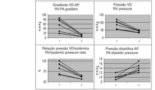
Quanto aos resultados hemodinâmicos imediatos após IPVP, verificou-se diminuição da pressão ventricular direita (94±27 para 44±7mmHg), do gradiente através do TSVD (65±28 para 11±4mmHg) e da relação VD/Ao (94±27 para 44±7%). A pressão diastólica na artéria pulmonar aumentou (10±1 para 14±2mmHg) (Figura 1).
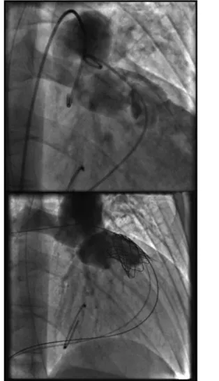
Os resultados angiográficos mostraram resolução da estenose e/ou insuficiência em todos os doentes (Figura 2).
Os cateterismos foram complicados por ruptura e extracção percutânea de balão(1), lesão transitória do plexo braquial(1) e lesão vascular do membro inferior em doente com varizes sem necessidade de cirurgia(1). Seguimento
Na avaliação ecocardiográfia mais recente, verificou-se em todos os doentes submetidos a IPVP uma diminuição das dimensões e
manu-calcification, coronary artery coursing adja-cent to the conduit and weight at the lower limit. Pre-stenting of the conduit (total 13 stents) was performed in all patients.
The immediate hemodynamic results fol-lowing PPVI showed a reduction in RV pres-sure (94±27 to 44±7 mmHg), RVOT gradient (65±28 to 11±4 mmHg) and RV/Ao ratio (94±27 to 44±7%), together with an increase in pulmonary artery diastolic pressure (10±1 to 14±2 mmHg) (Figure 1).
Catheterization was complicated by bal-loon rupture and percutaneous extraction(1), transient brachial plexus injury(1) and lower limb vascular lesion in a patient with varicose veins without need for surgery(1).
Follow-up
The most recent echocardiographic assess-ment showed a reduction in RV dimensions and maintenance of the RV pressure estimat-ed by the tricuspid regurgitant jet at the time of discharge, with progressive normalization of interventricular septal motion. Pulmonary regurgitation is mild or absent in all patients. After a median follow-up of 7.8 months (2.8-10.1), patients are currently in NYHA functional class I(5)or II(1).
Cardiac MRI and exercise testing will be performed in all patients one year after
PPVI. 1843
Recebido para publicação: ????????????? • Aceite para publicação: ?????????
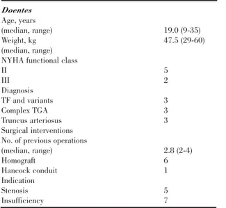
Doentes Idade, anos (mediana, limites) 19.0 (9-35) Peso, kg (mediana, limites) 47.5 (29-60) Classe funcional NYHA II 5 NYHA III 2 Diagnóstico TF e variantes 3 TGA complexa 3 Truncus arteriosus 3 Cirurgias Nº cirurgias prévias (mediana, limites) 2.8 (2-4) Homoenxerto 6 Conduto Hancock 1 Indicação Estenose 5 Insuficiência 7
Tabela 1. Dados dos doentes
Doentes
Age, years
(median, range) 19.0 (9-35)
Weight, kg 47.5 (29-60)
(median, range) NYHA functional class
II 5 III 2 Diagnosis TF and variants 3 Complex TGA 3 Truncus arteriosus 3 Surgical interventions No. of previous operations
(median, range) 2.8 (2-4) Homograft 6 Hancock conduit 1 Indication Stenosis 5 Insufficiency 7
Table 1. Patient characteristics
NYHA: New York Heart Association; TF: tetralogy of Fallot; TGA: transposition of the great arteries
NYHA: New York Heart Association; TF: tetralogia de Fallot; TGA: transposição das grandes artérias
tenção de pressão estimada no VD por insu-ficiência tricúspide à data da alta, com norma-lização progressiva do movimento do septo interventricular. A insuficiência pulmonar é em todos ligeira.
Com um tempo de seguimento mediano de 7.8 meses (2.8-10.1), estes doentes encontram-se actualmente em clasencontram-se funcional NYHA I(5) ou II(1).
Um ano após a IPVP, será efectuada RM cardíaca e prova de esforço a todos os doentes.
DISCUSSÃO
Os condutos VD-AP utilizados em recons-truções cirúrgicas do TSVD desenvolvem com frequência insuficiência e/ou estenose pro-gressivas, o que condiciona uma morbilidade e mortalidade significativas nestes doentes(3,7). A re-intervenção cirúrgica pode ser reali-zada com sucesso mas permanece um desafio pela presença de múltiplas operações prévias e não assegura a ausência de re-intervenções futuras(8,9).
As estenoses vasculares têm sido aborda-das com sucesso por cardiologia de interven-ção, recorrendo a angioplastia com balão com ou sem colocação de stent. A IPVP,
introdu-DISCUSSION
The RV-PA conduits used in surgical RVOT reconstruction frequently develop pro-gressive insufficiency and/or stenosis, result-ing in significant morbidity and mortality among these patients(3, 7).
Surgical re-intervention can be successful but it remains a challenge due to multiple pre-vious operations and does not guarantee that further intervention will not be necessary(8, 9).
Vascular stenosis is now successfully treated by interventional cardiology, using balloon angio-plasty with or without stenting. The introduction of PPVI ten years ago has brought considerable benefits to patients with RV-PA conduit dysfunc-tion, since it simultaneously resolves both insuf-ficiency and stenosis, which often coexist in these patients, as was found in our sample.
After a decade of experience and around a thousand such valve implantations in Europe and the USA, PPVI has immediate mortality of less than 1%, severe periprocedural mor-bidity of 4%, and medium-term survival of 96.6%(6, 10). In the largest series published (n=155), which included the first patients to undergo PPVI(10), 70% required no new inter-vention at 70 months. Most re-interinter-ventions in this series were due to valved stent fracture,
1844
Figura 1. Resultados hemodinâmicos imediatos da IPVP. VD, ventrículo direito; AP, artéria pulmonar. Figure 1. Immediate hemodynamic results following PPVI. PA: pulmonary artery; RV: right ventricle
Gradiente VD-AP RV-PA gradient
Pressão VD RV pressure
Relação pressão VD/sistémica RV/systemic pressure ratio
Pressão diastólica AP PA diastolic pressure
zida há uma década, trouxe um benefício apreciável para os doentes com condutos VD-AP disfuncionantes por resolver em simul-tâneo a insuficiência e estenose que com fre-quência co-existem, como é patente na nossa amostra.
Após uma década de experiência e cerca de um milhar de válvulas implantadas na Europa e Estados Unidos, a IPVP apresenta uma mortalidade imediata inferior a 1%, morbilidade grave peri-procedimento de 4% e sobrevida a médio prazo de 96.6%(6, 10). Na maior série publicada (n=155), que inclui os primeiros doentes a quem foi efectuada IPVP, 10 a ausência de intervenção a 70 meses é de 70%. Neste grupo, a maioria das re-interven-ções foi efectuada devido a fracturas do stent valvulado, motivo pelo qual a técnica passou a incluir a implantação prévia de outro stent no conduto (pré-stenting). Esta técnica foi tam-bém seguida pelos autores. Em 12% dos doentes foi implantada mais tarde uma segun-da válvula pulmonar (stent-in-stent). E final-mente, obteve-se uma preservação sustentada da competência valvular pulmonar.
Do ponto de vista clínico, a IPVP está as-sociada a uma melhoria da capacidade funcio-nal. Nos doentes com predomínio de insu-ficiência pulmonar, o seguimento por resso-nância magnética mostrou uma persistência da dilatação e disfunção ventricular direitas, sugerindo a irreversibilidade destas lesões e contrariando o conceito da benignidade tradi-cionalmente associado a esta fisiopatologia, seis e questionando a necessidade de resolução da insuficiência pulmonar em fases mais preco-ces de degradação ventricular direita.
CONCLUSÕES
A IPVP é segura e eficaz a restabelecer a competência e aliviar a estenose, prolongando assim a vida dos condutos VD-AP. A nossa série apresenta resultados precoces que acompanham as publicações internacionais.
Quando aplicável, a IPVP tornou-se a primei-ra escolha nos doentes com condutos VD-AP disfuncionantes.
which prompted a change in the technique to include prior implantation of another stent in the conduit (pre-stenting); this practice was fol-lowed in our series. A second pulmonary valve (stent-in-stent) was subsequently implanted in 12% of the patients. Sustained preservation of pulmonary valve competence was achieved.
From a clinical standpoint, PPVI is associ-ated with improved functional capacity. In patients with predominantly pulmonary insuf-ficiency, follow-up assessment by MRI showed persistence of RV dilatation and dys-function, suggesting that such damage is irre-versible, which is at variance with the tradi-tional view of the benign nature of this pathol-ogy(6) and indicates a need to resolve pul- 1845 Figura 2. Angiografia do tracto de saída do ventrículo direito durante a diástole, antes(A) e depois (B) da IPVP, notando-se a resolução da insuficiência pulmonar
Figura 2. Angiography of the right ventricular outflow tract during diastole, before (A) and after (B) PPVI, showing resolution of pul-monary insufficiency.
Embora o timing de intervenção se man-tenha controverso, o sucesso desta técnica po-de contribuir para promover a intervenção mais precoce, antes da instalação de alterações ir-reversíveis nas cavidades direitas.
Pedido de Separatas Address for Reprints: José Diogo Ferreira Martins Serviço de Cardiologia Pediátrica Hospital de Santa Marta, EPE Rua de Santa Marta, 50 1169-024 Lisboa
jdferreiramartins@gmail.com
monary insufficiency in the early stages of RV degeneration.
CONCLUSIONS
Percutaneous pulmonary valve implanta-tion is safe and effective in re-establishing valve competence and reducing stenosis, thus prolonging the life of RV-PA conduits. Our early results are similar to those reported in international series.
PPVI has become the first-line treatment for eligible patients with RV-PA conduit dys-function.
Although timing remains the subject of debate, the success of the technique may help to encourage earlier intervention to prevent irreversible changes in the right chambers.
1846
Recebido para publicação: ????????????? • Aceite para publicação: ?????????
1.Nordmeyer J, Coats L, Bonhoeffer P. Current experience with percutaneous pulmonary valve implantation. Semin Thorac Cardiovasc Surg 2006;18(2):122-5.
2.Khambadkone S, Bonhoeffer P. Nonsurgical pulmonary valve replacement: why, when, and how? Catheter Cardiovasc Interv 2004;62(3):401-8.
3.Gatzoulis MA, Balaji S, Webber SA, et al. Risk factors for arrhythmia and sudden cardiac death late after repair of tetralogy of Fallot: a multicentre study. Lancet 2000;356(9234):975-81. 4.Bonhoeffer P, Boudjemline Y, Saliba Z, et al. Percutaneous replacement of pulmonary valve in a right-ventricle to pulmonary-artery prosthetic conduit with valve dysfunction. Lancet 2000;356(9239):1403-5.
5.Khambadkone S, Coats L, Taylor A, et al. Percutaneous pul-monary valve implantation in humans: results in 59 consecutive patients. Circulation 2005;112(8):1189-97.
BIBLIOGRAFIA / REFERENCES
6.Lurz P, Bonhoeffer P, Taylor AM. Percutaneous pulmonary valve implantation: an update. Expert Rev Cardiovasc Ther 2009;7(7):823-33.
7.Wessel HU, Paul MH. Exercise studies in tetralogy of Fallot: a review. Pediatr Cardiol 1999;20(1):39-47; discussion 8.
8.Bielefeld MR, Bishop DA, Campbell DN, Mitchell MB, Grover FL, Clarke DR. Reoperative homograft right ventricular outflow tract reconstruction. Ann Thorac Surg 2001;71(2):482-7; discussion 7-8. 9.Warner KG, Anderson JE, Fulton DR, Payne DD, Geggel RL, Marx GR. Restoration of the pulmonary valve reduces right ven-tricular volume overload after previous repair of tetralogy of Fallot. Circulation 1993;88(5 Pt 2):II189-97.
10.Lurz P, Coats L, Khambadkone S, et al. Percutaneous pulmonary valve implantation: impact of evolving technology and learning curve on clinical outcome. Circulation 2008;117(15):1964-72.