Autopsy and Case Reports. ISSN 2236-1960. Copyright © 2020. This is an Open Access article distributed under the terms of the Creative Commons Attribution Non-Commercial License, which permits unrestricted a Centro Hospitalar Universitário do Porto, Serviço de Cirurgia Geral. Porto, Portugal.
b Centro Hospitalar e Universitário do Porto, Serviço de Anatomia Patológica. Porto, Portugal.
c Universidade do Minho, Escola de Medicina, Instituto de Investigação em Ciências da Vida e Saúde (ICVS). Braga, Portugal. d Laboratório associado ICVS/3B’s. Braga/Guimarães, Portugal.
Abdominal Actinomycosis misdiagnosed as liposarcoma
Eunice Vieira e Monteiro
a
, Joana Gaspar
a
, Claudia Paiva
a
,
Raquel Correia
a
, Vitor Valente
a
, André Coelho
b
, Nuno Jorge Lamas
b,c,d
How to cite: Vieira e Monteiro E, Gaspar J, Paiva C, et al. Abdominal actinomycosis misdiagnosed as liposarcoma. Autops
Case Rep [Internet]. 2020 Jan-Mar.;10(1):e2020137. https://doi.org/10.4322/acr.2020.137
ABSTRACT
Actinomycosis is an uncommon, endogenous, and chronic infection with varied and nonspecific clinical features such as abdominal, pelvic or cervical masses, ulcerative lesions, abscesses, draining fistula, fibrosis, and constitutional symptoms. The disease ensues when the bacteria disrupt the mucosal barrier, invade, and spread throughout interfascial planes. Currently, the diagnosis of actinomycosis is challenging because of its very low frequency and depending on the clinical presentation it may masquerade malignancies. Therapy consists initially in intravenous penicillin, followed by an oral regimen that may be extended until a year of treatment. A timely diagnosis is crucial to avoid extensive therapeutic attempt as surgery. However, a biopsy or drainage of abscesses and fistula’s tract may be required not only as a diagnostic procedure as part of the therapy. We report the case of a 72-year-old woman with an abdominal mass initially misdiagnosed as a liposarcoma. A second biopsy of a skin lesion of the abdominal wall made the diagnosis of actinomycosis, avoiding a major surgical procedure. The patient was treated with a long-term course of antibiotics with favorable outcome. Liposarcoma was ruled out after the patient’s full recovery with antibiotics and the misdiagnosis was credit to the overconfidence on the immunohistochemical positivity to MDM2.
Keywords:
Abdominal actinomycosis; liposarcoma; challenging diagnosis
INTRODUCTION
Actinomyces species, the etiological agent of actinomycosis, were originally classified as fungi1,2 but
are true bacteria.3 Actinomycosis is an uncommon,
chronic granulomatous disease caused by filamentous, gram-positive, anaerobic bacteria.
Actinomyces spp. is a low-pathogenicity member of the commensal flora of mucous membranes in humans (gastrointestinal tract, bronchi, and female genital tract) and are of low pathogenicity. The disease usually occurs in immunocompetent persons but may afflict those with diminished host defenses, though.4-8
The local tissue damage caused by trauma, recent surgery, irradiation, and loss of mucosal integrity by foreign bodies, perforated appendix or diverticulitis, neoplasia is recognized as predisposing factors.4,5,7-10 However, cases were reported without
any preceding mucosal injury.11 Some authors suggest
that inflammatory or malignant processes may contribute to the development of actinomycosis.4,7
Actinomycosis usually spreads locally in an indolent manner and may take months to years before the first symptoms appear.7,9,10
CASE REPORT
A 72-year-old woman was referred to a tertiary hospital for specific treatment after the diagnosis of abdominal liposarcoma. Her medical history included anorexia, asthenia, abdominal pain, general weakness and weight loss of 10 kg over the last 6 months. Her weight at the time of admission was 45 kg. Her past medical history was unremarkable. On physical examination, she presented a palpable mass of 15 cm in its longest axis, in the upper abdomen, mildly tender hard, and immobile. No muscular defense nor rebound pain was present. The hemogram showed leukocytes of 8,490/mm3 (reference range [RR]; 4 – 11000/mm3)
with neutrophilia, microcytic hypochromic anemia with hemoglobin of 9.7 g/dl (RR; 13-17 g/dl), C-Reactive Protein of 12.26 mg/L (RR; 0-5mg/L) and normal serum levels of alpha fetoprotein, carcinoembryonic antigen and carbohydrate antigen 19-9. Her serologies revealed nonimmune status for hepatitis B; hepatitis C, human immunodeficiency virus (HIV) and syphilis were negative; cytomegalovirus (CMV) and Epstein-Barr virus (EBV) showed positive IgG but negative IgM. The esophagogastroduodenoscopy disclosed a hiatal hernia, and the colonoscopy find out an infiltrative-appearing lesion in the hepatic angle and transverse colon, resembling a neoplasia, and diverticula in the left colon.
The contrast-enhanced computed tomography (CT) (Figure 1A) showed a heterogeneously enhanced
solid mass (17,5 x 12 x 6 cm) involving the colonic hepatic angle and the proximal transverse colon with effaced limits, invading the muscles of the abdominal wall. No evidence of regional or distant lymph node disease, and no pulmonary or hepatic lesions.
An ultrasound-guided percutaneous core needle biopsy was performed, which histopathologic examination showed well-differentiated adipocytes, fusiform cells with slight atypia with mononuclear and eosinophilic infiltration, rare mitoses and lack of necrosis. The immunohistochemical (IHC) study showed diffuse expression of Vimentin and S100 positivity in the adipocytes. The fusiform cells and some adipocytes showed nuclear pattern expression of MDM2. This morphologic and immunohistochemical study rendered the diagnosis of well-differentiated liposarcoma.
A month and a half later, she was hospitalized under the care of the multidisciplinary group of soft tissue tumors. At this time, she presented a palpable erythematous periumbilical lesion. At this time, the abdominal magnetic resonance imaging (Figure 1B) identified a similar image depicted at the CT along with the involvement the anterior abdominal wall including the skin.
Re-evaluating the case, and taking into account the emergence of the skin involvement depicted by the MRI, and the erythematous periumbilical skin lesion not present in the referral letter, an incisional
Figure 1. A – Abdominal enhanced CT scan, axial plane, showing soft tissue mass infiltrating the colon, mesenteric fat and abdominal wall (muscular plane); B – MRI, T1 weighed with fat suppression after gadolinium injection, showing tissue enhancement similar to the CT image; however, with the skin involvement.
biopsy of this lesion was undertaken. At this time, the histopathological report revealed abundant granulation tissue with mixed inflammatory infiltration and abscess in the reticular dermis with deep extension. In the core of the lesion fungi-like structures consistent with Actinomyces were found. (Figure 2). Thus, the diagnosis actinomycosis was made.
She started with intravenous 20 million units of benzyl-penicillin per day for 4 weeks. However, a doubt remained on the reliability of the histological diagnosis of liposarcoma. Could the infection by Actinomyces spp explain all the clinical findings or was there an association of liposarcoma and actinomycosis?
The patient was then discharged with amoxicillin 2 g two times a day for 10 months.
She attended periodically to the outpatient consultation and showed weight gain and progressive effacement of the palpable abdominal mass.
By the end of 10 months on oral amoxicillin, she repeated a CT scan (Figure 3) that revealed the absence of any residual tumor lesion. All the inflammatory process was resolved with the normal anatomical restoration of the involved structures, what permitted the antibiotic withdrawal.
After 6 months of follow-up since the antibiotics was withdrawn, the Actinomyces infection did not recur. Thus, liposarcoma was ruled out. We suspect that the
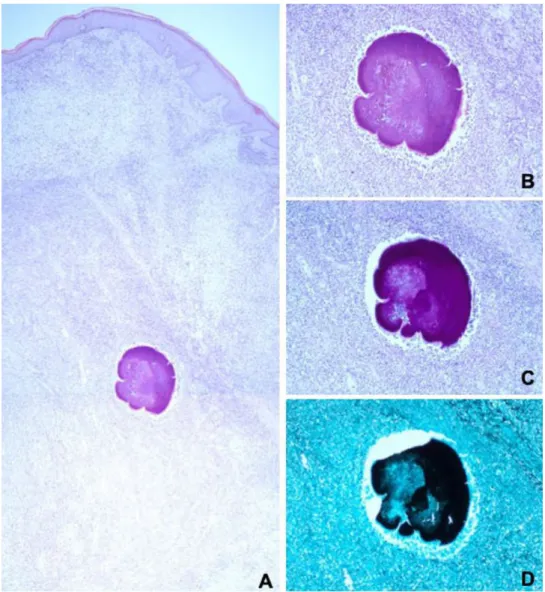
Figure 2. Photomicrographs of the skin biopsy. A – abundant granulation tissue with associated mixed inflammatory infiltrate extending until the deep dermis, with areas of abscess formation. In the middle of the lesion, there are microorganisms with morphological features characteristic of Actinomyces spp. There was no evidence of the presence of neoplastic tissue (H&E, 40X); B – Representative Actinomyces spp. microorganism found in the dermis, with associated polymorphic inflammatory infiltrate (H&E,100X); C – Representative Actinomyces spp. microorganism highlighted using the Periodic acid–Schiff–diastase stain (100X); D – Representative Actinomyces spp. microorganism highlighted using the Grocott’s stain (100X).
first biopsy did not reach the lesion and the diagnosis of well-differentiated liposarcoma relied mostly on the positivity of the IHC reaction for MDM2, which was further ascertained a false positive result.
DISCUSSION
In the pre-antibiotic era, actinomycosis was much more common than today, and it was usually misdiagnosed because of mimicking other conditions such as malignancy and tuberculosis.9
Actinomyces infection involves the abdomen in up to 20% of the cases, and has been called “the great imitator”.4,5,7,10-12 The most frequently affected
gastrointestinal sites are the appendix, cecum, and colon, but any part of the gastrointestinal tract can be involved.4-6 The disease is distributed worldwide,
involves all ages, and males seem to be more frequently affected than females, at a ratio of 3:1.5,8
Currently, Abdominal actinomycosis (AA) is a challenging diagnosis as it is a rare disease with a varying and nonspecific clinical presentation that may mimic other more common entities such as colorectal carcinoma or other malignant tumors, Crohn’s disease, appendicitis, diverticulitis, ulcerative colitis, pelvic inflammatory disease and intestinal tuberculosis.4,6,7
The diagnosis of AA is frequently made in surgical specimens, when surgery is undertaken in the pursuit of neoplasia or appendicitis. Less than 10% of the cases are diagnosed before surgery.4,5,7-10
A high index of suspicion is required in patients presenting with constitutional and nonspecific abdominal symptoms, abdominal swelling, or palpable abdominal mass. The course of the disease is usually indolent, accompanied by fever, abdominal pain, and weight loss. The unspecificity of the clinical features renders a wide range of differential diagnosis.4,9,10
Abdominal actinomycosis should be included as a differential diagnosis when an unusual mass or abscess presents on abdominal CT.9
Patients, diagnosed with abdominal actinomycosis, may present a palpable mass and eventually fistula to the skin. The presence of abscesses and fistula may be detected not only on the physical examination but also by different imaging exams. As the disease progresses, the fistula may become evident. The presence of sulfur granules on microbiologic or histological examination, though important, is not pathognomonic.4-10 The most
important CT finding is a large mass adjacent to the involved bowel. In the rectosigmoid junction, cystic masses are more common, whereas in the transverse or ascending colon purely solid masses are the predominant finding. Involvement of the rectum has been described and clinically resembles malignancy. There are also reports of splenic and esophageal actinomycosis.4,8,9
The invasive nature of the actinomycosis (contiguous spreading to the surrounding tissues) may also resemble malignancy. However, differently from the later, regional lymphadenopathy are rarely found.13
The hematogenous spread of actinomycosis is rare, but may occur to the liver (15%) and lungs may produce nodules, that bear a resemblance with metastatic malignancy.4,9,10
The gold standard diagnostic method is the isolation of Actinomyces spp. However, the microbiological isolation lacks sensitivity. It takes, at least, a week for the growth in anaerobic incubation media, and the sulfur granules are found in only 50% of the specimens.4-6,9,10,13 The microscopic finding of
the isolated Actinomyces in the culture media is a gram-positive, nonacid-fast, thin, branching filaments.
The tissue specimen for histopathologic examination and culture may be obtained either via percutaneously (core, tru-cut needle biopsy or fine needle aspiration) under ultrasound or CT guidance or open surgical resection (excisional or incisional biopsy).
As the conventional methods could be insufficient to identify Actinomyces spp., molecular methods, Figure 3. Abdominal CT undertaken after 10 months
of antibiotic therapy, showing resolution of the inflammatory process.
for example, 16 S ribosomal RNA gene sequencing, DNA–DNA hybridization, real-time polymerase chain reaction (PCR) and fluorescence in situ hybridization (FISH) can be of utmost importance for the correct diagnosis.8
We believe that the initial misdiagnosis of this case occurred because the first biopsy did not reach the lesion of interest, and sampled only the skin and subcutaneous tissue. The diagnosis dilemma, relied on the positivity for MDM2 immunohistochemical reaction. The specificity of MDM2 for the diagnosis of well-differentiated liposarcoma varies from 58,8% and 80%.14,15 Sensitivity and specificity, for this diagnosis,
increases when MDM2 and CDK4 are tested in conjunction, what did not occur in this case.15
Hopefully, the time elapsed between both hospitalizations was suffice for the emergence of a skin lesion that permitted the right diagnosis when the patient was re-evaluated, preventing an extensive surgery.5,9 The authors believed that the
actinomycosis explained all the clinical findings and that the histological interpretation of the first biopsy was misconceived. Actually, there was no abdominal liposarcoma, since the patient became asymptomatic and CT was normal at the end of the antibiotic therapy.
The use of penicillin for a prolonged period is the medical treatment of choice. Although therapy needs to be individualized, 18 to 24 million units of IV penicillin for 2 to 6 weeks, followed by oral therapy with penicillin or amoxicillin for 6 to 12 months. Alternatives for patients with penicillin allergy include tetracycline, erythromycin, doxycycline, and clindamycin.5-9 If there are many avascular spaces due
to severe tissue reaction, medical therapy sometimes needs longer duration.4,5,8-10,12,16,17
In most cases, surgery can be avoided, or a minor procedure may be necessary to aid the therapy. Some patients may require the drainage of an abscesses or excision of a fistula. Surgery may be necessary in case of refractoriness to medical therapy, in cases of relapse or when it is not possible to otherwise exclude cancer. Nevertheless, these patients must be on antibiotic treatment. As long as the masses, abscesses, and fistula disappear, surgical scheduling may be withdrawn.6,8,12
CT scan is an objective imaging technique to follow-up the effectiveness of the treatment. Notwithstanding, the MRI shows a higher sensitivity for detecting residual disease compared with the CT.16,17
In the present case, antibiotic treatment was enough to reach the cure. Although the prognosis of this infection is good with proper medical treatment, with or without surgery, actinomycosis still can lead to the death in cases of tough early diagnosis or/and severe infection.5
Our patient had no identifiable cause for the development of AA. Her medical history was unremarkable. Apparently, she had no immunity derangement, and also had not undergone endoscopic procedures. Moreover, the colonoscopy showed diverticula in the descending colon far from the colonic relation with the mass. Therefore, this case of AA was considered of unknown origin.18
CONCLUSION
Abdominal actinomycosis is an uncommon infectious disease that, in some cases, the clinical presentation may resemble a malignant disease.
A high index of suspicion is needed to avoid delayed diagnosis and unneeded treatment. There are many reports of actinomycosis that were initially diagnosed as tumors and inadvertently treated with extensive surgery.
AA should be considered as a differential diagnosis when an unusual mass or abscess presents on abdominal CT.
Preoperative histopathologic examination of tissue obtained by percutaneous or surgical biopsy of the mass should be considered in these cases to make the diagnosis and optimize therapy.
The goals of treatment are to eradicate the infection and prevent a recurrence. Prolonged treatment of high dose penicillin may be required to cure.
Surgery is only considered when malignancy cannot be excluded, or if removal of persistent fistula, drainage of abscess and excision of necrotic tissues is necessary for antibiotics to be effective. CT and MRI also aid to follow-up therapeutic effectiveness.
REFERENCES
1. Cope VZ. Visceral actinomycosis. BMJ. 1949;2(4640):1311-6. http://dx.doi.org/10.1136/bmj.2.4640.1311. PMid:15407913.
2. Bollinger O. Ueber sine neue Pilzkrankheit beim Rinde. Centralb f. d. med Wissensch. Berl. 1877;15:481. 3. Mandel GL, Douglas RG, Bennet JE. Mandell, Douglas,
and Bennett’s principles and practice of infectious diseases. 2nd ed. New York: Churchill Livingstone; 1985. 4. Târcoveanu E, Vasilescu A, Andronic D, et al. Abdominal
actinomycosis mimicking colon cancer. Chirurgia (Bucur). 2019;114(2):251-8. http://dx.doi.org/10.21614/ chirurgia.114.2.251. PMid:31060658.
5. Li J, Li Y, Zhou Y, Wang C, Wu B, Wan J. Actinomyces and alimentary tract diseases: a review of its biological functions and pathology. BioMed Res Int. 2018;2018:2018. PMid:30225251.
6. Castro LL, Cabral MMDÁ, Andrade RFM, Buzatti KC, Silva RG. Actinomycosis mimicking colonic neoplasia. J Coloproctol. 2012;32(3):312-5. http://dx.doi. org/10.1590/S2237-93632012000300017.
7. Lisa-Gracia M, Martín-Rivas B, Pajarón-Guerrero M, Arnáiz-García A. Abdominal actinomycosis in the last 10 years and risk factors for appendiceal actinomycosis: review of the literature. Turk J Med Sci. 2017;47(1):98-102. http:// dx.doi.org/10.3906/sag-1511-52. PMid:28263474. 8. Boyanova L, Kolarov R, Mateva L, Markovska R, Mitov
I. Actinomycosis: A frequently forgotten disease. Future Microbiol. 2015;10(4):613-28. http://dx.doi.org/10.2217/ fmb.14.130. PMid:25865197.
9. Wong, V. K. & Turmezei, T. D. Actinomycosis. BMJ. 2011;343:1-7. http://dx.doi.org/10.1136/bmj.d6099. 10. Yang SS, Im YC. Severe abdominopelvic actinomycosis
with colon perforation and hepatic involvement mimicking advanced sigmoid colon cancer with hepatic metastasis: A case study. BMC Surg. 2018;18(1):1-8. http://dx.doi. org/10.1186/s12893-018-0386-3. PMid:30068330. 11. Nissotakis C, Sakorafas GH, Koureta T, Revelos K,
Kassaras G, Peros G. Actinomycosis of the appendix:
diagnostic and therapeutic considerations. Int J Infect Dis. 2008;12(5):562-4. http://dx.doi.org/10.1016/j. ijid.2007.12.015. PMid:18400543.
12. Brown JR. Human actinomycosis; study of 181 subjects. Hum Pathol. 1973;4(3):319-30. http://dx.doi. org/10.1016/S0046-8177(73)80097-8. PMid:4756858. 13. Heo SH, Shin SS, Kim JW, et al. Imaging of actinomycosis
in various organs: a comprehensive review. Radiographics. 2014;34(1):19-33. http://dx.doi.org/10.1148/ rg.341135077. PMid:24428279.
14. Aleixo PB, Hartmann AA, Menezes IC, Meurer RT, Oliveira AM. Can MDM2 and CDK4 make the diagnosis of well-differentiated/dedifferentiated liposarcoma? An immunohistochemical study on 129 soft tissue tumors. J Clin Pathol. 2009;62(12):1127-35. http://dx.doi. org/10.1136/jcp.2009.070201. PMid:19946100. 15. Clay MR, Martinez AP, Weiss SW, Edgar MA. MDM2
and CDK4 immunohistochemistry: should it be used in problematic differentiated lipomatous tumors? A new perspective. Am J Surg Pathol. 2016;40(12):1647-52. http://dx.doi.org/10.1097/PAS.0000000000000713. PMid:27508976.
16. Ko S, Ng S, Lee T, Lo CW. Retroperitoneal actinomycosis with intraperitoneal spread. Stellate pattern on CT. Clin Imaging. 1996;20(2):133-6. http://dx.doi. org/10.1016/0899-7071(94)00083-2. PMid:8744824. 17. Hawnaur JM, Reynolds K, McGettigan C. Magnetic
resonance imaging of actinomycosis presenting as pelvic malignancy. Br J Radiol. 1999;72(862):1006-11. http://dx.doi.org/10.1259/bjr.72.862.10673954. PMid:10673954.
18. Sung HY, Lee IS, Kim SI, et al. Clinical features of abdominal actinomycosis: 15-year experience of a single institute. J Korean Med Sci. 2011;26(7):932-7. http://dx.doi.org/10.3346/jkms.2011.26.2011;26(7):932-7.932. PMid:21738348.
Author contributions: All the authors where involved with the patient care. Vieira e Monteiro E wrote
the manuscript. Lamas NJ and Coelho A were the pathologists responsible for the pathological diagnosis of actinomycosis and provided the histological pictures. All authors collectively proofread the final version of the manuscript and approved for publication.
The authors retain an informed consent signed by the patient authorizing the data publication
Conflict of interest: None
Financial support: None
Submitted on: September 23rd, 2019
Accepted on: November 13th, 2019
Correspondence
Eunice Vieira e Monteiro
Centro Hospitalar Universitário do Porto Largo do Prof. Abel Salazar – Porto – Portugal 4099-001
Phone: +35 1914440936