Orientador de dissertação:
Professor Rui Oliveira
Coordenador de seminário de dissertação:
Professor Rui Oliveira
Tese submetida como requisito parcial para a obtenção do grau de:
Mestre em Psicobiologia
2010
Adult neurogenesis in a new model specie, the
cichlid fish Oreochromis mossambicus.
Dissertação de Mestrado realizada sob a orientação de
professor Rui Oliveira, apresentada no Instituto Superior de
Psicologia Aplicada para obtenção de grau de Mestre na
especialidade de Psicobiologia conforme despacho da
DGES nº 6037 / 2007 publicado em Diário da Republica 2ª
séria de 23 de Março, 2007
ii
Acknowledgments
I would like to thank to my supervisor, Professor Rui Oliveira. He made this thesis possible giving me access to all the resources, and the most important, the scientific input that I needed to make this work real and above all, for keeping me motivated even when I’m not.
I would also like to thank to Professor Gunther Zupanc from Jacobs University, Bremen, for having me during four months in his lab. He taught me all the techniques used in the present work.
To David Gonçalves for his corrections and for everything that he taught me during the last years, I have learned a lot... Can’t thank you enough!!
To Ushi for all the support that she gave me in Bremen, not only in the Lab!!!
To Ruxandra for the help with the confocal microscopy.
To Leonor, she helped me to get inspiration in moment where inspiration was in fairway kingdom!!!
To Marta she is always there with a positive and funny thing to say. She always believes!!
To Miguel for the reviewing that he did in the state of the art section. Now he knows everything that he always wanted to know about adult neurogenesis and he never had the courage to ask …
To all my colleagues, they are always available for one more presentation and for the daily support!!!
To my family, friends and David for all the patience, whether I be in a good mood, tired, angry, or stressed… they are always there for me !!!!!!
iv
Resumo
Em comparação com outros vertebrados, os peixes teleósteos têm um enorme potencial para produzir células novas no cérebro de animais adultos. Em contraste com os mamíferos, onde o processo de neurogénese adulta encontra-se restrito a duas áreas cerebrais, a zona subventricular (SVZ) e a zona subgranular parte do giro dentado do hipocampo, em peixes teleósteos foram descritas mais de 10 regiões neurogénicas. Através da marcação de células mitóticas com 5-bromo-2’-deoxiuridina (BrdU), foram caracterizadas as zonas proliferativas da Tilapia de Moçambique (Oreochromis mossanbicus). Nesta espécies, foram encontradas zonas proliferativas em regiões específicas do bolbos olfactivo, telecéfalo, região pré-optica, hipotálamo, tálamo, tecto óptico, torus longitudinalis, nas três divisões do cerebelo, valvula
cerebelli, corpus cerebelli, e lobus caudalis e na região da medula, abrangendo assim toda a
extensão cerebral. A localização destas zonas proliferativas parece ser extremamente conservada ao longo da taxonomia e até o número total de células produzidas parece ser mantido com pouca variação. Com um tempo de sobrevivência de 2 horas, foram encontrados na tilapia um total de 80.000 células novas em comparação com as 100.000 descritas para o peixe eléctrico Apteronotus leptorhynchus. Os nossos resultados sugerem que a actividade mitótica em regiões discretas do
cérebro adulto são uma característica primitiva que tem sido conservada ao longo da evolução.
Palavras Chave: Tilápia de Moçambique, Neurogénese Adulta, Bolbos olfactivos, Telencéfalo, Tecto óptico, Cerebelo, BrdU, Peixes teleósteos
v
Abstract
Compared to other vertebrate species, fish exhibit an enormous potential to produce new cells in the adult brain. In contrast to mammals, where proliferation zones are restricted to two brain areas, the sub ventricular zone (SVZ), and the subgranular zone (SGZ), part of the dentate gyrus of hippocampus, in teleost species more than 10 neurogenic regions have been described. By labeling mitotically dividing cells with 5-bromo-2'-deoxyuridine (BrdU), we have characterized the proliferation zones in the Mozambique tilapia (Oreochromis mossambicus). Proliferation zones were located in specific brain regions of the olfactory bulb, telencephalon, preoptic area, hypothalamus, thalamus, optic tectum, torus longitudinalis, in all three subdivisions of the cerebellum, the valvula cerebelli, the corpus cerebelli, and the lobus caudalis cerebelli and in the region of the medulla oblongata. These proliferation zones appeared to be extremely conserved across taxonomy and even the total number of new generated cells seems to be preserved. After 2 hours survival time we found a total of approximately 80.000 new cells for tilapia compared to 100.000 new cells described for Apteronotus leptorhynchus. Our results suggest that the presence of mitotic activity in specific brain regions is a primitive feature that has been conserved through evolution.
Key words: Mozambique tilapia, adult neurogenesis, olfactory bulb, telencephalon, optic tectum,
vi
Table of Contents
Acknowledgments ... ii
Resumo ... iv
Abstract ... v
List of Figures ... viii
List of Tables... x
Abbreviation list ... xi
Introduction ... 1
Materials and methods ... 2
Animals ... 2
Intraperitoneal injection of 5-bromo-2'-deoxyuridine ... 3
Intracardial perfusion and tissue preparation ... 3
BrdU immunohistochemistry ... 4
Microscopic examination and data analysis ... 4
Results ... 5 Proliferation zones ... 5 Olfactory bulb ... 5 Telencephalon ... 10 Diencephalon... 13 Optic tectum ... 14
Nucleus recessi lateralis ... 15
Cerebellum ... 16
vii
Lateral part of the valvula cerebelli ... 16
Medial part of the valvula cerebelli ... 17
Corpus cerebelli ... 17 Lobus caudalis ... 18 Discussion ... 20 Cell proliferation ... 21 Telencephalon ... 21 Olfactory bulb ... 21
Dorsal and ventral telencephalon ... 23
Optic tectum ... 24
Cerebellum ... 26
Conclusion ... 27
Bibliography ... 28
Annex I ... Erro! Marcador não definido. State of the art ... 32
The development of the nervous system ... 32
The importance of the cell cycle ... 32
Adult neurogenesis ... 34
Adult neurogenesis a historical view ... 34
Adult neurogenesis in the mammalian brain ... 35
Adult neurogenesis in non-mammalian vertebrates ... 37
viii
List of Figures
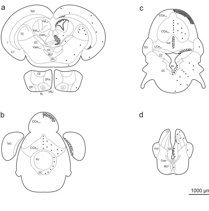
Fig 1 - Distribution and semi-quantitative analysis of proliferation zones in the forebrain (a-c) and midbrain (d-f) of Mozambique tilapia. Mitotic zones were revealed by the analysis of BrdU labeling in coronal sections at a post-administration survival time of 2 hours. Proliferative areas are indicated by black dots. Each dot represents approximately 50 BrdU-labeled cells.
Fig 2 - Distribution and semi-quantitative analysis of proliferation zones in the hindbrain of Mozambique tilapia. Mitotic zones were revealed by the analysis of BrdU labeling in coronal sections at a post-administration survival time of 2 hours. Proliferative areas are indicated by black dots. Each dot represents approximately 50 BrdU-labeled cells.
Fig 3 - Confocal images of BrdU-labeled cells in coronal sections of the olfactory bulb and telencephalon in adult tilapia at 2 hours post-administration survival time. a: Olfactory bulb (BO). Relatively few labeled cells were found in this brain region. Most of these cells were located in the glomerular layer (BOgl), whereas only a few were observed in the granular layer (BOgra). The
labeled cells in BOgl tended to occupy ventral and lateral regions (arrowhead). b: Anterior
subdivision of the dorsal telencephalon (DMa). This brain region was distinguished by a high density of BrdU-labeled cells in a narrow zone at the ventricle (V). c: Medial part of the ventral subdivision of the ventral telencephalon (VVm). The majority of labeled cells were concentrated at the rostral pole of this structure, from which the section was taken. DLa, anterior subdivision of the dorsolateral telencephalon. d: Dorsal part of the ventral telencephalon (Vd). The labeled cells in this brain area are exclusively found in the immediate vicinity of the ventricle. DMvv, ventral subdivision of the ventral part of the dorsomedial telencephalon.
Fig 4 - Confocal images of BrdU-labeled cells in coronal sections of the diencephalon in adult tilapia at 2 hours post-administration survival time. a: Anterior part of the periventricular preoptic nucleus (PPa). BrdU labeled cells were found over the entire dorsoventral extent of the nucleus.
ix
b: Periventricular nucleus of the posterior tuberculum (TPP). Throughout this area, labeled cells displayed a remarkably high packing density. CP, central posterior thalamic nucleus.
Fig 5 - Confocal images of BrdU-labeled cells in coronal sections of the optic tectum (TeO) in adult tilapia at 2 hours post-administration survival time. a: Rostral level of TeO. Most of the new cells were concentrated in the innermost layer (Layer 1). This layer intrudes the dorsolateral aspect of the torus longitudinalis (TL). The site of the intrusion is distinguished by the presence of a dense cluster of labeled cells (arrowhead). b: Caudal level of TeO. The vast majority of the new cells were concentrated, at a uniform and high areal density, in Layer 1.
Fig 6 - Confocal images of BrdU-labeled cells in coronal sections of the recessus lateralis (RL) in adult tilapia at 2 hours post-administration survival time. a: Lateral part of nucleus recessi
lateralis (nRLl). A high areal density of BrdU-labeled cells was found at the ventral and ventrolateral aspects of this structure. b: Medial part of the nucleus recessi lateralis (nRLm). Labeled cells were equally distributed throughout the medial border of this structure. ILvm, ventromedial subdivision of the inferior lobe of the hypothalamus.
Fig 7- Confocal images of BrdU-labeled cells in coronal sections of the cerebellum in adult tilapia at 2 hours post-administration survival time. a: Lateral part of the valvula cerebelli (Val). The majority of the new generated cells were located in the molecular layer of Val (Valmol). By
contrast, in the granular layer of this structure (Valgra) very few cells were observed. The
distribution of the cells in Valmol was restricted to the dorsal and medial aspect of this structure,
with the cells being mostly round and ovoid. Some of these cells, indicated by arrow, are shown at higher magnification in a'. At the lateral aspect of Valmol, a stripe of elongated cells was found
along the dorsoventral extent. Some of these cells, indicated by arrowhead, are shown at higher magnification in a''. b: Medial part of the valvula cerebelli (Vam). Virtually all of the BrdU-labeled cells were located in the molecular layer of Vam (Vammol). Their nuclei displayed an
elongated morphology and were orientated parallel to the midline. Part of these cells are shown at higher magnification in b'. c: Corpus cerebelli (CCe). The majority of labeled cells were found in the molecular layer of this structure (CCemol) immediately beneath the pial surface. They
x
stretched over several hundreds of micrometers laterally in each hemisphere. In addition, BrdU-labeled cells occurred in the midline region of the dorsal molecular layer and extended from the dorsal pial surface to the dorsal aspect of the granular layer of the CCe (CCegra). Part of the latter
cellular population, indicated by arrow, is shown in c'.
List of Tables
Table 1- Quantitative analysis of the proliferation zones in all brain regions of tilapia. Proliferative zones were revealed by the analysis of BrdU labeling at 2 hours survival time.
xi
Abbreviation list
A anterior thalamic nucleus AC anterior commisure BO olfactory bulb
BOgl glomerular layer of the olfactory bulb
BOgra granular layer of the olfactory bulb
CC crista cerebellaris CCe corpus cerebelli
CCegra granular layer of corpus cerebelli
CCemol molecular layer of corpus cerebelli
CE central nucleus of the inferior lobe Ch horizontal commissure
CM corpus mamillare
CP central posterior thalamic nucleus CVEn ventral rhombencephalic commissure D dorsal telencephalon
DA anterior part of the dorsal telencephalon DC dorsocentral telencephalon
DCa anterior subdivision of the dorsal central telencephalon
DCad dorsal part of the anterior subdivision of the dorsocentral telencephalon DCm medial subdivision of the dorsocentral telencephalon
DD dorsal division of the dorsal telencephalon DFl nucleus diffusus lateralis of the inferior lobe
DFld dorsal subdivision of nucleus diffusus lateralis of the inferior lobe DFlv ventral subdivision nucleus diffusus lateralis of the inferior lobe DFm nucleus diffusus medialis of the inferior lobe
DL dorsolateral telencephalon
DLa anterior subdivision of the dorsolateral telencephalon DLd dorsal subdivision of the dosolateral telencephalon DLp posterior subdivision of the dorsolateral telencephalon DLv ventral subdivision of the dorsolateral telencephalon DM dorsomedial telencephalon
DMa anterior subdivision of the dorsomedial telencephalon
DMdd dorsal part of the dorsal subdivision of the dorsomedial telencephalon DMdv ventral part of the dorsal subdivision of the dorsomedial telencephalon DMv ventral subdivision of the dorsomedial telencephalon
DMvd dorsal part of the ventral subdivision of the dorsomedial telencephalon DMvv ventral subdivision of the ventral part of the dorsomedial telencephalon
xii DP dorsal posterior thalamic nucleus Dp posterior part of dorsal telencephalon EG eminentia granularis
G glomerular nucleus GC central gray
H habenula
HV ventral zone of the periventricular hypothalamus IL inferior lobe of the hypothalamus
ILdl dorsolateral subdivision of the hypothalamus
ILvm ventromedial subdivision of the inferior lobe of the hypothalamus IPn interpeduncular nucleus
LCe lobus caudalis
LCegra granular layer lobus caudalis
LH lateral hypothalamic nucleus LVII lobus facialis
LX lobus vagi M
MLF medial longitudinal fascicle mol molecular layer
nIII nucleus oculomotorius nRL nucleus recessi lateralis
nRLl lateral part of nucleus recessi lateralis nRLm medial part of the nucleus recessi lateralis nRP nucleus recessi posterioris
nVd descending trigeminal nucleus nX
P preoptic region
PGm medial part of the preglomerular nucleus PP periventricular preoptic nucleus
PPa anterior part of the periventricular preoptic nucleus PPm medial part of the periventricular preoptic nucleus PPp posterior part of the periventricular preoptic nucleus RL recessus lateralis
RV rhombencephalic ventricle S
Sc suprachiasmatic nucleus TA nucleus anterior tuberis TeO optic tectum
TGN tertiary gustatory nucleus
xiii TL torus longitudinalis
TLA torus lateralis
TLAi inferior subdivision of the torus lateralis tO optic tract
tOd tractus opticus dorsalis
tolfm medial part of the olfactory tract tOv tractus opticus ventralis
TP nucleus posterior tuberculum
TPP periventricular nucleus of the posterior tuberculum TS torus semicircularis
tTB tectobulbar tract V ventral telencephalon V ventricle
Val lateral part of valvula cerebelli
Valgra granular layer of the lateral part of valvula cerebelli
Valmol molecular layer of the lateral part of the valvula cerebelli
Vam medial part of valvula cerebelli
Vamgra granular layer of the medial part of the valvula cerebelli
Vammol molecular layer of the medial part of the valvula cerebelli
Vd dorsal part of the ventral telencephalon VM ventromedial thalamic nucleus
Vs supracommissural part of the ventral telencephalon
VVm medial part of the ventral subdivision of the ventral telencephalon XY
1
Introduction
It is now well established that neurogenesis occurs throughout adulthood in the mammalian brain (Gage, 2000). In mammals this process occurs only in two brain areas located at the telencephalon, the sub ventricular zone (SVZ) lining the lateral ventricles, from where the newly generated cells migrate along the so-called rostral migratory stream into the olfactory bulb (Altman, 1969; Alvarez-buylla, Ling, & Yu, 1994; Alvarezbuylla, Ling, & Yu, 1994) and the subgranular zone (SGZ), part of the dentate gyrus of hippocampus. Since this discovery, a lot of research has been directed towards understanding the physiology and the molecular mechanisms underlying neuronal differentiation. However, neurogenesis within the mature brain is not restricted to mammals, and affects other vertebrates in a large extent, including birds, reptiles, amphibians and fishes (Chapouton, Jagasia, & Bally-Cuif, 2007).
Like in the adult mammalian brain, in non-mammalian brains the majority of the mitotic cells are found in discrete areas called ‘‘proliferation zones” and most of these zones are located near the surfaces of ventricles or related systems. In birds, constitutive adult proliferation and neurogenesis are found dispersed along the telencephalic lateral ventricle, particularly in the ventral and dorsal aspects of the lateral wall of this structure (Alvarez-buylla et al., 1994; Alvarez-buylla, Theelen, & Nottebohm, 1990; Goldman & Nottebohm, 1983). In reptiles, post-natal proliferation have been found in several brain areas such as the olfactory bulb, striatum, dorsoventricular ridge, cortex, nucleus sphericus and cerebellum (Font, Desfilis, Perez-Canellas, & Garcia-Verdugo, 2001; Gárcia-Verdugo, Llahi, Ferrer, & Lopezgarcia, 1989; López-García, Molowny, Garciaverdugo, & Ferrer, 1988; Marchioro et al., 2005; Pérez-Sánchez, Molowny, Garciaverdugo, & Lopezgarcia, 1989). In amphibian brain, there is only one study that describes the proliferative spots (Raucci et al., 2006). In this group adult proliferation has been detected in the telencephalon, preoptic region, thalamus, hypothalamus, midbrain and cerebellum (Chetverukhin & Polenov, 1993; Dawley, Fingerlin, Hwang, John, & Stankiewicz, 2000; Polenov & Chetverukhin, 1993; Raucci et al., 2006). In teleost fishes, neurogenesis is abundant in all the brain extension from rostral to caudal with more than 10 neurogenic regions (the olfactory bulb; telencephalon; thalamus; epithalamus; preoptic region; hypothalamus; tectum; cerebellum; rhombencephalon; and spinal cord) identified and approximately 100 neurogenic
2
areas in Apteronotus leptorhynchus (Ekström, Johnsson, & Ohlin, 2001; Grandel, Kaslin, Ganz, Wenzel, & Brand, 2006; Meyer, 1978; Zikopoulos, Kentouri, & Dermon, 2000; Zupanc, Hinsch, & Gage, 2005; Zupanc & Horschke, 1995).
As we can become aware in these brief summary across taxonomy there are large differences in neurogenic potential between ‘‘lower” and ‘‘higher” vertebrates. In “higher” vertebrates, neurogenic regions are restricted to telencephalon and, as we came across taxonomy, more brain areas with neurogenic potencial emerge and the “neurogenic brain” is achieved in fishes, the lower vertebrate of the chordata phyla.
The adult proliferation pattern in the teleost brain has been studied in detail only in three species: the gymnotiform weakly electric fish, Apteronotus leptorhynchus; the gasterosteiform three-spined stickleback, Gasterosteus aculeatus; and the cypriniform zebrafish, Danio rerio (Ekström et al., 2001; Grandel et al., 2006; Zupanc et al., 2005; Zupanc & Horschke, 1995). Despite the phylogenetic distance between these groups, the distribution of the new generated cells within the proliferative zones seems to be well conserved across species. In the present study we present a detailed characterization of the neurogenic areas in another teleost fish, the Perciform, Mozambique tilapia, Oreochromis mossambicus (Peters, 1852; teleostei, Cichlidae) including a detailed mapping of the proliferations zones in the entire brain. The results of this investigation will bring new insights to a comparative approach on fish neurogenesis since will allow us to compare the mitotic areas in a perciform, with fish from distant superorders and see how preserved these proliferative zones are.
Materials and methods
Animals
A total of three tilapia, O. mossambicus were used in this study. The fish were kept in 300 L
tanks under a 12-h light/12-h dark photoperiod at a temperature of approximately 26-28ºC, pH
3
one was a male. In females, the total length was 10.5 cm and 11.6 cm respectivally, and 17, 0 g
and 18.3 g for body weight. The male had of total length of 14.6 cm and 36.8 g of body weight.
The relative gonadal weight (= fresh weight of gonads divided by body weight) was 0.0476 and
0.0513 for females and 0.0115 for the male. All of the individuals were mature fish, as
determined by post-mortem gonadal inspection and as indicated by the relative gonadal weight.
Intraperitoneal injection of 5-bromo-2'-deoxyuridine
For labeling of S-phase cells, the fish were anesthetized in 2% urethane (Sigma) in aquarium water and injected intraperitoneally with 50 μL of labeling reagent [aqueous solution of 5-bromo-2'-deoxyuridine (BrdU; 3 mg/mL) and 5-fluoro-2'-deoxyuridine (0.3 mg/ mL)], as supplied with the cell proliferation kit (Amersham). After a recovery period in oxygenated aquarium water, the fish were transferred to isolation tanks.
Intracardial perfusion and tissue preparation
After 2 hours post administration of BrdU, the fish were deeply anesthetized by immersion into a 1.5% solution of ethyl 3-aminobenzoate methanesulfonate salt (‘MS-222'; Sigma) and intracardially perfused with a flush solution containing 0.2 M phosphate buffer (PB; pH 7.4), 0.9% NaCl, 5 mg/100 mL heparin sodium salt (research grade, 150,000 IU/g; AppliChem), and 1 mL/100 mL 2% lidocaine. After all blood had been washed out, the perfusion was continued for 30 min with 2% paraformaldehyde (AppliChem) in 0.2 M PB. The brains were removed from the skull, postfixed in fixative solution for 1h at 4ºC, and cryoprotected in 1 M sucrose in phosphate-buffered saline (PBS) overnight at the same temperature. After embedding the brains in a 1:1 mixture of AquaMount (Lerner Laboratories) and Tissue Tek O.C.T. Compound (Sakura), 16-μm thick coronal sections were cut on a cryostat and mounted onto gelatin/chrome-alum-coated slides.
4
BrdU immunohistochemistry
For detection of BrdU-labeled cells, the sections were dried, washed with 0.1 M Tris-buffered saline (TBS; pH 7.5) and incubated in 50% formamide at 65ºC for 2 h. After one rinse with 2x SSC for 30 min, the sections were incubated in 2 M HCl at 37ºC for 40 min. The acid was neutralized by two rinses for 10 min each in 0.1 M borate buffer, pH 8.5, followed by six rinses in TBS for 20 min each. After blocking the slides for 1 h in TBS containing 1% bovine serum albumin (BSA; fraction 5, pH 5.2; AppliChem), 1% teleostean gelatin (Sigma), 3% normal sheep serum (Sigma), and 0.3% Triton X-100 (AppliChem) (here referred to as ‘Blocking Solution 1’) at room temperature (RT), the sections were incubated overnight at 4ºC with rat-anti-BrdU primary antibody (Oxford Biotechnology Cat. No. OBT0030), diluted 1:200 in Blocking Solution 1. The sections were washed three times for 10 min with TBS, blocked for 30 min with Blocking Solution 2 (which was identical to Blocking Solution 1, except that instead of normal sheep serum, normal donkey serum was used) and incubated for 90 min at RT with secondary antibody, Cy3-conjugated donkey anti-rat IgG (H+L) (Dianova; Cat. No. 712-165-150), diluted 1:1000 in Blocking Solution 2. Following three washes for 10 min each in TBS and three washes of 3 min in 0.1 M PBS, pH 7.4, the sections were counterstained with DAPI (2µg/ml) for 3 min at RT and then rinsed in PBS three times for three minutes each. Slides were embedded in polyvinyl alcohol containing n-propyl gallate.
Microscopic examination and data analysis
The sections were examined under a Zeiss Axioskop epifluorescence microscope (Carl Zeiss). Brain areas were identified using the DAPI counterstaining and a partial tilapia brain map (Pepels, Meek, Bonga, & Balm, 2002). The brain sections were draw by means of a camera lucida, and the images were scanned using a HP scanjet 7400C and further processed in Corel Draw 11. For confocal microscopy, a Zeiss LSM 510 META laser scanning microscope was used. Optical sections were taken using 10x and 20x objectives, with a pinehole opening of one Airy at a resolution of 1.024 x 1.024 pixels, using LSM5 (version 3.2; Carl Zeiss) software.
5
Results
Proliferation zones
Intraperitoneal injection of the thymidine analogue BrdU, followed by a survival time of 2 hours, revealed BrdU-labeled cells in numerous brain regions throughout the neuraxis (Figs. 1, 2; Table 1). In the following, we will describe those among these brain regions that displayed relatively high areal densities of labeled cells, compared to neighboring regions, and thus qualify to be called ‘proliferation zones.’
Olfactory bulb
In the olfactory bulb, BrdU labeling exhibited a differential distribution: the vast majority of the labeled cells were located in the glomerular layer (BOgl), whereas rather few cells were observed
in the granular layer (BOgra) (Figs. 1a, 3a). Within the glomerular layer, labeled cells tended to
occupy ventral and lateral regions. Typically, the labeled nuclei were ovoid, with the major axis (10-13 μm) displaying roughly twice the length of the minor axis (3-8 μm).
7
Fig 1 - Distribution and semi-quantitative analysis of proliferation zones in the forebrain (a-c) and midbrain (d-f) of Mozambique tilapia. Mitotic zones were revealed by the analysis of BrdU labeling in coronal sections at a post-administration survival time of 2 hours. Proliferative areas are indicated by black dots. Each dot represents approximately 50 BrdU-labeled cells.
Fig 2 - Distribution and semi-quantitative analysis of proliferation zones in the hindbrain of Mozambique tilapia. Mitotic zones were revealed by the analysis of BrdU labeling in coronal sections at a post-administration survival time of 2 hours. Proliferative areas are indicated by black dots. Each dot
8 represents approximately 50 BrdU-labeled cells.
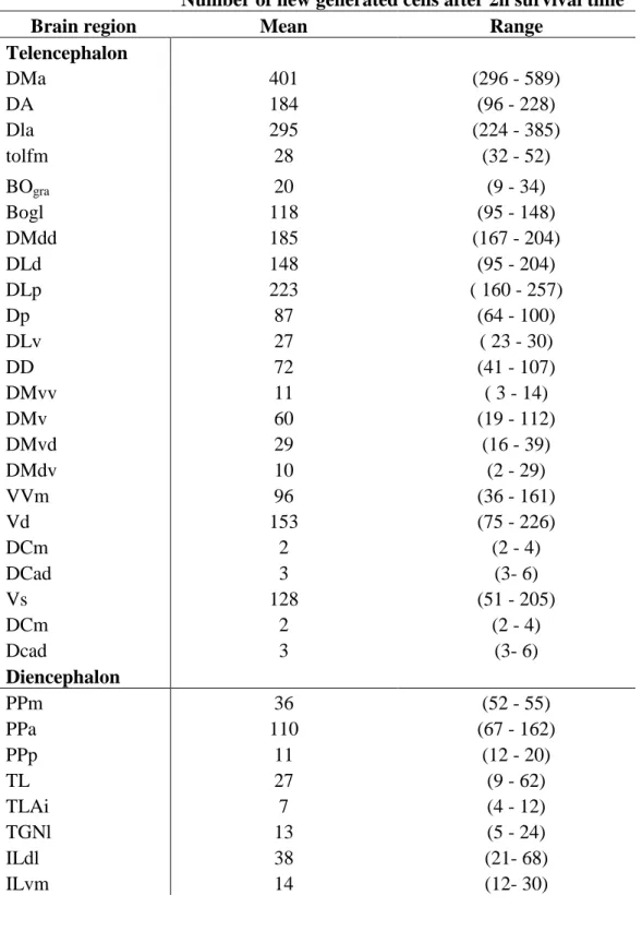
Table 1- Quantitative analysis of the proliferation zones in all brain regions of tilapia. Proliferative zones were revealed by the analysis of BrdU labeling at 2 hours survival time.
Number of new generated cells after 2h survival time
Brain region Mean Range
Telencephalon DMa 401 (296 - 589) DA 184 (96 - 228) Dla 295 (224 - 385) tolfm 28 (32 - 52) BOgra 20 (9 - 34) Bogl 118 (95 - 148) DMdd 185 (167 - 204) DLd 148 (95 - 204) DLp 223 ( 160 - 257) Dp 87 (64 - 100) DLv 27 ( 23 - 30) DD 72 (41 - 107) DMvv 11 ( 3 - 14) DMv 60 (19 - 112) DMvd 29 (16 - 39) DMdv 10 (2 - 29) VVm 96 (36 - 161) Vd 153 (75 - 226) DCm 2 (2 - 4) DCad 3 (3- 6) Vs 128 (51 - 205) DCm 2 (2 - 4) Dcad 3 (3- 6) Diencephalon PPm 36 (52 - 55) PPa 110 (67 - 162) PPp 11 (12 - 20) TL 27 (9 - 62) TLAi 7 (4 - 12) TGNl 13 (5 - 24) ILdl 38 (21- 68) ILvm 14 (12- 30)
9 PGm 10 (5 - 18) LH 24 (1 - 61) HV 12 (2 - 34) TA 53 (2 - 120) VM 18 (6 - 32) A 56 (25 - 94) nRP 65 (46 - 82) nRLm 237 (145 - 354) TPP 237 (195 - 264) CP 22 (20 - 25) DP 20 (6 - 40) G 47 (30 - 63) DFld 20 (16 - 26) DFlv 15 (7 - 27) nRLl 265 (249 - 283) DFm 36 (15 - 51) TP 19 ( 3 - 51) CM 13 (7- 20) TP 19 (3 - 51) nX 25 (16 - 37) PGm 10 (5 - 18) Ch 50 (31 - 60) H 58 (40 - 92) DFl 30 (13 - 47) Sc 49 (29 - 119) CE 13 (7 - 17) Mesencephalon TeO layer 1 2176 (1845 - 2665) TeO layer 2 82 (72 - 101) TeO layer 3 72 (62 - 77) TeO layer 4 106 (82 - 121) TeO layer 5 117 (87 - 140) TeO layer 6 186 (169 - 215) TeO layer 7 54 (37 - 74) tOd 41 (19 - 66) tOv 39 (28 - 59) TS 247 (222 - 295) TL 300 (163 - 455) tO 102 (80 - 131) Rombencephalon
10 Valgra 30 (11 - 45) GC 342 (217- 503) Vammol 1285 (847 - 1769) nIII 5 (1- 8) MLF 4 (5 - 2) Ipn 5 (4 - 11) Valmol 1797 (1236 - 2478) CCemol 5090 (4474 - 6165) CCegra 575 (181 - 1171) mol 827 (725 - 1023) LCegra 775 (550 - 1090) CC 120 (116 - 125) EG 39 (36 - 46) LVII 52 (42 - 68) nVd 32 (10 - 86) MLF 52 (31- 65) LX 238 (122 - 411) CVEn 12 (9 - 15) Z 141 (77 - 184) M 569 (440 - 800) S 100 (38 - 158) Telencephalon
The rostral beginning of the dorsal telencephalon was distinguished by a high areal density of BrdU-labeled cells in a narrow zone at the ventricular wall of the anterior subdivision of the dorsomedial telencephalon (DMa) (Figs. 1a, 3b). These cells stretched throughout the rostro-caudal extent of this structure. Further rostro-caudally, the density of the cells in DMa decreased, while at the dorsal aspects of this structure BrdU-labeled cells became contiguous with labeled cells that appeared in the area just beneath the pial surface of the dorsal part of the dorsal subdivision of the dorsomedial telencephalon (DMdd), the anterior part of the dorsal telencephalon (DA), the dorsal subdivision of the dorsolateral telencephalon (DLd), the ventral subdivision of dorsolateral telencephalon (DLv), and the anterior subdivision of the dorsolateral telencephalon
11
(DLa) (Fig. 1b). The density of labeled cells in DLv and in the dorsal part of DLa was low, relative to the densities observed in DMdd, DA, DLd, and the ventral part of DLa.
The morphology of the labeled nuclei in the different regions of the dorsal telencephalon was rather uniform, being composed of two populations. One consisted of predominantly round nuclei, with diameters of approximately 6-7 μm, while the nuclei of the other population displayed an ovoid profile, with the lengths of the major axis and minor axis ranging between 7 and 10 μm, and 5 and 7 μm, respectively.
In the ventral telencephalon, the most rostrally located structure that displayed BrdU-labeled cells was the medial part of the ventral subdivision of the ventral telencephalon (Vvm) (Figs. 1b, 3c). At its rostral pole, the areal density of BrdU-labeled cells was remarkably high, but gradually decreased at more caudal levels. The labeled nuclei in Vvm were predominantly round, with diameters of 7-8 μm.
Two other structures within the ventral telencephalon with relatively large numbers of BrdU-labeled cells were the dorsal part of the ventral telencephalon (Vd) (Figs. 1b, 3d) and the supracommissural part of the ventral telencephalon (Vs) (Fig. 1c). The labeled cells in Vs appeared as a continuation of the labeling in Vd at the level of the anterior commissure. The populations of labeled nuclei differed somewhat between the two structures. Vd exhibited mostly ovoid nuclei (major axis approximately 10 μm, minor axis approximately 5 μm), whereas Vs contained, in addition to the ovoid nuclei, also round nuclei with diameters typically ranging between 7 and 8 μm.
12
Fig 3 - Confocal images of BrdU-labeled cells in coronal sections of the olfactory bulb and telencephalon in adult tilapia at 2 hours post-administration survival time. a: Olfactory bulb (BO). Relatively few labeled cells were found in this brain region. Most of these cells were located in the glomerular layer (BOgl), whereas only a few were observed in the granular layer (BOgra). The labeled cells in BOgl tended to occupy ventral and lateral regions (arrowhead). b: Anterior subdivision of the dorsal telencephalon (DMa). This brain region was distinguished by a high density of BrdU-labeled cells in a narrow zone at the ventricle (V). c: Medial part of the ventral subdivision of the ventral telencephalon (VVm). The majority of labeled cells were concentrated at the rostral pole of this structure, from which the section was taken. DLa, anterior subdivision of the dorsolateral telencephalon. d: Dorsal part of the ventral telencephalon (Vd). The labeled cells in this brain area are exclusively found in the immediate vicinity of the ventricle. DMvv, ventral subdivision of the ventral part of the dorsomedial telencephalon.
13
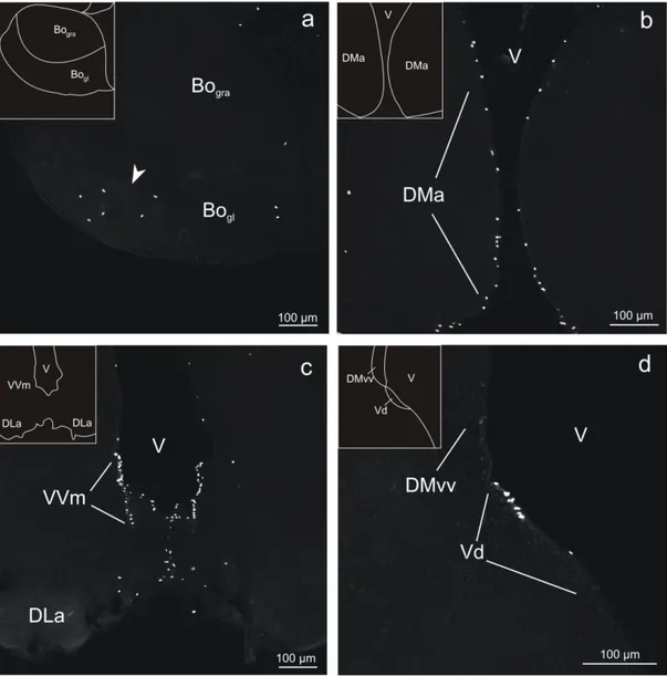
Diencephalon
At the transition of telencephalon and diencephalon, BrdU labeling was most conspicuous in the anterior part of the periventricular preoptic nucleus (PPa), where labeled cells could be found over the entire dorsoventral extent (Figs. 1c, 4a). Typically, their nuclei were round, with diameters of 7-10 μm.
Periventricular nucleus of the posterior tuberculum
In the periventricular nucleus of the posterior tuberculum (TPP), labeled cells were restricted in medio-lateral direction to a narrow zone, less than 50 μm wide, in the immediate vicinity of the wall of the third ventricle, but occurred throughout the dorso-lateral extent, spanning approximately 1000 μm, of this brain nucleus (Figs. 1e, 4b). Within this area, labeled cells displayed a remarkably high packing density. Most of the labeled nuclei were round or ovoid and had typically diameters of 7-10 μm and lengths of the major and minor axes of 7-10 μm and 5-7 μm, respectively.
14
hours post-administration survival time. a: Anterior part of the periventricular preoptic nucleus (PPa). BrdU labeled cells were found over the entire dorsoventral extent of the nucleus. b: Periventricular nucleus of the posterior tuberculum (TPP). Throughout this area, labeled cells displayed a remarkably high packing density. CP, central posterior thalamic nucleus.
Optic tectum
The optic tectum (TeO) was characterized by a large number of BrdU-labeled cells (Figs. 1d-f; 2a, b). The majority of these cells were concentrated in two areas. The first concentration of cells was observed at the dorsomedial end of the innermost layer (Layer 1) (Figs. 1e, 5a). There, Layer 1 intrudes the dorsolateral aspect of the torus longitudinalis (TL) in a conspicuous way. The second concentration of labeled cells, distinguished by a high areal density, was found in Layer 1 at the caudal pole of the tectum (Figs. 2b, 5b). In the remaining six layers of TeO (Layers 2-7), the areal density of cells was rather low, and no marked differential distribution of cells between these layers, or within the individual layers, could be detected. In the proliferation zone at the caudal pole of the tectum, a variety of nuclear morphologies were encountered, including round, ovoid, and elongated nuclei. Typically, the round nuclei had diameters of 7-10 μm. Many of the elongated cells were remarkably slim, with the major axis (approximately 10 μm) often showing four times the length of the minor axis (approximately 2-3 μm). By contrast, in Layers 2-7 most of the cells were round (diameter approximately 7-10 μm).
15
Fig 5 - Confocal images of BrdU-labeled cells in coronal sections of the optic tectum (TeO) in adult tilapia at 2 hours post-administration survival time. a: Rostral level of TeO. Most of the new cells were concentrated in the innermost layer (Layer 1). This layer intrudes the dorsolateral aspect of the torus longitudinalis (TL). The site of the intrusion is distinguished by the presence of a dense cluster of labeled cells (arrowhead). b: Caudal level of TeO. The vast majority of the new cells were concentrated, at a uniform and high areal density, in Layer 1.
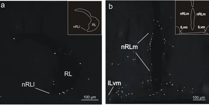
Nucleus recessi lateralis
In the lateral part of the nucleus recessi lateralis (nRLl), a high areal density of BrdU-labeled cells was found (Figs. 1e, f). Most of these cells were located at the ventral and ventrolateral aspects of this structure (Fig. 6a). Typically, labeled nuclei were predominantly ovoid, with the major axis of approximately 10 μm and the minor axis of 4-7 μm.
Labeled cells also occurred at relatively high areal densities in the medial part of the nucleus recessi lateralis (nRLm), where they were concentrated in the immediate vicinity of the ventricular wall (Figs. 1e, 6b). In contrast to nRLl, in nRLm two major morphologies of labeled cells were encountered - one displaying a round shape of the nucleus (diameter 7-8 μm), whereas the other was characterized by an ovoid nuclear profile (major axis approximately 10 μm; minor axis approximately 5 μm).
16
Fig 6 - Confocal images of BrdU-labeled cells in coronal sections of the recessus lateralis (RL) in adult tilapia at 2 hours post-administration survival time. a: Lateral part of nucleus recessi lateralis (nRLl). A high areal density of BrdU-labeled cells was found at the ventral and ventrolateral aspects of this structure. b: Medial part of the nucleus recessi lateralis (nRLm). Labeled cells were equally distributed throughout the medial border of this structure. ILvm, ventromedial subdivision of the inferior lobe of the hypothalamus.
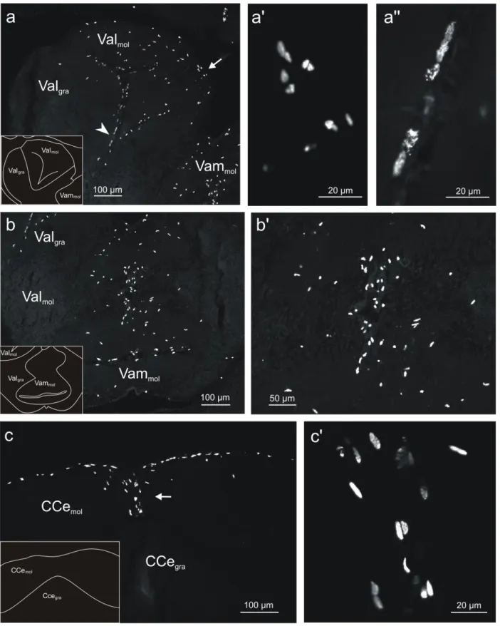
Cerebellum
The largest number of BrdU-labeled cells, relative to all other brain structures, was found in the three subdivisions of the cerebellum - the valvula cerebelli, the corpus cerebelli, and the lobus caudalis.
Valvula cerebelli
The valvula cerebelli consists of two parts, the lateral part (Val) and the medial part (Vam). Each of these two parts can be subdivided into a molecular layer (Valmol and Vammol, respectively) and
a granular layer (Valgra and Vamgra, respectively).
Lateral part of the valvula cerebelli
In the lateral part of the valvula, a huge number of BrdU-labeled cells were present, and almost all of them were found in the molecular layer (Figs. 2a, 7a). These cells were largely restricted to the dorsal and medial aspects of the molecular layer (Fig. 7a’), with one exception. From the dorsal area, a stream of labeled elongated cells emerged and traveled in ventral direction through the molecular layer to the associated granular layer (Fig. 7a”). Virtually all labeled nuclei were aligned parallel to the main direction of this stream. These cells were elongated, with the major axis (10-20 μm) of the nuclei typically being 2-3 times longer than the minor axis (approximately 5 μm). The remaining cells in the molecular layer of the lateral part of the valvula formed a mixed population, consisting of both round cells (diameter of the nucleus approximately 5-10
17
μm) and ovoid cells (major axis of the nucleus: approximately 10 μm; minor axis of the nucleus: approximately 5 μm).
Medial part of the valvula cerebelli
Like in the lateral part of the valvula, in the medial part of the valvula virtually all BrdU-labeled cells were located in the molecular layer, and only a very few cells were observed in the granular layer (Figs. 2a, 7b). Most of the labeled cells within the molecular layer were concentrated in a region stretching up to 150 μm laterally from the midline (Fig. 7b’). A distinct feature of the BrdU-labeled nuclei was their elongated shape. Typically, the major axis (10-20 μm) was 2-3 times longer than the minor axis ( 3-8 μm). Overall, the elongated nuclei displayed a non-random orientation, with the major axis of many nuclei being perpendicular to the midline.
Corpus cerebelli
The corpus cerebelli (CCe) extends from the caudal pole of TeO to the caudal end of the rhombencephalon and thus covers roughly half of the dorsal surface of the brain. Like the valvula, CCe is distinguished by its molecular layer (CCemol) and granular layer (CCegra).
At the rostralmost levels of CCe, the vast majority of labeled cells were found in the dorsal molecular layer (Figs. 2b, c; 7c). These cells were spread over relatively wide areas within this region, exhibiting somewhat higher densities at the midline and immediately below the pial surface. More caudally, these cells became restricted to two major areas. The first area was situated below the dorsal pial surface, stretching in each hemisphere approximately 500 μm laterally from the midline. The second area was defined by a stream of cells at the midline in the dorsal molecular layer, running from the pial surface in ventral direction until the dorsal tip of the granular layer was reached (Fig. 7c’). These labeled cells formed two populations, defined by their ovoid and elongated nuclear profiles, respectively. The ovoid cells displayed lengths of the major and minor axes of approximately 10 μm and 5 μm, respectively. The nuclei of the elongated cells were characterized by a length of the major axis of approximately 15 μm and of the minor axis of approximately 5 μm. The cells of each of the two populations exhibited a
non-18
random orientation, with the major axis of the nucleus predominantly parallel to the pial surface in case of the ovoid cellular population, and parallel to the midline in case of the elongated cellular population. The relatively few labeled cells in the granule layer were observed throughout this structure, but they were more concentrated in the region of the midline. Their nuclear profiles were rather uniform, with the major axis of approximately 10 μm and the minor axis of approximately 5 μm.
Lobus caudalis
The lobus caudalis (LCe) consists of a granular layer dorsal to the rhombencephalic ventricle. In the rostral half of LCe, where the granular layer is restricted to a small area at the dorsolateral corner of the rhombencephalic ventricle, BrdU-labeled cells were restricted to this area, and their number was very low (Fig. 2c). In the caudal half, where the granular layer of LCe enlarges by an expansion in both lateral and dorsal direction, the labeled cells were distributed over the entire area defined by the granular layer, and their numbers increased markedly, compared to the numbers encountered at more rostral levels. Most of the labeled cells were either round, with typical diameters of 7-8 μm, or ovoid, with typical lengths of the major axis of 7-12 μm and of the minor axis of 5-7 μm.
Dorsally to LCe, a molecular layer is situated which is bordered dorsally by CCegra and
laterally by the eminentia granularis (EG). Its association with a specific granular layer is unclear and we, therefore, refer to this as ‘mol’. Within this molecular layer, a high areal density of labeled cells were found at the midline, stretching throughout its dorsoventral extent (Fig. 2c). Most of the cells were ovoid (major axis, 10 μm; minor axis, 7-8 μm).
19
Fig 7- Confocal images of BrdU-labeled cells in coronal sections of the cerebellum in adult tilapia at 2 hours post-administration survival time. a: Lateral part of the valvula cerebelli (Val). The majority of the new generated cells were located in the molecular layer of Val (Valmol). By contrast, in the granular layer
20
of this structure (Valgra) very few cells were observed. The distribution of the cells in Valmol was restricted to the dorsal and medial aspect of this structure, with the cells being mostly round and ovoid. Some of these cells, indicated by arrow, are shown at higher magnification in a'. At the lateral aspect of Valmol, a stripe of elongated cells was found along the dorsoventral extent. Some of these cells, indicated by arrowhead, are shown at higher magnification in a''. b: Medial part of the valvula cerebelli (Vam). Virtually all of the BrdU-labeled cells were located in the molecular layer of Vam (Vammol). Their nuclei displayed an elongated morphology and were orientated parallel to the midline. Part of these cells are shown at higher magnification in b'. c: Corpus cerebelli (CCe). The majority of labeled cells were found in the molecular layer of this structure (CCemol) immediately beneath the pial surface. They stretched over several hundreds of micrometers laterally in each hemisphere. In addition, BrdU-labeled cells occurred in the midline region of the dorsal molecular layer and extended from the dorsal pial surface to the dorsal aspect of the granular layer of the CCe (CCegra). Part of the latter cellular population, indicated by arrow, is shown in c'.
Discussion
The present study clearly demonstrated the existence of multiples proliferation zones in the brain of the adult Mozambique tilapia. Mitotic activity was particularly pronounced in the olfactory bulb, in the Dorsal, Medial and Ventral subdivisions of Telencephalon, preoptic area and periventricular nucleus of the posterior tuberculum of the Diencephalon, optic tectum of Mesencephalon, and in the three subdivisions of the cerebellum.
Analysis of the distribution of the new generated cells shows that mitotic areas appeared normally in the vicinity of the ventricular zones. However, in actinopterygian groups, for instances teleost fish, during embryonic development telencephalon undergoes a process of eversion instead of evagination, as occur in amniote groups. This developmental difference produces a notable morphological divergence in telencephalic hemispheres, with internal ventricles in non-actinopterygian, and massive telencephalic hemispheres flanking a single ventricular cavity in actinopterygians (Northcutt, 1981). These morphological differences, together with the capability of increasing the brain weight and size throughout their lifetime, give fish the possibility to produce an incredible amount of new cells in the adult brain.
21
Cell proliferation
Stem cells are defined by their ability to self-renew and the capacity of their progenitor to adopt multiple lineages. In addition to self-renewal, expansion of progenitor population by repeated entry into cell cycle is a fundamental feature of neurogenesis. As a result, the analysis of cell cycle activity is a critical component of neurogenesis. DNA replication during the S phase of the cell cycle has been used as an opportunity to label proliferating cells by supplying a thymidine analogue that can be incorporated into the cell (Gage, kempermann, & Song, 2008). The thymidine analogue is retained in the post-mitotic cell to be subsequently detected in histological sections. In our study we used the 5-bromo-2'-deoxyuridine (BrdU). This thymidine analogue revealed proliferation zones in all the major subdivisions of the adult brain of tilapia, with approximately 100 proliferative areas. After 2 hours survival time, the estimated time that BrdU is metabolically available to label mitotic active cells (Zupanc & Horschke, 1995), we found a total of approximately 80.000 new cells and 91 proliferative zones (Table1). The analysis of BrdU-positive cells also revealed different distribution patterns throughout the brain structures.
In the following section, we compare our results with findings obtained in other species. For this detailed comparison, we will focus on specific brain regions that are of special interest from a comparative point of view and were also the largest fractions of the brain with mitotic activity: olfactory bulb, dorsal telencephalon, optic tectum and cerebellum.
Telencephalon
Olfactory bulb
The results of the present investigation have shown that in the olfactory bulb of tilapia the majority of BrdU-labbeled cells were found in the glomerular layer (BOgl). Similar results have been observed in zebrafish and gilthead sea bream (Byrd & Brunjes, 2001; Zikopoulos et al., 2000; Zupanc et al., 2005). However, in other teleost species, for instance, goldfish, mediterranean barbell, commum carp and rainbow trout (Alonso, Lara, Vecino, Covenas, & Aljon, 1989), the proliferation zone was found in the innermost layer of the olfactory bulb, the
22
granular cell layer. These results showed that both layers, glomerular and granular respectively, have mitotic ability, and depending on the species cell proliferation can occur in the glomerular or granular cells layers.
In our study, BrdU-positive cells revealed a parallel orientation with the midline of the ventricle, what apparently indicate a migratory behaviour to the glomerular layer of the olfactory bulb. A previous study in zebrafish corroborate this idea, showing that after 4 weeks of post-BrdU administration the great majority of cells are located in the internal cellular layer (Byrd & Brunjes, 2001). In reptiles, the cells generated in the vicinity of the ventricle migrate as well into the granule cell layer (López-García et al., 1988), and in mammals part of newly generated neurons in the olfactory bulb are granule neurons, with only a small proportion acquiring a periglomerular neuronal fate (Saghatelyan, de Chevigny, Schachner, & Lledo, 2004). All this evidence suggest that that the new cells will migrate to the granular cells layer where they differentiate into granule neurons. However we need further investigation with longer survival times to confirm this hypothesis.
Another interesting thing to mention is the characteristics of the new cells. In species where the proliferation spot is the glomerular layer, the cells need to migrate to the granular cell layer and they need an appropriate stimuli and environment to induce migration. In contrast, in species where the mitotic zone is the granular cell layer, there is no need for migration or stimulation. We can hypothesize also that the brain environment and the number of generated cells is different according to the proliferation zones. We can expect higher concentration of cells in species where migration has to occur, compared to species where migration is not required, given that during the migration process, a high number of cells normally died.
Nevertheless, as the olfactory bulb is one of the two brain regions where adult neurogenesis has been described in mammals, it is especially interesting to study this phenomenon in more detail.
23
Dorsal and ventral telencephalon
The results of 2 hours post-BrdU administration experiments in telencephalon areas were remarkably similar to those found in other teleost fishes (Ekström et al., 2001; Zikopoulos et al., 2000; Zupanc et al., 2005; Zupanc & Horschke, 1995). For tilapia, we observed a high proliferative activity in the ventricular zone. This proliferation initiated at the rostral beginning of the dorsal telencephalon (DA), and became contiguous with the dorsomedial (DMa, DMdd), and dorsolateral telencephalon (DLa, DLd, DLv and DLp), covering all the dorsal telencephalic areas.
In ventral telencephalon (Vvm, Vd and Vs), the density of BrdU-labeled cells was remarkably high. These mitotic areas, in dorsal and ventral telencephalon, correspond very well to those observed for the brown ghost, stickleback, zebrafish, salmon and gilthead sea bream (Ekström et al., 2001; Lema, Hodges, Marchetti, & Nevitt, 2005; Zikopoulos et al., 2000; Zupanc et al., 2005; Zupanc & Horschke, 1995).
The uniformity of the results obtained across different teleost species shown that mitotic activity is extremely conserved across fish taxonomy, and if we go further to other vertebrate groups, i.e. mammals, birds, reptiles and amphibians, we can also observe that telencephalic subdivisions are the only brain regions where constitutive adult neurogenesis has been well-documented, indicative of the significance of mitosis in this brain region (Kaslin, Ganz, & Brand, 2009).
Several studies showed the importance of adult neurogenesis in the telencephalon. In mammals, the existent data suggest that neurogenesis and neuronal replacement are related with the acquisition of new information (Ming & Song, 2005). In birds, this phenomenon seems to be correlated as well with memory formation. Several studies of food-hoarding, social change, and reproductive cycle, indicate that the number of new neurons increases markedly as the memory load increases (Barnea, 2009). On the basis of neuroanatomic evidence and results of functional studies (Northcutt, 2008; Wullimann & Mueller, 2004), the dorsolateral telencephalon in fish is thought to be homologous to the mammalian hippocampus, and the dorsomedial telencephalon
24
homologous to the amygdala, both areas associated with memory formation (Broglio et al., 2005).
Despite the fact that in fish there is no study that correlates directly cell proliferation and memory formation, there are several works that indicate such correlation. Initial studies in rainbow trout (O.mykiss) showed that the telencephalon area is significantly reduced in fish reared in hatcheries compared to those reared in the wild (Marchetti & Nevitt, 2003), and Lema
et al (2005) showed that different environments induce differences in mitotic activity rates in
coho salmon (Oncorhynchus kisutch). These results suggest that the environment plays a key role in the regulation of brain development in adult fishes, via changes in mitotic activity.
A considerable amount of experimental data, also involves the dorsolateral telencephalon in allocentric spatial memory (Rodriguez et al., 2002), a process that produces a brain representation of the spatial environment with geometric relationships independent of the subject own position (O'keefe & Nadel, 1979). In goldfish, a significant increase in protein synthesis in the dorsolateral telencephalon neurons was observed in fish trained in a spatial learning task, and lesions in the same region induced a dramatic impairment in place learning (Vargas, Rodriguez, Lopez, Arias, & Salas, 2000).
Based on the facts presented above, we can infer that also in fishes the formation of new cells has a relation with the acquisition of new information, as we can state for the so-called “higher” vertebrates. However, further studies have to be conducted in this direction.
Optic tectum
Our findings revealed that the higher concentration of labeled cells in the optic tectum was found in Layer 1, at the caudal pole of the tectum, and once more our results are consistent with previous results published for other teleost species, such as zebrafish (Marcus, Delaney, & Easter, 1999; Zupanc et al., 2005), Goldfish (Meyer, 1978; Raymond & Easter, 1983), brown ghost (Zupanc & Horschke, 1995), gilthead sea bream (Zikopoulos et al., 2000) and stickleback (Ekström et al., 2001).
25
The optic tectum, the homologous structure of the superior colliculus in mammals, is characterized by a specialized cytoarchitecture and by abundant connectivity with other sensory and motor centers (Vanegas, 1984). Its general organization and function have been conserved across phylogeny in minimum details, such as the pattern of intrinsic and extrinsic connectivity and the tectoreticular projections (Torres, Pérez-Péreza, Herreroa, Ligeroa, & Nunez-Abadesa, 2002).
The analysis of adult neurogenesis in the visual system covered so far a broad range of non related teleost species and a similar pattern emerge among them. Germinal cells are normally absent at the rostral aspect of the tectum, and their density increases in caudal direction on both the dorsomedial and ventrolateral edges of this structure. The two arms of the tectum joined at the caudal pole of this structure, where the density of mitotic cells reaches maximum values (Raymond & Easter, 1983). This pattern of cell birth seems to be related with developmental and organization features of the optic tectum. During embryonic development, this brain structure has a pronounced rostral-to-caudal gradient of maturation, such that the first cells to differentiate are at the rostrolateral pole and the last ones are at the caudomedial edge (Cowan, Martin, & Wenger, 1968).
The data from embryonic development correspond to the matrix of proliferation in the adult brain, indicating that the caudal pole of this structure retain the ability of mitotic division, as we can confirm once more with the present results for tilapia. Kirsche (1967, in Raymond & Easter, 1983) also propose that the proliferation zones in adult animals exist within brain areas that are the last to develop or their development persist within the brain maturation process. Regarding to organizational aspects, the continual integration of new neurons to the visual system must preserve the topography of retinal projection (Straznicky & Gaze, 1971), therefore the caudal pole of the tectum is the finest place for this renewal. These results together suggest that apart from the ecological and ethological constraints among the different species, the commonality of the proliferation zones in visual and nonvisual (electric fish) species does not appear to be related with visual inputs because the proliferation areas are constant between species.
26
Cerebellum
Among all the brain regions studied for tilapia, the main cell production was found in the three subdivision of the cerebellum: valvula cerebelli, corpus cerebelli, and the lobus caudalis. An identical result was obtained in all other teleost species examined in detail, including guppy (Kranz & Richter, 1970) brown ghost (Zupanc & Horschke, 1995), gilthead sea bream (Zikopoulos et al., 2000), three-spined stickleback (Ekström et al., 2001) and zebrafish (Zupanc et al., 2005).
Beyond the proliferation zones, the distribution of new cells seems to be conserved among the different species. In all the species studied so far, the majority of new cells for corpus and valvula cerebelli, was generated in the molecular layers. For tilapia, in the dorsal aspect of corpus cerebelli immediately below the dorsal pial surface, the new cells stretched in each hemisphere approximately 500 μm laterally from the midline. This same result was found in zebrafish, where the cells stretched 100 µm laterally in either hemisphere (Zupanc et al., 2005), in brown ghost cells stretching up to 200-300 µm laterally on each side of the brain (Zupanc & Horschke, 1995) and in the stickleback (Ekström et al., 2001). The µm of stretching are possibly correlated with the brain size and almost no variability can be found.
In valvula cerebelli, the general pattern of proliferation was largely restricted to the dorsal and medial aspects of the molecular layer. From the dorsal area, a stream of elongated cells emerged traveling in ventral direction to the associated granular layer. The elongated morphology of these cells suggest migratory activities. This result could represent a specie-specific difference. In other teleost species, valvula cerebelli has shown consistent results in the distribution of the new cells. In brown ghost, stickleback, zebrafish and gilhead sea bream, the concentration of cells were restricted to the midline of the valvula (Zupanc et al., 2005; Zupanc & Horschke, 1995), and for the latter species, new cells could also be found in the medio-lateral axis (Zikopoulos et al., 2000). These differences in proliferative zones can be related with the differences in embryonic development of the cerebellum.
In lobus caudalis, in contrast with the others cerebellum structures, BrdU-labeled cells were distributed over the entire area defined by the granular layer. The labeled cells increased
27
gradually from rostral to caudal aspects of this structure and the same pattern was also described for other teleost species (Ekström et al., 2001; Zikopoulos et al., 2000; Zupanc et al., 2005; Zupanc & Horschke, 1995).
The consistence of the proliferative zones is probably related with the embryonic development. Developmental studies in trout, Salmo gairdneri (Pouwels, 1978), identified three matrix zones in the cerebellar wall that retain mitotic characteristics permanently, matrix M, L and P. The matrix M is located at the paramedian region of the cerebellar wall, the matrix L surrounds the lateral recessus of the fourth ventricle and the matrix P connects the left and right matrix zone L. Nevertheless, developmental studies in tilapia are necessary to confirm the matrix zones that persist from early development, and see if these matrixes are able to explain the small differences found in this particular species.
Conclusion
In conclusion, we have demonstrated the presence of restricted proliferation zones in all brain subdivisions of adult Mozambique tilapia. The proliferation zones are mostly restricted to ventricular areas, as described previously for other species, and mitotic active cells were only rarely observed in deep brain tissue.
The adult proliferation zones in tilapia show once more that they are extremely well conserved. In the present study, the data obtained for tilapia (perciform) was compared with other perciforms (gilthead seabram), with cypriniformes (zebrafish and guppy), gymnotiformes (weakly electric fish), and gasterosteiformes (three-spined stickleback), and the results were consistent among all the different superorders. Even when comparisons were made with amniotic groups (reptiles, birds and mammals), these proliferative zones were still conserved. Our results suggest that the presence of mitotic activity in specific brain regions is a primitive feature that has been conserved through evolution.
28
Bibliography
Alonso, J. R., Lara, J., Vecino, E., Covenas, R., & Aljon, J. (1989). Cell proliferation in the olfactory bulb of adult freshwater teleosts. Journal of anatomy, 163, 15.
Altman, J. (1969). Autoradiographic and Histological Studies of Postnatal Neurogenesis .4. Cell Proliferation and Migration in Anterior Forebrain, with Special Reference to Persisting Neurogenesis in Olfactory Bulb. Journal of Comparative Neurology, 137(4), 433-&. Alvarez-buylla, A., Ling, C. Y., & Yu, W. S. (1994). Contribution of Neurons Born during
Embryonic, Juvenile, and Adult Life to the Brain of Adult Canaries - Regional Specificity and Delayed Birth of Neurons in the Song-Control Nuclei. Journal of
Comparative Neurology, 347(2), 233-248.
Alvarez-buylla, A., Theelen, M., & Nottebohm, F. (1990). Proliferation Hot-Spots in Adult Avian Ventricular Zone Reveal Radial Cell-Division. Neuron, 5(1), 101-109.
Alvarezbuylla, A., Ling, C. Y., & Yu, W. S. (1994). Contribution of Neurons Born during Embryonic, Juvenile, and Adult Life to the Brain of Adult Canaries - Regional Specificity and Delayed Birth of Neurons in the Song-Control Nuclei. Journal of
Comparative Neurology, 347(2), 233-248.
Barnea, A. (2009). Interactions between environmental changes and brain plasticity in birds.
General and Comparative Endocrinology, 163(1-2), 128-134.
Broglio, C., Gomez, A., Duran, E., Ocana, F. M., Jimenez-Moya, F., Rodriguez, F., et al. (2005). Hallmarks of a common forebrain vertebrate plan: Specialized pallial areas for spatial, temporal and emotional memory in actinopterygian fish. Brain Research Bulletin, 66(4-6), 277-281.
Byrd, C. A., & Brunjes, P. C. (2001). Neurogenesis in the olfactory bulb of adult zebrafish.
Neuroscience, 105(4), 793-801.
Chapouton, P., Jagasia, R., & Bally-Cuif, L. (2007). Adult neurogenesis in non-mammalian vertebrates. Bioessays, 29(8), 745-757.
Chetverukhin, V. K., & Polenov, A. L. (1993). Ultrastructural Autoradiographic Analysis of Neurogenesis in the Hypothalamus of the Adult Frog, Rana-Temporaria, with Special Reference to Physiological Regeneration of the Preoptic Nucleus .1. Ventricular Zone Cell-Proliferation. Cell and Tissue Research, 271(2), 341-350.
Cowan, W. M., Martin, A. H., & Wenger, E. (1968). Mitotic Patterns in Optic Tectum of Chick during Normal Development and after Early Removal of Optic Vesicle. Journal of
Experimental Zoology, 169(1), 71-&.
Dawley, E. M., Fingerlin, A., Hwang, D., John, S. S., & Stankiewicz, C. A. (2000). Seasonal cell proliferation in the chemosensory epithelium and brain of red-backed salamanders, Plethodon cinereus. Brain Behavior and Evolution, 56(1), 1-13.
Ekström, P., Johnsson, C. M., & Ohlin, L. M. (2001). Ventricular proliferation zones in the brain of an adult teleost fish and their relation to neuromeres and migration (secondary matrix) zones. Journal of Comparative Neurology, 436(1), 92-110.
29
Font, E., Desfilis, E., Perez-Canellas, M. M., & Garcia-Verdugo, J. M. (2001). Neurogenesis and neuronal regeneration in the adult reptilian brain. Brain Behavior and Evolution, 58(5), 276-295.
Gage, F. H. (2000). Mammalian neural stem cells. Science, 287(5457), 1433-1438.
Gage, F. H., kempermann, G., & Song, H. J. (2008). Adult Neurogenesis (pp. 673). New York: Cold Spring Harbor.
Gárcia-Verdugo, J. M., Llahi, S., Ferrer, I., & Lopezgarcia, C. (1989). Postnatal Neurogenesis in the Olfactory Bulbs of a Lizard - a Tritiated-Thymidine Autoradiographic Study.
Neuroscience Letters, 98(3), 247-252.
Goldman, S. A., & Nottebohm, F. (1983). Neuronal Production, Migration, and Differentiation in a Vocal Control Nucleus of the Adult Female Canary Brain. Proceedings of the
National Academy of Sciences of the United States of America-Biological Sciences, 80(8), 2390-2394.
Grandel, H., Kaslin, J., Ganz, J., Wenzel, I., & Brand, M. (2006). Neural stem cells and neurogenesis in the adult zebrafish brain: Origin, proliferation dynamics, migration and cell fate. Developmental Biology, 295(1), 263-277.
Kaslin, J., Ganz, J., & Brand, M. (2009). Proliferation, neurogenesis and regeneration in the non-mammalian vertebrate brain. Philosophical Transactions of the Royal Society
B-Biological Sciences, 363(1489), 101-122.
Kranz, D., & Richter, W. (1970). [Autoradiographic studies on the synthesis of DNA in the cerebellum and medulla oblongata of teleosts of various ages]. Z Mikrosk Anat Forsch,
82(2), 264-292.
Lema, S. C., Hodges, M. J., Marchetti, M. P., & Nevitt, G. A. (2005). Proliferation zones in the salmon telencephalon and evidence for environmental influence on proliferation rate.
Comparative Biochemistry and Physiology a-Molecular & Integrative Physiology, 141(3), 327-335.
López-García, C., Molowny, A., Garciaverdugo, J. M., & Ferrer, I. (1988). Delayed Postnatal Neurogenesis in the Cerebral-Cortex of Lizards. Developmental Brain Research, 43(2), 167-174.
Marchetti, M. P., & Nevitt, G. A. (2003). Effects of hatchery rearing on brain structures of rainbow trout, Oncorhynchus mykiss. Environmental Biology of Fishes, 66(1), 9-14. Marchioro, M., Nunes, J. M. D. M., Ramalho, A. M. R., Molowny, A., Perez-Martinez, E.,
Ponsoda, X., et al. (2005). Postnatal neurogenesis in the medial cortex of the tropical lizard Tropidurus hispidus. Neuroscience, 134(2), 407-413.
Marcus, R. C., Delaney, C. L., & Easter, S. S. (1999). Neurogenesis in the visual system of embryonic and adult zebrafish (Danio rerio). Visual Neuroscience, 16(3), 417-424.
Meyer, R. L. (1978). Evidence from Thymidine Labeling for Continuing Growth of Retina and Tectum in Juvenile Goldfish. Experimental Neurology, 59(1), 99-111.
Ming, G. L., & Song, H. J. (2005). Adult neurogenesis in the mammalian central nervous system.
Annual Review of Neuroscience, 28, 223-250.
Northcutt, R. G. (1981). Evolution of the Telencephalon in Non-Mammals. Annual Review of
Neuroscience, 4, 301-350.
Northcutt, R. G. (2008). Forebrain evolution in bony fishes. Brain Research Bulletin, 75(2-4), 191-205.
O'keefe, J., & Nadel, L. (1979). The Cognitive Map as a Hippocampus. Behavioral and Brain