w w w . e l s e v i e r . c o m / l o c a t e / b j i d
The Brazilian Journal of
INFECTIOUS DISEASES
Original article
Interferon-gamma release assay performance in
northeastern Brazil: influence of the
IFNG
+ 874 A>T
polymorphism
Valdirene Leão Carneiro
a,b,∗,1, Maria Teresita Bendicho
a,b, Rosalina Guedes Santos
a,1,
Marilda Casela
a,1, Eduardo M. Netto
a,d, Scarlet Torres Moraes Mota
c,
Iza Cristina Araújo Pina
c, Roberto Meyer Nascimento
a, Songeli Menezes Freire
a,1,2,
Theolis Barbosa
a,c,e,1,2aUniversidade Federal da Bahia, Instituto de Ciências da Saúde, Salvador, BA, Brazil
bUniversidade do Estado da Bahia, Departamento de Ciências da Vida, Salvador, BA, Brazil
cFundac¸ão Oswaldo Cruz, Instituto Gonc¸alo Moniz, Salvador, BA, Brazil
dFundac¸ão José Silveira, Instituto Brasileiro para a Investigac¸ão da Tuberculose, Salvador, BA, Brazil
eBrazilian Network for Research in Tuberculosis – REDE TB, Brazil
a r t i c l e
i n f o
Article history:
Received 3 January 2018 Accepted 20 April 2018 Available online 19 May 2018
Keywords:
Tuberculosis infection Genetic variation Sensitivity
a b s t r a c t
Introduction:Latent tuberculosis infection diagnosis based on the release of interferon-gamma in cultures of peripheral blood cells stimulated withMycobacterium tuberculosis antigens has replaced the tuberculin skin test in many countries with low tuberculosis prevalence. The IFN-␥production can be influenced by genetic polymorphisms, of which theIFNG+ 874 (rs62559044) locus is the most studied. We investigated the possible influence of theIFNG+ 874 A/T polymorphism on interferon-gamma test performance.
Methods:Patients diagnosed with pulmonary tuberculosis (75), volunteers with positive tuberculin skin test (70) and healthy volunteers with negative tuberculin skin test and no his-tory of contact with tuberculosis (57) were evaluated regarding theIFNG+ 874 genotype and the IFN-␥levels in whole blood cultures performed using an interferon-gamma commercial kit (QuantiFERON-TB®Gold In-Tube).
Results:IFN-␥production was not influenced by theIFNG+ 874 genotype, regardless of anti-gen or mitoanti-gen-based stimulation, which suggests that other anti-genes may influence IFN-␥ production in response to mycobacteria. TheIFNG+ 874 polymorphism was found to exert no influence over QFT-IT test sensitivity in our study.
∗ Corresponding author.
E-mail addresses: [email protected](V.L. Carneiro), [email protected](M.T. Bendicho),[email protected] (R.G. Santos), [email protected](M. Casela),[email protected](E.M. Netto),[email protected](S.T. Mota),izacristina [email protected] (I.C. Pina),[email protected](R.M. Nascimento),[email protected](S.M. Freire),[email protected](T. Barbosa).
1 Member of INCT-DT/MCT/CNPq.
2 These authors contributed equally to this work.
https://doi.org/10.1016/j.bjid.2018.04.004
Conclusions: TheIFNG+ 874 polymorphism was not shown to influence QuantiFERON-TB® Gold In-Tube test performance in an admixed population from northeastern Brazil.
© 2018 Sociedade Brasileira de Infectologia. Published by Elsevier Editora Ltda. This is an open access article under the CC BY-NC-ND license (http://creativecommons.org/ licenses/by-nc-nd/4.0/).
Introduction
Mycobacterium tuberculosis(Mtb) infection is present in
one-third of the world’s population. While some of these individuals are in a subclinical stage of tuberculosis (TB), most infected people will remain asymptomatic throughout their lives.1These individuals are considered to have latent tuberculosis (LTBI) and only a fraction will develop active TB. This latent pool complicates disease control, as reactivation can occur at any time and chemoprophylaxis is administered only in individuals of known recent contact with an index case.2,3LTBI screening is performed using tests based on an immune response to mycobacterial antigens. The tuberculin skin test (TST) has been used for over a century and offers good sensitivity and specificity in endemic conditions. How-ever, it may present cross-reactivity to sensitization with the BCG vaccine as well as environmental mycobacteria.4,5
IFN-␥release assays (IGRAs) were first described in 20006,7 and present an alternative to TST.8To date, most studies interpret IGRAs similarly to TST, except in populations vaccinated with BCG after childhood, or in individuals receiving multiple doses of this vaccine, as well as in locations where environmental mycobacteria infections are common.4,5 In these situations, IGRAs do seem to present a better positive predictive value due to greater specificity.9,10Cost-effectiveness is another cri-terion to select IGRA as the test of choice, e.g. in countries with low TB prevalence.11–13
Both TST and IGRA tests are not able to distinguish active from latent infection. This is partly because these tests are based on the immune response, and those at increased risk to develop active TB are immunocompromised individ-uals who also present the highest rates of false-negative results. Moreover, the antigens expressed by Mtb during latent infection may not be the same as those expressed during the active replication stage.2,14 As a result, a lower speci-ficity of TB diagnosis is expected in countries with high TB burdens.15–17
Studies focusing on the impact of genetic background with regard to IGRA testing are surprisingly scarce. IFN-␥ production may be influenced by the presence of polymor-phisms in the gene that encodes this cytokine, of which the most studied is theIFNG+ 874 A/T polymorphism. The IFNG+ 874 A/T polymorphism determines the emergence of an NF-B binding site that can increase the transcription of the human IFN-␥gene.18 The AA genotype at this locus has been linked to TB susceptibility and low IFN-␥ pro-duction, including in the Brazilian population.19 In a study among South African families in a hyperendemic area for
tuberculosis, the influence of heredity on quantitative responses to IFN-␥release was estimated to be between 43% and 58%, depending on the nature of the antigen used as a stimulus20 It is possible, therefore, that the IFNG+ 874 A/T polymorphism influences the sensitivity and specificity val-ues of IGRA assays. In this context, the aim of this study was to evaluate the influence of theIFNG+ 874 A/T polymorphism inM. tuberculosisinfected individuals residing in an endemic area.
Methods
Recruitment and study design
The present study included 202 volunteers, divided into three groups: (i) patients with a diagnosis of pulmonary tuber-culosis by positive culture and/or BAAR staining who were naïve to antituberculosis treatment (TB, n= 75); (ii) volun-teers with a positive TST greater than 10 mm, with known contact with TB patients, who tested negative under cul-ture and BAAR staining (TST+, n= 70); (iii) volunteers with a negative TST, an absence of tuberculosis symptoms and no history of contact with individuals with tuberculosis (TST−, n= 57). All individuals were recruited from two local reference institutions for TB diagnosis located in the city of Salvador, Bahia-Brazil. Written informed consent was obtained from all participants. This study was approved by the Institutional Review Board of the Bahiana School of Medicine and Public Health (registered under protocol: CAAE 57662016.8.1001.5662) and complied with the Declaration of Helsinki, as well as with Brazilian regulations pertaining to research ethics involving human beings (Resolution: CNS 466/2012).
Latent TB infection diagnosis
Individuals were considered positive by TST when presenting skin induration measuring at least 10 mm. QFT-IT was repeated for samples with discordant TST and IGRA results.
Genotyping and IFN-production
Following whole blood sample collection, genomic DNA was extracted using a PureLink® Genomic DNA Kit (Invitrogen, Carlsbad, CA, USA) in accordance with manufacturer’s instruc-tions. Genotyping for the IFN-␥polymorphism was performed using a Cytokine Genotyping Tray (One Lambda, Inc., Canoga Park, CA-USA) according to the manufacturer’s protocols. The Human Th1/Th211plex FlowCytomix Multiplex Kit (eBio-science, Vienna, Austria) was employed in accordance with manufacturer’s instructions to measure IFN-␥in pg/mL.
To correlate genotype and IFN-␥ production in the supernatant of samples, we analyzed both the total IFN-␥ pro-duction in each culture condition (pg/mL) and the differences between the IFN-␥levels (IU/mL) in the Ag tube and the Nil tube of the volunteers enrolled, stratified by genotype.
Statistical analyses
Variables were described as frequencies, percentages and median [interquartile range, in square brackets] values. The Chi-square test was used to evaluate the study population for the Hardy-Weinberg equilibrium, and to compare genotype and allele frequencies among groups. The Mann–Whitney test was used to compare IFN-␥production levels among differ-ent IFNG+ 874 A/T genotypes. The Pearson Chi-square test was used to compare proportions of each genotype among individuals with positive versus negative QFT-IT. Statistical significance was considered with an alpha error of 5%. Kappa index, odds ratio (OR) and sensitivity were calculated, with a confidence interval (CI) of 95%. Statistical analyses were per-formed using Prism software version 5 (GraphPad Inc., San Diego, CA) and R Project version 2.15.2 (GNU Project, Boston, MA).
Results
Clinical characteristics and frequency distribution of IFNG + 874 A/T polymorphism genotypes
The frequency distribution of the IFNG+ 874 A/T polymor-phism genotypes was in Hardy–Weinberg equilibrium for all groups (p= 0.78). TheIFNG+ 874 AA genotype was more fre-quently found among individuals with TB (chi-squared = 10.94, p= 0.004) when compared with TST−individuals (Table 1). The frequency of the A allele was also higher in TB patients than in TST- volunteers (chi-squared = 17.6;p= 0.001). Age and BCG scar positivity were similar among the different groups stud-ied and among theIFNG+ 874 A/T polymorphism genotypes.
The association between SNP IFNG + 874 A/T and IFN- production
No significant differences were observed with respect to unstimulated IFN-␥production among the different groups
and genotypes studied (Fig. 1A). IFN-␥production in response to polyclonal phytohemagglutinin (PHA) stimulation was lower among patients with pulmonary TB (3848 [1143–9120] pg/ml) in comparison to TST+ (11,080 [7462–17,248] pg/ml, p< 0.0001) and TST−individuals (10,852 [6782–13,684] pg/ml, p< 0.0001). Healthy uninfected individuals with theIFNG+ 874 AA genotype exhibited lower IFN-␥production upon stimula-tion with PHA when compared toIFNG+ 874 TT/TA genotype individuals (p= 0.012, Fig. 1B). This difference was not sig-nificant when the IFN-␥levels were normalized by the total CD3+ cell counts (median values of 4.22 [2.84–9.12] for AA genotype individuals versus 6.24 [4.08–7.95] for those with TT/TA genotypes,p= 0.3866), but these data were available for only 33 individuals of this group. Irrespective of genotype, no significant differences were seen in IFN-␥production under stimulation with PHA among the infected individuals (Fig. 1B). IFN-␥production upon mycobacterial antigen stimulation was not affected by genetic background at the IFNG+ 874 locus (Fig. 1C). Similarly, no significant differences were detected when analyzing QFT-IT test results (UI/mL), i.e. mycobacte-rial antigen-stimulated versus unstimulated IFN-␥production (Fig. 1D).
Influence of the IFNG + 874 A/T polymorphism on QFT-IT test results
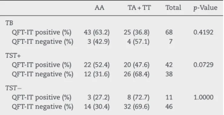
No significant differences were observed with respect to the proportions of the different IFNG+ 874 A/T polymorphism genotypes when comparing positive and QFT-IT-negative individuals (p= 0.20,p= 0.50 and p= 0.28 for TST−, TST+ and TB, respectively,Table 2).
QFT-IT was found to detect TB with sensitivity of 90.7% (81.97–95.41) and specificity of 80.7% (68.66–88.87). In the TB group, the QFT-IT test presented similar sensitivity in patients of the AA genotype of the IFNG+ 874 polymorphism (93.5%, 82.5–97.8%) in comparison to those of the TT/TA genotype (86.2%, 69.5–94.5%). The odds of detecting TB were similar for AA genotype and TT/TA genotype individuals (OR = 2.27, 0.435–13.03). Among AA genotype individuals, 3/46 TB patients presented false-negative QFT-IT results, while 4/29 individuals of either TT or TA genotype had false-negative QFT-IT results. TST and QFT-IT presented regular agreement (kappa = 0.34), with similar results when stratified by IFNG+ 874 A/T polymorphism genotypes (0.33 for the AA genotype and 0.28 for the TT/TA genotype).
Discussion
The QFT-IT diagnostic values obtained in our study were lower than previously reported in other studies,10,21yet con-sistent with previous findings in Brazil,22as well as in other countries.23–25QFT-IT sensitivity and specificity to detect TB was similar to values previously reported in countries with moderate disease burden.8,11,26
As previously reported, the frequency of the AA genotype and the A allele was higher in TB patients than in TST− volunteers.19
Table 1 – Frequency distribution of genotypes and alleles ofIFNG+ 874 A/T polymorphism according to age, sex and ethnic background.
TB TST+ TST−
AA (n= 46) TA/TT (n= 30)
A (n= 118) T (n= 34) AA (n= 34) TA/TT (n= 36)
A (n= 97) T (n= 43) AA (n= 17) TA/TT (n= 41)
A (n= 65) T (n= 51)
Male (%) 26 (66.7) 13 (33.3) 63 (80.8) 15 (19.2) 17 (60.7) 11 (39.3) 42 (75.0) 14 (25.0) 8 (34.8) 15 (65.2) 26 (56.5) 20 (43.5) Female (%) 20 (54.1) 17 (45.9) 55 (74.3) 19 (25.7) 17 (40.5) 25 (59.5) 55 (65.5) 29 (34.5) 9 (25.7) 26 (74.3) 39 (55.7) 31 (44.3) White (%) 4 (100.0) 0 (0.0) 8 (100.0) 0 (0.0) 3 (37.5) 5 (62.5) 10 (62.5) 6 (37.5) 4 (-26.7) 11 (73.3) 16 (53.3) 14 (46.7) Non-White
(%)
26 (54.2) 22 (45.8) 71 (74.0) 25 (26.0) 31 (50.0) 31 (50.0) 87 (70.2) 37 (29.8) 13 (30.2) 30 (69.8) 49 (57.0) 37 (43.0)
Fig. 1 – IFN-production by pulmonary TB, TST+ and TST−individuals according toIFNG+ 874 genotype. (A) Non-stimulated IFN-␥production (NIL); (B) PHA-stimulated IFN-␥production as measured in the supernatant (PHA); (C) Mycobacterial
antigen-stimulated IFN-␥production (Ag); (D) Difference in units (UI/mL) between mycobacterial antigen-stimulated and
unstimulated IFN-␥production.
PHA stimulation according to genotype, with the lowest production found in IFNG+ 874 AA genotype individuals, as expected.18 This difference was not significant when IFN-␥ levels were normalized by number of CD3+ lym-phocytes. By contrast, no differences in IFN-␥ production were found among the different genotypes within the infected groups evaluated, regardless of stimulation with either Mtb or PHA, which is consistent with previously reported findings,27 yet stands in contrast to other reports indicating that AA genotype TB patients had lower IFN-␥ production in mycobacterial antigen-stimulated cultures.28–30 This seems to suggest that, both in the population stud-ied, as well as in mycobacterial antigen-stimulated cultures, the IFNG+ 874 locus had no influence over IFN-␥ levels
under mycobacterial stimulation (in vivo or in vitro), as similar levels were detected regardless of genotype. The estimated heritability of the IFN-␥ response to ESAT-6 was 58%, whereas the estimated heritability for BCG was 43%.19,31 It has been suggested that other genetic factors may influence anti-mycobacterial IFN-␥ production in Mtb infection/disease.32,33 Despite the fact that two loci have recently been associated with IFN-␥production in response to ESAT-6 and BCG, the IFNG gene is not present in these regions.34
Table 2 – QFT-IT test results according toIFNG+ 874 A/T genotype.
AA TA + TT Total p-Value
TB
QFT-IT positive (%) 43 (63.2) 25 (36.8) 68 0.4192 QFT-IT negative (%) 3 (42.9) 4 (57.1) 7
TST+
QFT-IT positive (%) 22 (52.4) 20 (47.6) 42 0.0729 QFT-IT negative (%) 12 (31.6) 26 (68.4) 38
TST−
QFT-IT positive (%) 3 (27.2) 8 (72.7) 11 1.0000 QFT-IT negative (%) 14 (30.4) 32 (69.6) 46
IFN-␥production in TB patients with varied clinical presenta-tions showed a lower percentage of patients with detectable IFN-␥production among those of the AA genotype. This stands in contrast to our findings, as the proportion of responsive AA genotype individuals was similar to that of other genotypes evaluated.27
A recent Canadian study investigated possible influences of
theIFNG+ 874 polymorphism on QFT-IT results. Using these
authors’ published results, we calculated TB patient QFT-IT test sensitivity at 80% (64.1–90.0) among AA genotype individuals, compared to 75% (53.1–88.8) for TT/TA genotype individuals.35However, when combining these authors’ previ-ously published results with the present findings, the number of individuals evaluated was insufficient to demonstrate a rel-evant difference in sensitivity when comparing among the IFNG+ 874 genotypes.
Conclusions
IFNG+ 874 polymorphism had no influence over QFT-IT test performance in an admixed population from northeastern Brazil. Additional study is required to further elucidate any possible effect on QFT-IT test results arising from other genetic variations.
Conflicts of interest
The authors declare no conflicts of interest.
Financial support
Conselho Nacional de Desenvolvimento Científico e Tec-nológico (INCT-DT/MCT/CNPq 573839/2008-5) and Fundac¸ão de Amparo à Pesquisa do Estado da Bahia (PET035/2013).
Acknowledgements
To Yasmin S. de Azevedo Oliveira, Ana Paula Torres, Alice S. Martins dos Santos and Cleidiane Borges Daltro in con-tributing for technical support. To the team of the Laboratory of Immunology and Molecular Biology (Institute of Health Sciences/UFBA) for technical assistance. To the scientific coordination of Instituto Brasileiro para a Investigac¸ão da Tuberculose for their help with the logistics regarding the
field work. To Andris Walter for expert language review of the manuscript.
r e f e r e n c e s
1. WHO. Global tuberculosis report; 2016. Available from: http://apps.who.int/iris/bitstream/10665/250441/1/978924156 5394-eng.pdf[cited 11.07.17].
2. Barry CE, Boshoff HI, Dartois V, Dick T, Ehrt S, Flynn J, et al. The spectrum of latent tuberculosis: rethinking the biology and intervention strategies. Nat Rev Microbiol. 2009;7:845–55. 3. Dheda K, Schwander SK, Zhu B, Van Z-S, Richard N, Zhang Y.
The immunology of tuberculosis: from bench to bedside. Respirology. 2010;15:433–50.
4. Huebner RE, Schein MF, Bass JB Jr. The tuberculin skin test. Clin Infect Dis. 1993:968–75.
5. Weir RE, Fine PE, Floyd S, Stenson S, Stanley C, Branson K, et al. Comparison of IFN-␥responses to mycobacterial antigens as markers of response to BCG vaccination. Tuberculosis. 2008;88:31–8.
6. Arend SM, Geluk A, van Meijgaarden KE, van Dissel JT, Theisen M, Andersen P, et al. Antigenic equivalence of human T-cell responses toMycobacterium tuberculosis-specific RD1-encoded protein antigens ESAT-6 and culture filtrate protein 10 and to mixtures of synthetic peptides. Infect Immun. 2000;68:3314–21.
7. Andersen P, Munk ME, Pollock JM, Doherty TM. Specific immune-based diagnosis of tuberculosis. Lancet. 2000;356:1099–104.
8. Menzies D, Pai M, Comstock G. Meta-analysis: new tests for the diagnosis of latent tuberculosis infection: areas of uncertainty and recommendations for research diagnosis of latent tuberculosis infection. Ann Intern Med.
2007;146:340–54.
9. Trajman A, Steffen RE, Menzies D. Interferon-gamma release assays versus tuberculin skin testing for the diagnosis of latent tuberculosis infection: an overview of the evidence. Pulm Med. 2013. Available from:
https://www.hindawi.com/journals/pm/2013/601737/abs/ [cited 09.07.17].
10. Pai M, et al. Alternatives to the tuberculin skin test: interferon-␥assays in the diagnosis of Mycobacterium tuberculosis infection. Indian J Med Microbiol. 2005;23:151. 11. Pai M, Menzies D. The new IGRA and the old TST: making good
use of disagreement. Am Thorac Soc. 2007. Available from: http://www.atsjournals.org/doi/full/10.1164/rccm.200701-024ED[cited 09.07.17].
12. Pai M, Denkinger CM, Kik SV, Rangaka MX, Zwerling A, Oxlade O, et al. Gamma interferon release assays for detection of Mycobacterium tuberculosisinfection. Clin Microbiol Rev. 2014;27:3–20.
13. Mu ˜noz L, Santin M. Interferon-␥release assays versus tuberculin skin test for targeting people for tuberculosis preventive treatment: an evidence-based review. J Infect. 2013;66:381–7.
14. Berry MPR, Blankley S, Graham CM, Bloom CI, O’Garra A. Systems approaches to studying the immune response in tuberculosis. Curr Opin Immunol. 2013;25:579–87.
15. Pai M, O’Brien R. Serial testing for tuberculosis: can we make sense of T cell assay conversions and reversions? PLoS Med. 2007;4:e208.
16. Pai M. Spectrum of latent tuberculosis—existing tests cannot resolve the underlying phenotypes. Nat Rev Microbiol. 2010;8:242.
clinical utility in high-burden vs. low-burden settings. Curr Opin Pulm Med. 2009;15:188–200.
18. Pravica V, Perrey C, Stevens A, Lee J-H, Hutchinson IV. A single nucleotide polymorphism in the first intron of the human IFN-␥gene: absolute correlation with a polymorphic CA microsatellite marker of high IFN-␥production. Hum Immunol. 2000;61:863–6.
19. de Albuquerque AC, Rocha LQ, de Morais Batista AH, Teixeira AB, Dos Santos DB, Nogueira NAP. Association of
polymorphism + 874 A/T of interferon-␥and susceptibility to the development of tuberculosis: meta-analysis. Eur J Clin Microbiol Infect Dis. 2012;31:2887–95.
20. Cobat A, Gallant CJ, Simkin L, Black GF, Stanley K, Hughes J, et al. High heritability of antimycobacterial immunity in an area of hyperendemicity for tuberculosis disease. J Infect Dis. 2010;201:15–9.
21. Machado A Jr, Emodi K, Takenami I, Finkmoore BC, Barbosa T, Carvalho J, et al. Analysis of discordance between the tuberculin skin test and the interferon-gamma release assay. Int J Tuberc Lung Dis. 2009;13:446–53.
22. de Souza FM, do Prado TN, dos Santos Pinheiro J, Peres RL, Lacerda TC, Loureiro RB, et al. Comparison of interferon-␥ release assay to two cut-off points of tuberculin skin test to detect latentMycobacterium tuberculosisinfection in primary health care workers. PLOS ONE. 2014;9:e102773.
23. Diel R, Goletti D, Ferrara G, Bothamley G, Cirillo D, Kampmann B, et al. Interferon-␥release assays for the diagnosis of latent Mycobacterium tuberculosisinfection: a systematic review and meta-analysis. Eur Respir J. 2011;37:88–99.
24. Ringshausen FC, Nienhaus A, Schablon A, Schlösser S, Schultze-Werninghaus G, Rohde G. Predictors of persistently positiveMycobacterium-tuberculosis-specific interferon-gamma responses in the serial testing of health care workers. BMC Infect Dis. 2010;10:220.
25. Harada N, Nakajima Y, Higuchi K, Sekiya Y, Rothel J, Mori T. Screening for tuberculosis infection using whole-blood interferon-␥and Mantoux testing among Japanese healthcare workers. Infect Control Hosp Epidemiol. 2006;27:442–8. 26. Pai M, Zwerling A, Menzies D. Systematic review: T-cell-based
assays for the diagnosis of latent tuberculosis infection: an update. Ann Intern Med. 2008;149:177–84.
27. Sallakcı N, Coskun M, Berber Z, Gürkan F, Kocamaz H, Uysal G, et al. Interferon-␥gene + 874T–A polymorphism is
associated with tuberculosis and gamma interferon response. Tuberculosis. 2007;87:225–30.
28. López-Maderuelo D, Arnalich F, Serantes R, Gonzalez A, Codoceo R, Madero R, et al. Interferon-␥and interleukin-10 gene polymorphisms in pulmonary tuberculosis. Am J Respir Crit Care Med. 2003;167:970–5.
29. Selvaraj P, Alagarasu K, Harishankar M, Vidyarani M, Rajeswari DN, Narayanan PR. Cytokine gene polymorphisms and cytokine levels in pulmonary tuberculosis. Cytokine. 2008;43:26–33.
30. Hu Y, Wu L, Li D, Zhao Q, Jiang W, Xu B. Association between cytokine gene polymorphisms and tuberculosis in a Chinese population in Shanghai: a case–control study. BMC Immunol. 2015;16:8.
31. Cobat A, Poirier C, Hoal E, Boland-Auge A, de La Rocque F, Corrard F, et al. Tuberculin skin test negativity is under tight genetic control of chromosomal region 11p14-15 in settings with different tuberculosis endemicities. J Infect Dis. 2014;211:317–21.
32. Ansari A, Talat N, Jamil B, Hasan Z, Razzaki T, Dawood G, et al. Cytokine gene polymorphisms across tuberculosis clinical spectrum in Pakistani patients. PLoS ONE. 2009;4:e4778. 33. Conceic¸ão EL, Nascimento-Sampaio FS, Schwingel PA,
Oliveira ES, Rocha MS, Vieira I, et al. Revisiting the
heterogeneous IFN-␥response of Bacille of Calmette-Guérin (BCG)-revaccinated healthy volunteers in a randomized controlled trial: effect of the body mass index and of the IFNG + 874 A/T polymorphism. PLOS ONE. 2016;11:e0160149. 34. Jabot-Hanin F, Cobat A, Feinberg J, Grange G, Remus N, Poirier
C, et al. Major loci on chromosomes 8q and 3q control interferon␥production triggered by Bacillus Calmette-Guerin and 6-kDa early secretory antigen target, respectively, in various populations. J Infect Dis. 2016;213:1173–9.