RESUMO: Em áreas onde a tuberculose humana e a tuberculose bovina coexistem, a diferenciação entre M. bovis e M. tuberculosis é importante para monitorar a disseminação de M. bovis entre bovi‑ nos e destes para os seres humanos. Objetivou‑se neste estudo iso‑ lar e identificar M. bovis em bovinos com diagnóstico positivo pelo teste de tuberculinização no estado da Paraíba, nordeste do Brasil. Foram submetidos 32 bovinos positivos ao teste de tuberculiniza‑ ção comparativa, dos quais foram colhidas amostras de qualquer órgão com lesões sugestivas de tuberculose, e, nos casos em que não foram observadas lesões sugestivas, foram colhidas amostras de linfonodos. As amostras foram submetidas a exame histopato‑ lógico, cultivo micobacteriológico, coloração de Ziehl‑Neelsen e diagnóstico molecular. Apresentaram lesões sugestivas de tuber‑ culose 21 animais (65,6%). Com relação à distribuição das lesões de acordo com a região corporal, 77,7% localizavam‑se na cavi‑ dade torácica, 12,4% na cabeça e 9,9% na cavidade abdominal. De 55 amostras submetidas ao cultivo de micobactérias, em 31 (56,4%) foram isoladas micobactérias, sendo que em 13 (41,9%) foi identificado M. bovis, e nas 18 restantes (58,1%) foi identifi‑ cado Mycobacterium spp. Conclui‑se que o isolamento e a identi‑ ficação de M. bovis e Mycobacterium spp. em bovinos indicam que os seres humanos estão expostos ao risco de infecção. Isso reforça a necessidade de intensificação e otimização de medidas de pre‑ venção e controle previstas no Programa Nacional de Controle e Erradicação da Brucelose e Tuberculose Bovina. Sugere‑se a reali‑ zação de estudos de isolamento e identificação de micobactérias em outros estados do Nordeste.
PALAVRAS‑CHAVE: bovino; imunodiagnóstico; isolamento e identificação molecular; micobactérias.
ABSTRACT: In areas where human tuberculosis and bovine tuberculosis coexist, differentiation between M. bovis and M. tuberculosis is important for monitoring the spread of M. bovis among cattle and from cattle to humans. The objective of this study was to isolate and identify M. bovis in bovines with positive diagnosis identified on tuberculin test in the State of Paraíba, Northeastern Brazil. Thirty‑two bovines that tested positive in the comparative tuberculin test were used, from which samples of any organ with lesions suggestive of tuberculosis were collected, as well as lymph nodes, when no gross lesions were observed. Samples were submitted to histopathological exam, mycobacterial culture, Ziehl‑Neelsen staining and molecular diagnosis. Twenty‑one (65.6%) animals presented lesions suggestive of tuberculosis. As to body region 77.7% of lesions were found in the thoracic cavity, 12.4% in the head and 9.9% in the abdominal cavity. Among 55 samples submitted to mycobacterial culture, mycobacteria were isolated in 31 (56.4%), being 13 (41.9%) identified as M. bovis and 18 (58.1%) as Mycobacterium spp. Conclusion is that isolation and identification of M. bovis and Mycobacterium spp. in cattle suggests that humans are exposed to the risk of infection. This reinforces the need for intensification and optimization of prevention and control measures foreseen in the Brazilian National Program for the Control and Eradication of Bovine Brucellosis and Tuberculosis. Mycobacteria isolation and identification surveys are, therefore, encouraged in other Northeastern states.
KEYWORDS: bovine; immunodiagnosis; isolation and molecular identification; mycobacteria.
Isolation and identification of
Mycobacterium bovis
in bovines with positive reaction to the tuberculin
test in the state of Paraíba, northeast Brazil
Isolamento e identificação de
Mycobacterium bovis
em bovinos positivos
no teste de tuberculinização no estado da Paraíba, nordeste do Brasil
Joelson Marcolino Ramos1, Marcos Bryan Heinemann2, José Soares Ferreira Neto2, Antonio Francisco
de Souza Filho2 , Nicolás Céspedes Cárdenas2, Antônio Flávio Medeiros Dantas1, Clebert José Alves1,
Sérgio Santos de Azevedo1*
1Universidade Federal de Campina Grande – Patos (PB), Brazil 2Universidade de São Paulo – São Paulo (SP), Brazil
INTRODUCTION
Bovine tuberculosis is a chronic zoonotic disease caused by Mycobacterium bovis, which belongs to the Mycobacterium tuberculosis complex (BRASIL, 2006). Its diagnosis can be made by direct and indirect methods. Direct methods involve detection and identification of the etiologic agent in biological material, and the combination of isolation in culture medium and molecular identification with genotyping has contributed to a better understanding of the epidemiology of M. bovis infections, which provides greater efficiency to control pro‑ grams (CAZOLA et al., 2015).
Although tuberculosis in humans is mostly caused by M. tuberculosis, 3.1% of the cases of human tuberculosis across the world are caused by M. bovis (EL SAYED et al., 2015). However, in general, infection in humans is not con‑ firmed by agent isolation and identification, making it impos‑ sible to identify the possible source of infection. In addition, human diseases caused by M. tuberculosis and M. bovis are indistinguishable by clinical, radiological and pathological methods (ROCHA et al., 2011). The distinction of the vari‑ ous members of the M. tuberculosis complex is essential for the epidemiological investigation of bovine cases (OCEPEK et al., 2005). Therefore, the differentiation between M. bovis and M. tuberculosis is important for the identification of pos‑ sible source of infection and routes of transmission, which are fundamental for an effective control and eradication of the infirmity (ZANINI et al., 2001; RODRIGUEZ et al., 2004). Transmission of M. bovis to humans occurs through ingestion of meat, raw milk and dairy products from infected cattle or contact with secretions of fistulized abscesses and aerosols.
Official data indicate 1,015 cases of human tuberculosis in 2014 in the State of Paraíba (BRASIL, 2015). The differ‑ entiation between M. bovis and M. tuberculosis is important so as to monitor the spread of M. bovis among cattle and trans‑ mission to humans in areas where both types of tuberculosis coexist. Thus, the objective of this study was to isolate and identify M. bovis in cattle tested positive for tuberculin in the State of Paraíba, northeast Brazil.
MATERIAL AND METHODS
Thirty‑two bovines, aged two to ten years old, with posi‑ tive reaction in comparative tuberculin test, were assessed. They were from eight rural properties with mixed live‑ stock characteristics and no history of tuberculosis, located in the municipalities of Cacimba de Areia, Patos and São Mamede, in mesoregion of Sertão, state of Paraíba, north‑ east Brazil, from March to November 2014. The properties were named A (Coordinates: 07o08’41.7”S 37o07’16.7”W), B (07o08’39.1”S 37o06’42.6”W), C (06o56’41.7”S 37o10’22.6”W),
D (06o58’37.3”S 37o16’38.7”W), E (06o58’36.0”S 37o17’21.9”W), F (07o00’24.3”S 37o16’15.6”W), G (07o01’46.5”S 37o16’26.4”W) and H (06o57’53.5”S 37o20’32.2”W). The comparative tuberculin test was carried out as per the standards established in the technical man‑ ual of the Brazilian National Program for the Control and Eradication of Brucellosis and Tuberculosis (PNCEBT, acro‑ nym in Portuguese) (BRASIL, 2006), and the euthanasia and necropsy of animals testing positive followed PNCEBT stan‑ dards in collaboration with the Agricultural and Livestock Defense Agency of the State of Paraíba.
Samples were collected from any organ with lesions sug‑ gestive of tuberculosis. In addition, samples of parotid, sub‑ lingual, retropharyngeal, mediastinal and mesenteric lymph nodes were collected in cases without lesions. A portion of each sample was frozen for further use in mycobacteria isola‑ tion and identification, while the other portion was fixed in 10% formaldehyde for histopathological examination. The 10% formalin‑fixed samples were routinely cleaved and processed for the preparation of histopathological slides stained with Hematoxylin and Eosin (HE), according to the technique by BEHMER et al. (1976).
Tissue fragments were decontaminated by the heterotro‑ phic plate count (HPC) method (AMBROSIO et al., 2008) for mycobacteria isolation, with duplicate inoculation in Stonebrink‑Leslie and Lowenstein‑Jensen media and incuba‑ tion at 37°C for 90 days. Colonies suggestive of mycobacteria were collected for Ziehl‑Neelsen staining and DNA extraction using thermolysis (MAZARS et al., 2001). Only samples identi‑ fied as acid‑alcohol resistant bacillus (AARB) by Ziehl‑Neelsen staining were submitted to molecular identification.
and 72°C for 3 minutes; 33 cycles of 94°C for 30 seconds, 61°C for 2 minutes, and 72°C for 3 minutes; 1 cycle of 94°C for 30 seconds, 61°C for 2 minutes, and 72°C for 10 min‑ utes. Samples with amplification of 1030 bp were identified as belonging to the genus Mycobacterium, and samples with fragments of 372 bp were considered part of the M. tubercu-losis complex (WILTON; COUSINS, 1992).
All DNA samples with consistent amplification for M. tuberculosis using TB Multiplex‑PCR were amplified with primers RD4 (RD4‑1 5’‑ATGTGCGAGCTGAGCGATG‑3’; RD4‑2 5’‑TGTACTATGCTGACCCATGCG‑3’; and RD4‑3 5’‑AAAGGAGCACCATCGTCCAC‑3’) for the identifica‑ tion of M. bovis (WARREN et al., 2006). Reactions with 25 μL were performed, containing the dNTP reaction buffer (1.25 mM each), 20 pmol of each oligonucleotide, 50 mM KCl, 10 mM Tris‑HCl (pH 8.3), 1.5 mM MgCl2, primers, 1.25 units of Taq polymerase, and 5μL of the genomic DNA studied. AN5 strain for M. bovis and H37Rv for M. tubercu-losis were used as positive controls. The PCR cycles used were: initial denaturation at 95°C for 15 minutes, 45 cycles of 94°C for 1 minute, 62°C for 1 minute, and 72°C for 1 minute, and 1 cycle of 72°C for 10 minutes. Samples with amplification of 268bp were identified as M. bovis, and 172 bp as other mycobacteria of the M. tuberculosis complex.
RESULTS AND DISCUSSION
In this study, among 32 bovines, only two (6.3%) presented submandibular edema with clinical signs suggestive of tuber‑ culosis. M. bovis infection in bovines progresses slowly and clinical signs are uncommon. In early stages, depending on the location of lesions, the animals may present progressive cachexia, superficial and/or deep lymph node hyperplasia, dys‑ pnea, cough, mastitis and infertility, among other symptoms
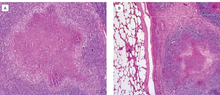
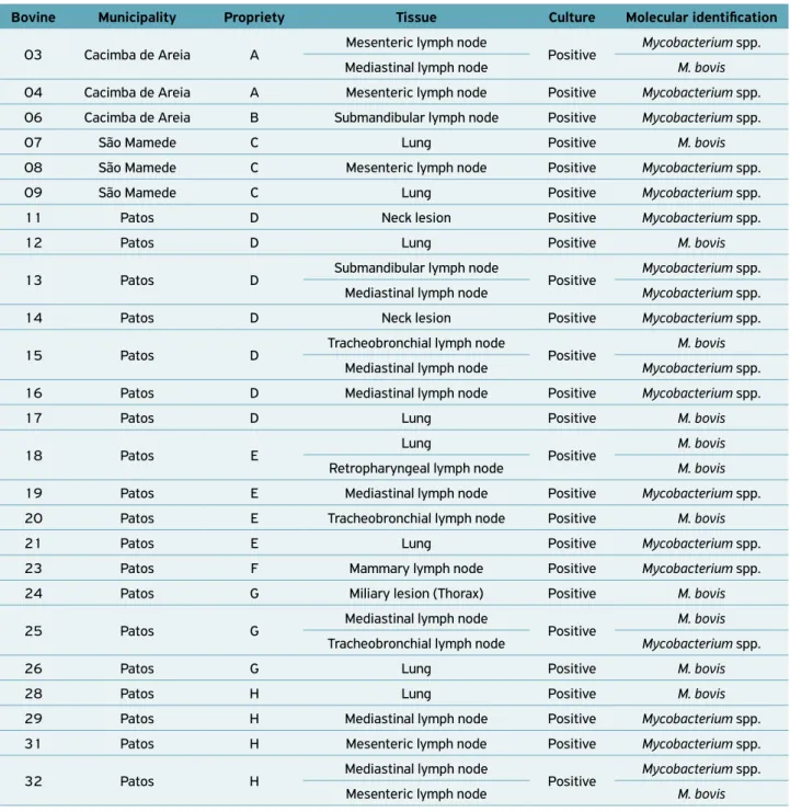
(HEINEMANN et al., 2008). Therefore, effective ante‑mortem surveillance for bovine tuberculosis should be based mainly on the detection of infected animals at early stages using sensitive immunodiagnostic tests (RUA‑DOMENECH et al., 2006). Furthermore, at necropsy, 21 (65.6%) animals presented lesions suggestive of tuberculosis. In fact, the recognition of macro‑ scopic lesions associated with tuberculosis, especially during routine sanitary inspections at slaughterhouses, is an important tool for infection diagnosis, contributing to the identification of foci in herds (CAZOLA et al., 2015). In addition, this is one of PNCEBT’s strategies of action in collaboration with the official sanitary inspection service, which is fundamen‑ tal for warranting the supply of products posing low‑risk to public health and, consequently, consumer protection (BIFFA et al., 2010; CAZOLA et al., 2015). In other countries such as the USA, Australia and Spain, where inspection services in slaughterhouses and programs for the eradication of tubercu‑ losis are satisfactory, the prevalence of the disease was reduced (KANTOR; RITACCO, 2006; RUA‑DOMENECH, 2006). In this study, nodular granulomatous lesions with focal and disseminated calcified and caseous appearance, varying in size and shape (Figs. 1A and 1B), were macroscopically observed. Moreover, a granulomatous inflammatory process was microscopically observed at different stages of evolution, with an extensive granuloma characterized by central area coagulative necrosis, homogeneous eosinophilic material, fragmented nuclei, nuclear remnants and foci of mineraliza‑ tion, surrounded by inflammatory infiltrates predominantly of macrophages and epithelioid cells, encapsulated by abun‑ dant fibrous connective tissue associated with several layers of mononuclear cells (Figs. 2A and 2B).
Similar findings were observed by MENDES et al. (2013), who used bovine animals slaughtered at a Federal Inspection Service in the State of Santa Catarina and found microscopic lesions characterized by central caseification necrosis composed of homogeneous eosinophilic material, cell debris and a variable
A B
Figure 1. (A) Mediastinal lymph node showing caseous nodules. (B) Lung with disseminated caseous nodules of varying size
amount of mineralization surrounded by large amounts of mac‑ rophages and multinucleated Langhans giant cells, connective tissue and neutrophils in some cases. Similarly, FRANÇA et al. (2013) assessed cattle slaughtered in Bahia and described tuber‑ culous lesions with a slightly different structure of adjacent tis‑ sue and nodules with amorphous mass filled with encapsulated caseous material, sometimes with coalescing multifocal areas.
A total of 121 samples with macroscopic lesions sugges‑ tive of tuberculosis were collected and distributed as follows: 80 (66.1%) lymph node samples (41 mediastinal, 9 mesen‑ teric, 7 submandibular, 13 tracheobronchial, 8 retropharyn‑ geal, and 2 mammary) 37 (30.6%) from the lung, one (0.83%) from the liver and three (2.5%) from miliary lesion in mus‑ cle. As to body region, 94 (77.7%) lesions were found in the thoracic cavity, 15 (12.4%) in the head and 12 (9.9%) in the abdominal cavity. PROAÑO‑PÉREZ et al. (2011) evaluated the distribution of lesions in organs from cattle slaughtered in Ecuador as indicator of the possible transmission route, and verified macroscopic lesions in mediastinal lymph nodes (51.3%), tracheobronchial lymph nodes (23.7%), retropha‑ ryngeal lymph nodes (9.2%), liver (11.8%) and other areas (3.9%). In Ethiopia, 84% of the visible lesions were found in the lungs and thoracic lymph nodes (TEKLUL et al., 2004). Moreover, BIFFA et al. (2012) evaluated 337 carcasses with lesions suggestive of tuberculosis and observed that they were more frequent in the lungs and respiratory lymph nodes (50.9%), followed by mesenteric and intestinal lymph nodes (16.5%). In Brazil, ALZAMORA FILHO et al. (2014) reported lesions suggestive of tuberculosis in the pulmonary parenchyma, head and mediastinal lymph nodes in 75% (135/180) of findings
in sanitary inspection. CAZOLA et al. (2015) reported that, among 13 bovines with positive reaction to the tuberculin test, seven (53.8%) had at least one lesion suggestive of tuberculo‑ sis in retropharyngeal, parotid and pulmonary lymph nodes or in the lung, and six (46.2%) had no lesions suggestive of the disease. These results corroborate the findings of the pres‑ ent study and reinforce the role of the respiratory route in the transmission of mycobacteria. The lower frequency of lesions in the abdominal cavity can be attributed to the fact that, in adult cattle, the oral route is secondary to respiratory, which justifies the greater frequency of lesions in thoracic cavity lymph nodes (PALMER; WALTERS, 2006; TAYLOR et al., 2007).
For the isolation and identification of mycobacteria, 55 samples were used, of which 31 (56.4%) presented lesions suggestive of tuberculosis and 24 (43.6%) had no macroscopic lesions. In total, mycobacteria were isolated in 31 (56.4%) sam‑ ples. In 13 (41.9%) samples, M. bovis was identified, while in the other 18 (58.1%) samples, Mycobacterium spp. was present (Table 1). From an epidemiological point of view, it is very important to identify the species of mycobacteria and the pos‑ sible source of infection for humans, since the disease caused by M. tuberculosis and M. bovis in humans is indistinguishable by clinical, radiological and pathological methods (ROCHA et al., 2011; WEDLOCK et al., 2002). However, informa‑ tion about the prevalence of M. bovis infection in humans is scarce, since in most cases the isolation and identification of the agent is not carried out, which makes it impossible to identify the source of infection. On the other hand, it is esti‑ mated that about 3.1% of cases of human tuberculosis around the world are caused by M. bovis (EL SAYED et al., 2015).
Figure 2. (A) Mediastinal lymph node showing central area of necrosis, with homogeneous eosinophilic material, fragmented nuclei, nuclear remnants and foci of mineralization. Adjacent to the area of necrosis, there is granulomatous inflammation consisting of macrophages, epithelioid cells and numerous lymphocytes. There is also a discrete proliferation of fibrous connective tissue. Obj. 10X, HE. Bar scale = 100 μm; (B) Lung with extensive granuloma characterized by central area of coagulative necrosis,
surrounded by inflammatory infiltrates predominantly of macrophages and epithelioid cells encapsulated by abundant fibrous connective tissue associated with several layers of mononuclear cells. Obj. 5X, HE. Bar scale = 200 μm.
Thus, the combination of mycobacterial isolation from bovine tissues and molecular identification contributes to a better understanding of the epidemiology of M. bovis infections, thus increasing the efficiency of disease control programs (CAZOLA et al., 2015).
The identification of Mycobacterium spp. in 18 (58.1%) samples with positive isolation, most isolates coming from mediastinal (6; 33.3%) and mesenteric (4; 22.2%) lymph nodes, was an important part of this study. These isolates are likely to be nontuberculous mycobacteria (NTMs), which are present in the environment and can be transmitted to
both animals and humans through inhalation or inges‑ tion, resulting in permanent or temporary colonization of the respiratory and digestive tracts (PRIMM et al., 2004). Mycobacterium gordonae, M. fortuitum, M. intracellulare, M. flavescens, M. duvalii, M. haemophilum, M. immunogenum, M. lentiflavum, M. mucogenicum, M. novocastrense, M. para-fortuitum, M. smegmatis, M. terrae, and M. vaccae are among the NTM species that have already been identified in animals with positive tuberculin test in Brazil (FRANCO et al., 2013). In another study also conducted with animals testing positive for tuberculin, the following species were identified through
Table 1. Results of culture analysis and identification of samples using TB Multiplex‑PCR for amplification of M. tuberculosis complex and RD‑4 primers for identification of M. bovis in the state of Paraíba, from March to November 2014.
Bovine Municipality Propriety Tissue Culture Molecular identification
03 Cacimba de Areia A Mesenteric lymph node Positive Mycobacterium spp.
Mediastinal lymph node M. bovis
04 Cacimba de Areia A Mesenteric lymph node Positive Mycobacterium spp.
06 Cacimba de Areia B Submandibular lymph node Positive Mycobacterium spp.
07 São Mamede C Lung Positive M. bovis
08 São Mamede C Mesenteric lymph node Positive Mycobacterium spp.
09 São Mamede C Lung Positive Mycobacterium spp.
11 Patos D Neck lesion Positive Mycobacterium spp.
12 Patos D Lung Positive M. bovis
13 Patos D Submandibular lymph node Positive Mycobacterium spp.
Mediastinal lymph node Mycobacterium spp.
14 Patos D Neck lesion Positive Mycobacterium spp.
15 Patos D Tracheobronchial lymph node Positive M. bovis
Mediastinal lymph node Mycobacterium spp.
16 Patos D Mediastinal lymph node Positive Mycobacterium spp.
17 Patos D Lung Positive M. bovis
18 Patos E Lung Positive M. bovis
Retropharyngeal lymph node M. bovis
19 Patos E Mediastinal lymph node Positive Mycobacterium spp.
20 Patos E Tracheobronchial lymph node Positive M. bovis
21 Patos E Lung Positive Mycobacterium spp.
23 Patos F Mammary lymph node Positive Mycobacterium spp.
24 Patos G Miliary lesion (Thorax) Positive M. bovis
25 Patos G Mediastinal lymph node Positive M. bovis
Tracheobronchial lymph node Mycobacterium spp.
26 Patos G Lung Positive M. bovis
28 Patos H Lung Positive M. bovis
29 Patos H Mediastinal lymph node Positive Mycobacterium spp.
31 Patos H Mesenteric lymph node Positive Mycobacterium spp.
32 Patos H Mediastinal lymph node Positive Mycobacterium spp.
sequencing: M. engbaekii, M. arupense, M. nonchromogenicum and M. heraklionense (BOLAÑOS et al., 2018).
Currently, the increase in number of cases of individuals infected with human immunodeficiency virus (HIV) leads to the rise in number of cases of emerging and reemerging diseases, especially those caused by opportunistic etiological agents such as Mycobacterium spp. In this context, direct contact with sources of infection and consumption of contaminated meat, milk and dairy products pose a serious risk of agent transmission to indi‑ viduals with HIV and other immunosuppressive conditions.
CONCLUSIONS
Conclusion is that the isolation and identification of M. bovis and Mycobacterium spp. in bovines with positive reaction to tuberculin test in the state of Paraíba, northeast Brazil, suggests
that humans are exposed to the risk of infection. This result reinforces the need for intensification and optimization of pre‑ vention and control measures outlined in the PNCEBT, such as the incentive to certification of controlled and free rural properties for tuberculosis and measures of measures of animal movement and fairs control. Moreover, particular attention should be given to sanitary inspection of slaughterhouses to identify outbreaks of the disease, as well as to isolate and classify mycobacteria in other states of the northeast region of Brazil.
ACKNOWLEDGMENTS
To the National Council for Scientific and Technological Development (CNPq) for the financial support (number 302222/2016‑2), and to the Agricultural and Livestock Defense Agency of the State of Paraíba.
REFERENCES
ALZAMORA FILHO, F.; VASCONCELOS, S. E. G.; GOMES, H. M.; CAVALCANTE, M. P.; SUFFY, P. N.; COSTA, J. N. Múltiplas estirpes de isolados de Mycobacterium bovis identificados por tipagem molecular em bovinos abatidos em matadouros‑frigoríficos.
Pesquisa Veterinária Brasileira, Seropédica, v.34, n.2, p.103‑108, 2014.
AMBROSIO, S. R.; OLIVEIRA, E. M. D.; RODRIGUEZ, C. A. R.; FERREIRA NETO, J. S.; AMAKU, M. Comparison of three decontamination methods for Mycobacterium bovis isolation.
Brazilian Journal of Microbiology, São Paulo, v.39, n.2, p.241‑244, 2008.
BEHMER, O. A.; TOLOSA, E. M. C.; FREITAS NETO, A. G. Manual de
técnicas para histologia normal e patológica. Ed. Art. São Paulo: EDUSP, 1976. 259 p.
BIFFA, D.; BOGALE, A.; GODFROID, J.; SKJERVE, E. Factors associated with severity of bovine tuberculosis in Ethiopian cattle. Tropical Animal Health and Production, Edinburgh, v.44, n.5, p.991–998, 2012.
BIFFA, D.; BOGALE, A.; SKJERVE, E. Diagnostic efficiency of abattoir meat inspection service in Ethiopia to detect carcasses infected with Mycobacterium bovis: Implications for public health.
BMC Public Health, London, v.10, n.462, p.1‑12, 2010.
BOLAÑOS, C.A.D.; FRANCO, M.M.J.; SOUZA FILHO, A.F.; IKUTA, C.Y.; BURBANO‑ROSERO, E.M.; FERREIRA NETO, J.S.; HEINEMANN, M.B.; MOTTA, R.G.; PAULA, C.L.; MORAIS, A.B.C.; GUERRA, S.T.; ALVES, A.C.; LISTONI, F.J.P.; RIBEIRO, M.G. Nontuberculous mycobacteria in milk from positive cows in the intradermal comparative cervical tuberculin test: implications for human tuberculosis infections. Revista do Instituto de Medicina Tropical,
São Paulo, v.60, p.1‑8, 2018.
BRASIL. MINISTÉRIO DA AGRICULTURA, PECUÁRIA E ABASTECIMENTO ‑ MAPA. Programa Nacional de controle e erradicação da brucelose e da tuberculose animal (PNCEBT). Brasília: MAPA, 2006. 188 p. (Manual técnico).
______. MINISTÉRIO DA SAÚDE ‑ MS. Secretária de Vigilância em Saúde. Departamento de Vigilância Epidemiológica. Sistema de Informação de Agravos de Notificação – Sinan. Available from: <http:// portalsaude.saude.gov.br/images/pdf/2015/setembro/24/ Casos‑novos‑tuberculose‑1990‑2014‑base‑jun‑2015.pdf>. Accessed on: Jan. 10 2016.
CAZOLA, D.O.; JORGE, K.S.G.; ZUMÁRRAGA, M.J.; SOUZA‑FILHO, A.F.; FLÁBIO, R.; ARAÚJO, F.R.; ANA LUIZA, A.R.; OSÓRIO, A.R.O. Identificação e genotipagem de Mycobacterium bovis em bovinos positivos no teste intradérmico para tuberculose em Mato Grosso do Sul. Pesquisa Veterinária Brasileira, Seropédica, v. 35, n. 2, p.141‑147, 2015.
EL‑SAYED, A.; EL‑SHANNAT, S.; KAMEL, M.; CASTAÑEDA‑VAZQUEZ, M.A.; CASTAÑEDA‑VAZQUEZ, H. Molecular epidemiology of Mycobacterium bovis in humans and cattle. Zoonoses and Public Health, Indianapolis, v.63, n.4, p.251‑264, 2015.
FRANÇA, L.R.; CRUZ, J.F.; NEVES, V.B.F.; CERQUEIRA, R.B. Prevalência e histopatologia de lesões sugestivas de tuberculose em carcaça de bovinos abatidos no Sudoeste da Bahia. Revista Brasileira Saúde e Produção Animal, Salvador, v.14, n.4, p.721‑733, 2013.
FRANCO, M.M.J.; PAES, A.C.; RIBEIRO, M.G.; PANTOJA, J.C.F.; SANTOS, A.C.B.; MIYATA, M.; LEITE, C.Q.F.; MOTTA, R.G.; LISTONI, F.J.P. Occurrence of mycobacteria in bovine milk samples from both individual and collective bulk tanks at farms and informal markets in the southeast region of São Paulo, Brazil. BMC Veterinary
HEINEMANN, M.B.; MOTA, P.M.P.C.; LOBATO, F.C.F.; LEITE, R.C.; LAGE, A.P. Tuberculose bovina: uma introdução à etiologia, cadeia epidemiológica, patogenia e sinais clínicos. Cadernos Técnicos de Veterinária e Zootecnia, Belo Horizonte, v.59, p.1‑12, 2008.
KANTOR. I.N.; RITACCO, V. An update on bovine tuberculosis programmes in Latin American and Caribbean countries. Veterinary Microbiology, Geneva, v.112, n. 2‑4, p.111‑118, 2006.
MAZARS, E.; LESJEAN, S.; BANULS, A.; GILBERT, M.; VICENT, V.; GICQUEL, B.; TIBAYRENC, M.; LOCHT, C.; SUPPLY, P. High‑resolution minisatellite‑based typing as a portable approach to global analysis of Mycobacterium tuberculosis molecular epidemiology. Proceedings of the National Academy of Sciences of the United States of America, Washington, v.98, n.4, p.1901‑1906, 2001.
MENDES, R.E.; SCHNEIDER, A.F.; WERLICH, D.E.; LUCCA, N.J.; LORENZETT, M.P.; PILATI, C. Estudo anatomopatológico em tecidos condenados pelo serviço de inspeção federal (SIF) por suspeita de tuberculose. Ciência Animal Brasileira, Goiânia, v.14, n.4, p.448‑453, 2013.
OCEPEK, M.; PATE, M.; ZOLNIR‑DOVC, M.; POLJAK, M. Transmission of Mycobacterium tuberculosis from human to cattle. Journal of Clinical Microbiology, Washington, v.43, n.7, p.3555–3557, 2005.
PALMER, M.V.; WATERS, W.R. Advances in bovine tuberculosis diagnosis and pathogenesis: what policy makers need to know.
Veterinary Microbiology, Geneva, v.112, n.2‑4, p.181‑190, 2006.
PRIMM, T.P.; LUCERO, C.A.; FALKINHAM, J.O. Health Impacts of Environmental Mycobacteria. Clinical Microbiology Reviews, Washington, v.17, n.1, p.98‑106, 2004.
PROAÑO‑PÉREZ, F.; BENITEZ‑ORTIZ, W.; DESMECHT, D.; CORAL, M.; ORTIZ, J.; RON, L.; PORTAELS, F.; RIGOUTS, L.; LINDEN, A. Post‑mortem examination and laboratory‑based analysis for the diagnosis of bovine tuberculosis among dairy cattle in Ecuador. Preventive Veterinary Medicine, Amsterdam, v.101, n.1‑2, p.65‑72, 2011.
ROCHA, A.; ELIAS, A.R.; SOBRAL, L.F.; SOARES, D.F.; SANTOS, A.C.; MARSICO, A.G.; HACKER, M.A.; CALDAS, P.C.; PARENTE, L.C.; SILVA, M.R.; FONSECA, L.; SUFFYS, P.; BOÉCHAT, N. Genotyping did not evidence any contribution of Mycobacterium bovis to human tuberculosis in Brazil. Tuberculosis, Edinburgh, v.91, n.1, p.14–21, 2011.
RODRIGUEZ, C.A.R.; ZUMÁRRAGA, M.J.; OLIVEIRA, E.M.D.; CATALDI, A.A.; ROMANO, M. I.; OTTO, H.H.; BONAFÉ, V.L.;
FERREIRA NETO, J.S. Caracterização molecular de isolados de Mycobacterium bovis do Estado de São Paulo Brasil, utilizando a técnica de spoligotyping. Arquivos do Instituto Biológico, São Paulo, v.71, n.3, p.277‑282, 2004.
RUA‑DOMENECH, R. Human Mycobacterium bovis infection in the United Kingdom: Incidence, risks, control measures and review of the zoonotic aspects of bovine tuberculosis. Tuberculosis, Edinburgh, v. 86, n. 2, p. 77‑109, 2006.
RUA‑DOMENECH, R.; GOODCHILD, A.T.; VORDERMEIER, H.M.; HEWINSON, R.G.; CHRISTIANSEN, K.H.; CLIFTON‑HADLEY, R.S. Ante mortem diagnosis of tuberculosis in cattle: a review of the tuberculin tests, gamma‑interferon assay and other ancillary diagnostic techniques. Research in Veterinay Science, London, v.81, n.2, p.190–210, 2006.
TAYLOR, G.M.; WORTH, D.R.; PALMER, S.; JAHANS, K.; HEWINSON, G. Rapid detection of Mycobacterium bovis DNA in cattle lymph nodes with visible lesions using PCR. Veterinary Research, London, v.3, n.12, p.1‑11, 2007.
TEKLU, A.; ASSEGED, B.; YIMER, E.;GEBEYEHU, M.; WOLDESENBET, Z. Tuberculous lesions not detected by routine abattoir inspection: the experience of the Hossana municipal abattoir, southern Ethiopia. Revue Scientifique et Technique (International Office of
epizootics), Hannover, v.23, n.3, p.957‑964, 2004.
WARREN, R.M.; GEY VAN PITTIUS, N.C.; BARNARD, M.; HESSELING, A.; ENGELKE, E.; DE KOCK, M.; GUTIERREZ, M.C.; CHEGE, G.K.; VICTOR, T.C.; HOAL, E.G.; VAN HELDEN, P.D. Differentiation of Mycobacterium tuberculosis complex by PCR amplification of genomic regions of difference. International Journal of Tuberculosis and Lung Disease, Paris, v.10, n.7, p.818–822, 2006.
WEDLOCK, D.N.; SKINNER, M.A.; LISLE, G.W.; BUDDLE, B.M. Review Control of Mycobacterium bovis infections and the risk to human populations. Microbes and Infection, Amsterdam, v.4, n.4, p.471–480, 2002.
WILTON, S.; COUSING, D. Detection and identification of multiple mycobacterial pathogens by DNA amplification in a single tube. Genome Research, Cold Spring Harbor, v.1, n.4, p.269‑273, 1992.
ZANINI, M.S.; MOREIRA, E.C.; LOPES, M.T.P.; OLIVEIRA, R.S.; LEÃO, S.C.; FIORAVANTI, R.I.; ROXO, E.; ZUMÁRRAGA, M.; ROMANO, M.I.; CATALDI, A.; SALAS, C.E. Mycobacterium bovis: Polymerase Chain Reaction identification in bovine lymphonode biopsies and genotyping in isolates from Southeast Brazil by Spolygotyping and Restriction Fragment Length Polymorphism. Memórias do Instituto Oswaldo Cruz, Rio de Janeiro, v.96, n.6, p.809‑813, 2001.