This study aimed to evaluate the in vitro antifungal susceptibility of Candida species
of head-and-neck-irradiated patients (Group 1), non-institutionalized (Group 2) and institutionalized elders (Group 3) using Etest® methodology. Candida was isolated from saliva and presumptively identified by CHROMagar Candida®, confirmed by morphological criteria, carbohydrate assimilation (API 20C AUX®) and genetic typing (OPE 18). The collection was made from 29, 34 and 29 individuals (Groups 1, 2 and 3, respectively) with 67 isolates. Etest® strips (ketoconazole, itraconazole, fluconazole, amphotericin B and flucytosine) on RPMI (Roswell Park Memorial Institute) agar, on duplicate, were used to evaluate susceptibility. ATTC (American Type Culture Collection) 10231 (Candida albicans) was used as quality control. Among the 67 isolates of Candida species, most
were susceptible to azoles, flucytosine and amphotericin B. None of the isolates showed resistance and dose-dependent susceptibility to amphotericin B. There were nine strains resistant to itraconazole, six to fluconazole and two to ketoconazole and ten dose-dependent, mainly to flucytocine. The highest MIC (minimum inhibitory concentration) to
C. albicans, C. tropicalis, C. parapsilosis was 2.671 μg.mL-1, 8.104 μg.mL-1, 4.429 μg.mL-1,
all for flucytosine. C. krusei and C. glabrata were associated with higher MIC for azoles and C. glabrata with higher MIC to flucytosine. In summary, susceptibility to all tested
antifungal agents was evident. The isolates were more resistant to itraconazole and dose-dependent to flucytosine. A comparison of C. albicans in the three groups showed
no outliers. Higher MIC was associated with C. krusei and C. glabrata.
Antifungal Susceptibility
In Vitro
Determined by the Etest
®
for
Candida
Obtained from the Oral Cavity of
Irradiated and Elderly Individuals
Edimilson Martins de Freitas1, Larissa Cavalcanti Monteiro², Michelle Bonfim
da Silva Fernandes1, Hercílio Martelli Junior1, Paulo Rogério Ferreti Bonan²,
Sergio Avelino Mota Nobre1
1UNIMONTES - State University of Montes Claros, Montes Claros, MG, Brazil
²UFPB - Federal University of Paraiba, João Pessoa, PB, Brazil
Correspondence: Paulo Rogério Ferreti Bonan, Universidade Federal da Paraíba, Cidade Universitária, Bairro Castelo Branco, 58051-900 João Pessoa, PB, Tel: +55-83-3216-7200. e-mail: [email protected]
Key Words:Candida, susceptibility,
Etest®, radiotherapy.
Introduction
Emergent resistant populations of Candida spp.
treated with antifungals have been frequently reported, demonstrating the need to determine the levels of susceptibility of clinical isolates (1,2). Invasive fungal infections are important infections and their prevalence is increasing in immunocompromised patients, such as oncologic patients (3). Candidiasis presentation, in the course of radiotherapy for head and neck, is variable, including erythematous and pseudomembranous forms. The occurrence of oral candidiasis in patients undergoing radiation treatment is mainly related to the subsequent qualitative and quantitative changes in the salivary gland after radiation (4). During the course of radiotherapy, the diversification of Candida species as well as the growing incidence of colonization by species such as C. albicans and C. tropicalis are increased (4-8). There are few studies investigating thoroughly the effectiveness/resistance of antifungal agents in irradiated head and neck cancer patients and resistance to certain antifungals (6,8).
Etest® is a reliable method to verify the sensitivity/
resistance of Candida species to antifungal agents. The
assays using this method involve investigating in vitro responses using antimicrobial strips on RPMI (Roswell Park Memorial Institute)culture media (9).
Using the Etest methodology, this study aimed to measure the susceptibility/resistance profiles of isolates of Candida sampled from the oral cavity of patients irradiated on the head and neck, and institutionalized and non-institutionalized elderly individuals.
Material and Methods
Origin of Biological Samples
Isolates of fungi were collected from in the oral cavity of individuals categorized into three groups: Group 1: 29 individuals undergoing radiation therapy on head and neck and undergoing cervicofacial field radiotherapy (10th and 23rd sessions), with daily doses from 180 to 200
100
E.M. de Freitas et al.
conditions predisposing to candidiasis (broad spectrum antimicrobials, xerostomia, immunosuppression), grouped by age (over 60 years old). Of them, 20 (58.8%) were males and 14 (41.2%) were females with a mean age of 66.35 years (median: 64.5 years). Group 3 was the reference group and consisted of 29 elderly residents of a nursing home, without the candidiasis predictive conditions of Group 2. Of them, 7 (24.1%) were males and 22 (75.9%) were females with a mean age of 73.69 years (median: 74 years). All groups presented mostly partial or totally edentulous individuals without prosthetic rehabilitation.
Isolation, Cultivation, Identification and Conservation of Yeast
The isolation of yeasts was performed using saliva samples collected from the oral mucosa and tongue with a sterile swab. After collection, each swab was placed in a sterile tube containing 1.5 mL of saline (NaCl, 0.85%), placed in a cooler (≈20 °C) and delivered to the laboratory. The content of organic material retained on the swab was detached into the saline solution, and 100 mL aliquots were distributed on 90 mm duplicate plates containing CHROMagar Candida® and incubated at 37 °C, allowing for the selective growth and morphological differentiation of Candida species (10).
The colonies grown on the selective medium were presumptively identified and isolated in Sabouraud agar supplemented with 1% chloramphenicol. Each colony of Candida spp. isolate was catalogued and then stored at -2 °C in YPD (Yeast Peptone Dextrose) broth (1% yeast extract, peptone 2% and 2% dextrose) with glycerol (40% v/v). Sixty-seven different strains were obtained. Among these, 36 isolates were from Group 1, 20 isolates from Group 2, and 11 isolates were from Group 3.
The taxonomic confirmation of isolates was performed using morphological criteria, referencing structures such as blastoconidia, chlamydosphore and pseudohyphae, as well as carbohydrate assimilation using the API 20 C AUX®
identification test (BioMérieux Company, Marcy-l’Étoile, France) and genotyping by RAPD-PCR using the OPE 18 primer (11). When it was necessary to differentiate C. albicans and C. dubliniensis from other species, growth was assessed at different temperatures (12,13). Some isolates were not consistent with the taxonomy using the applied methods; these cases were treated as Candida spp.
Evaluation of Antifungal Susceptibility
The MIC (minimum inhibitory concentration) was measured using Etest strips (Probac Brazil, São Paulo, SP, Brazil) (14-17). The antifungal agents used for susceptibility testing in this study were azoles (fluconazole (FL), ketoconazole (KE) and itraconazole (IT)), amphotericin B
(APB; polyenic) and flucytosine (FC; pyrimidine).
Preparation of Inoculates
Colonies identified with 24 h of culture in Sabouraud agar supplemented with chloramphenicol (1% v/v) were suspended in saline (NaCl 0.85%), which was calibrated by turbidimetry, using as reference 0.5 on the McFarland scale. A 0.1 mL aliquot of the inoculum calibrated suspension was spread on Petri dishes (150 mm) using a sterile swab in RPMI-1620 agar, alternated with glucose (0.2%) and MOPS (0.165 M, pH 7.0) (Probac Brazil) (18). The procedure was performed as recommended by the manufacturer (AB Biodisk, Solna, Sweden). The reading of MIC values was done between 24 and 48 h after incubation at 37 °C. The reference strain ATCC 10231 - C. albicans was used for quality control.
MIC Readings
After incubation, when growth was visible (24 and 48 h), the value of MIC was read at the point of intersection between the halo and the Etest strip. For APB, the MIC was read at the point of complete inhibition (100%) for FC and almost complete (95%) inhibition of growth. The MIC for azoles was read at the first point of significant inhibition (80% of visible growth). The MIC values (µg.mL-1) of the
Etest tape were interpreted as sensitive and resistant using the NCCLS reference (18).
Statistical Analysis
The statistical analysis was performed with SPSS 17.0 (SPSS, Chicago, IL, USA) using descriptive parameters, position and dispersion of values within each group of sampled individuals. For the most frequently encountered Candida species, the confidence interval (CI) was calculated with a significance level α=0.05.
Ethical Aspects
The work was conducted in accordance with the precepts established by resolution 466/12 of the Brazilian National Health Ministry of Health (Brazil), and by resolution 179/93 of the Code of Professional Dental Ethics.
Results
In Group 1 (irradiated patients), C. tropicalis (25.0%) was the most prevalent species, followed by C. parapsilosis and C. albicans (16.7% for both). In Group 2 (non-institutionalized elderly people), C. albicans was present in 45% of cases, followed by C. parapsilosis (20.0%) and C. guilliermondii (15.0%). In Group 3 (institutionalized elderly people), C. albicans was the only identified species.
Antifungal susceptibility of Candida study groups regarding susceptibility (S), dose-dependent
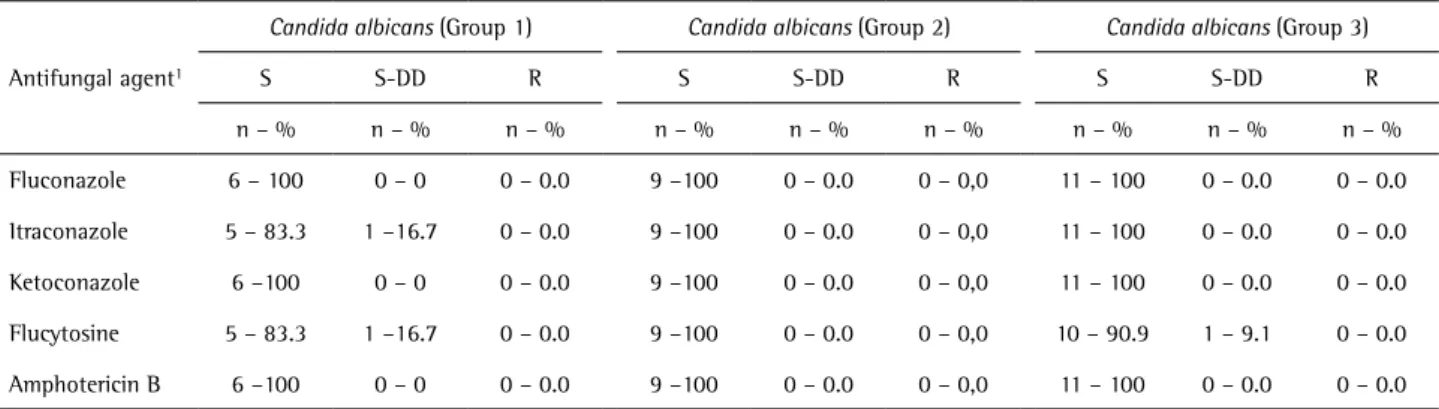
susceptibility (S-DD) and resistant (R). There was one S-DD species for IT and one for FC in Group 1 and one S-DD strain for FC in Group 3. All the other C. albicans strains were S for all antifungals. The MIC for susceptibility testing of the quality control strain (C. albicans ATCC 10231) for azoles (average of duplicate plates) was 0.625 µg.mL-1 for
FL, 0.007 µg.mL-1 for KE and 0.079 µg.mL-1 for IT. For FC, the
mean was 0.158 µg.mL-1 and for APB it was 0.023 µg.mL-1.
Based on the susceptibility test results with the Etest for 67 isolates of Candida species, most of them were susceptible to azoles, FC and APB. None of the isolates showed resistance or dose-dependent susceptibility to APB. Table 2 describes the overall conditions found for all species according to the Etest. This study found nine R to IT, six to FL and two to KE, and ten S-DD mainly to FC. Table 3 shows the MIC for antifungals, ranges, and lower and upper bounds, referring to species. For FL, the highest MIC was for C. krusei. For IT and KE, the highest MIC was for C. glabrata and C. krusei, respectively. The highest MIC for FC and APB were for C. krusei and C. spp., respectively.
Discussion
The reproducibility and reliability of the Etest is evident. In agreement with this, the present study tested strains of C. albicans isolated from the oral cavity of patients with fixed orthodontic appliances and their susceptibility to antifungal agents (FL, KE and APB), based on the Clinical and Laboratory Standards Institute (M44-A protocol) and by Etest, showing overall 100% agreement between the disk diffusion method and the Etest for FC and APB (19) .
A study conducted by Belazi et al. (17) with patients irradiated on the head and neck using the Etest showed FL resistance for some C. albicans strains and for isolates of C. krusei (MIC>32 µg.mL-1). The same strains were S-DD for IT. In this study, in Group 1 (irradiated patients) six strains (100%) of C. albicans were S for FL and one was S-DD for FC and IT. Only one strain (9.1%) of C. albicans (Group 3) was S-DD for FC and all C. albicans (Groups 2 and 3) were S for all the tested antifungals. These findings probably indicate different behaviors of this species in non-similar conditions. Considering all species, strains of C. krusei presented higher MIC values for FC, KE and FL, corroborating
Table 1. Levels of susceptibility of isolates of Candida albicans from the oral cavity of individuals in the three groups proposed by Wingeter et al. (21). (Sensitive - S; Sensitive dose-dependent - SDD and resistant - R)
Antifungal agent1
Candida albicans (Group 1) Candida albicans (Group 2) Candida albicans (Group 3)
S S-DD R S S-DD R S S-DD R
n – % n – % n – % n – % n – % n – % n – % n – % n – %
Fluconazole 6 – 100 0 – 0 0 – 0.0 9 –100 0 – 0.0 0 – 0,0 11 – 100 0 – 0.0 0 – 0.0
Itraconazole 5 – 83.3 1 –16.7 0 – 0.0 9 –100 0 – 0.0 0 – 0,0 11 – 100 0 – 0.0 0 – 0.0
Ketoconazole 6 –100 0 – 0 0 – 0.0 9 –100 0 – 0.0 0 – 0,0 11 – 100 0 – 0.0 0 – 0.0
Flucytosine 5 – 83.3 1 –16.7 0 – 0.0 9 –100 0 – 0.0 0 – 0,0 10 – 90.9 1 – 9.1 0 – 0.0
Amphotericin B 6 –100 0 – 0 0 – 0.0 9 –100 0 – 0.0 0 – 0,0 11 – 100 0 – 0.0 0 – 0.0
Table 2. Levels of susceptibility of isolates of Candida from the oral cavity of individuals in the three groups proposed by Wingeter et al. (21) (Sensitive - S; Sensitive doses dependent – S-DD and resistant - R)
Susceptibility
Antifungal agent1
Azoles Pirimidyne Polyenic
FL IT KE FC APB
n % n % n % n % n %
S 60 89.6 52 77.6 59 88.1 57 85.1 67 100.0
SDD 1 1.5 6 9.0 6 9.0 10 14.9 -
-R 6 9.0 9 13.4 2 3.0 - - -
-Total 67 100.0 67 100.0 67 100.0 67 100.0 67 100.0
102
E.M. de Freitas et al.
Table 3. MIC average and range for antifungals according to Candida species
Drugs Species n Mean
95% confidence interval for mean
Minimum Maximum
Lower bound Upper bound
FL
C. albicans 26 .09623 .08229 .1017 .035 .190
C. dubliniensis 3 .10967 .07116 .14818 .094 .125
C. tropicalis 10 .61950 .36604 .87296 .190 1.250
C. krusei 2 182.00 -1317.332 1681.332 64.000 300.000
C. glabrata 3 67.000 -123.59991 257.59991 9.000 154.000
C. parapsilosis 10 30.584 -37.13381 98.30361 .064 300.000
C.guilliermondii 4 75.266 -163.13484 313.667 .250 300.000
C. lusitaniae 3 .24667 -.08877 .58210 .110 .380
C. kefyr 3 .04100 -.00554 .08754 .020 .056
C. famata 1 .09400 . . .094 .094
C. spp 2 150.02 -1755.60861 2055.65561 .047 300.000
Total 67 22.118 4.18995 40.04718 .020 300.000
IT
C. albicans 26 .0523 .0376 .0671 .02 .19
C. dubliniensis 3 .0540 -.0333 .1413 .03 .09
C. tropicalis 10 .2518 -.0720 .5756 .02 1.50
C. krusei 2 5.2500 -29.6921 40.1921 2.50 8.00
C. glabrata 3 7.8333 -14.1177 29.7844 2.00 18.00
C. parapsilosis 10 .2070 -.0661 .4801 .02 1.25
C. guilliermondii 4 3.6438 -7.3459 14.6334 .06 14.00
C. lusitaniae 3 .0347 -.0123 .0816 .02 .06
C. kefyr 3 .0057 -.0044 .0157 .00 .01
C. famata 1 .0470 . . .05 .05
C. spp 2 2.1875 -20.8425 25.2175 .38 4.00
Total 67 .8840 .1642 1.6039 .00 18.00
KE
C. albicans 26 .00585 .00516 .00653 .001 .010
C. dubliniensis 3 .00633 .00116 .01150 .004 .008
C. tropicalis 10 .01600 .00916 .02284 .007 .040
C. krusei 2 .56500 -1.78565 2.91565 .380 .750
C. glabrata 3 .48567 -.19338 1.16471 .285 .797
C. parapsilosis 10 .11620 -.10608 .33848 .006 1.000
C. guilliermondii 4 .26100 -.52296 1.04496 .010 1.000
C. lusitaniae 3 .01133 .00560 .01707 .010 .014
C. kefyr 3 .00200 .00200 .00200 .002 .002
C. famata 1 .00800 . . .008 .008
C. spp 2 .43900 -5.10091 5.97891 .003 .875
Total 67 .09030 .03183 .14877 .001 1.000
FC
C. albicans 26 2.67119 -.52326 5.86564 .072 40.000
C. dubliniensis 3 13.3753 -43.90303 70.65369 .047 40.000
C. tropicalis 10 8.10400 -3.92172 20.12972 .040 40.000
C. krusei 2 40.000 40.00000 40.00000 40.000 40.000
C. glabrata 3 .13600 -.32879 .60079 .024 .352
C. parapsilosis 10 4.42950 -4.54942 13.40842 .047 40.000
C. guilliermondii 4 10.2585 -21.29739 41.81439 .064 40.000
C. lusitaniae 3 .05933 -.05068 .16935 .028 .110
C. kefyr 3 .77100 .14343 1.39857 .500 1.000
C. famata 1 .02300 . . .023 .023
C. spp 2 20.0005 -234.11724 274.11824 .001 40.000
Total 67 5.95327 2.63808 9.26845 .001 40.000
APB
C. albicans 26 .0503 .0267 .0738 .02 .32
C. dubliniensis 3 .0207 .0106 .0307 .02 .02
C. tropicalis 10 .0972 .0387 .1557 .02 .22
C. krusei 2 .1605 -.9767 1.2977 .07 .25
C. glabrata 3 .2193 .0874 .3513 .16 .25
C. parapsilosis 10 .1538 -.0593 .3669 .02 1.00
C. guilliermondii 4 .1558 -.0735 .3850 .03 .35
C. lusitaniae 3 .0713 -.0588 .2015 .02 .13
C. kefyr 3 .0870 .0170 .1570 .06 .11
C. famata 1 .0640 . . .06 .06
C. spp 2 .2435 -2.2533 2.7403 .05 .44
Antifungal susceptibility of Candida previous findings (17).
Moreover, considering the overall MIC, APB showed higher values and absence of R or S-DD, corroborating a previous result (20). In fact, S for APB determined by in vitro analysis is a common finding, although R has been found in few cases in another study (21). On bloodstream candidiasis, overall S rates were 98.0 % for APB and 98.7 % for FL (22). In our study, referring to FL, the rate was 89.6% to S. This difference could be explained by the different microbiota found in these studies, including different rates of prevalence.
Referring to IT and FL activities, the present study showed higher R overall in the evaluated species compared with other antifungals. In fact, a previous study that evaluated 160 strains of yeast-like fungi cultured from samples from the lower respiratory tract, blood, peritoneal
cavity and other areas showed more R to IT in Candida
species using the Etest (23). Another study investigating 159 clinical isolates of Candida species from patients with invasive candidiasis in a Kuala Lumpur hospital revealed six isolates R to FL, comprising two isolates of C.albicans, two of C. parapsilosis, one C. tropicalis and one C. glabrata; all of these isolates showed cross-resistance to IT (24).
Evaluating S to FC, a study of blood-borne candidiasis showed R to FC in two (0.8%) C. albicans isolates, seven (9.3%) C. tropicalis strains, three (1.6%) C. parapsilosis isolates and all ten (100%) of the C. krusei investigated isolates (25). Adding, in the present study, C. glabrata presented the highest MIC for IT. As previously shown, C. glabrata presents 100% susceptibility to APB and caspofungin and is the least susceptible to IT, posaconazole and voriconazole (23).
In summary, a different pattern of S-DD was observed to IT in C. albicans collected from irradiated patients. Nine R to IT, six to FL and two to KE, and ten S-DD mainly to FC were found. For FL, the highest MIC was for C. krusei. For IT and KE, the highest MIC was for C. glabrata and C. krusei, respectively. The highest MIC for FC and APB were for C. krusei and C. spp., respectively.
Resumo
Esse estudo objetivou avaliar a susceptibilidade antifúngica in vitro
de espécies de Candida obtidas de pacientes irradiados em cabeça e pescoço (Grupo 1), idosos não institucionalizados (Grupo 2) e idosos institucionalizados (Grupo 3) usando a metodologia Etest®. Candida foi isolada da saliva e identificada presuntivamente pelo teste CHROMagar Candida®, confirmada pelo critério morfológico, assimilação de carboidratos API 20C AUX® e identificação genética (OPE 18). A coleta foi feita em 29, 34 e 29 indivíduos (Grupos 1, 2 and 3, respectivamente) com 67 isolados. As fitas de Etest® (cetoconazol, itraconazol, fluconazol, anfotericina B and flucitosina) em meio ágar RPMI (Roswell Park Memorial Institute), em duplicata, foram utilizados para avaliar a susceptibilidade. A ATTC (American Type Culture Collection) 10231 (Candida albicans) foi
usada como controle de qualidade. Dos 67 isolados de espécies de Candida, a maioria foi susceptíveis aos azoles, flucitosina e anfotericina B. Nenhum
dos isolados mostrou resistência ou susceptibilidade dose-dependente a anfotericina B. Houve nove espécies resistentes ao itraconazol, seis ao fluconazol e duas ao cetoconazol e dez dose-dependentes, principalmente a flucitosina. Os maiores valores de MIC (mínima concentração inibitória) para C. albicans, C. tropicalis, C. parapsilosis foram, respectivamente, 2,671 μg.mL-1, 8,104 μg.mL-1, 4, 429 μg.mL-1, todos para a flucitosina. C. krusei
e C. glabrata foram associadas a um maior MIC para azoles e C. glabrata
com maior MIC para flucitosina. Em resumo, a susceptibilidade a todos os antifúngicos testados foi evidente. Os isolados foram mais resistentes ao itraconazol e dose dependentes para a flucitosina. A comparação para
C. albicans nos três grupos não mostrou diferença. Os maiores valores de MIC estavam relacionados a C. krusei e C. glabrata.
Acknowledgements
The authors would like to thank the Foundation for Research Support of Minas Gerais (FAPEMIG), Minas Gerais, Brazil, for financial support, and Marise Silveira for biostatistical services.
References
1. Costa KRC, Ferreira JC, Komesu MC, Candido RC. Candida albicans and Candida tropicalis in oral candidosis: quantitative analysis, exoenzyme activity, and antifungal drug sensitivity. Mycopathol 2009;167:73–79. 2. Sun J, Qi C, Lafleur MD, Qi Q. Fluconazole susceptibility and genotypic
heterogeneity of oral Candida albicans colonies from the patients with cancer receiving chemotherapy in China. Int J Oral Sci 2009;1:156–162. 3. Low CY, Rotstein C. Emerging fungal infections in immunocompromised
patients. F1000 Medicine Reports 2011;3:14.
4. Suryawanshi H, Ganvir SM, Hazarey VK, Wanjare VS. Oropharyngeal candidosis relative frequency in radiotherapy patient for head and neck cancer. J Oral Maxillofac Pathol. 2012;16:31-37.
5. Blozis GG, Robinson JE. Oral tissue changes caused by radiation therapy and their management. Dent Clin North Am 1968;6:643-656. 6. Redding SW, Dahiya MC, Kirkpatrick WR, Coco BJ, Patterson TF,
Fothergill AW, et al.. Candida glabrata is an emerging cause of oropharyngeal candidiasis in patients receiving radiation for head and neck cancer. Oral Surg Oral Med Oral Pathol Oral Radiol Endod. 2004;97:47-52.
7. Azizi A, Rezaei M. Prevalence of Candida species in the oral cavity of patients undergoing head and neck radiotherapy. J Dent Res Dent Clin Dent Prospects. 2009;3:78-81.
8. Panghal M, Kaushal V, Kadayan S, Yadav JP. Incidence and risk factors for infection in oral cancer patients undergoing different treatments protocols. BMC Oral Health 2012;12:22.
9. Sanitá PV, Mima EGO, Pavarina AC, Jorge JH, Machado AL, Vergani CE. Susceptibility profile of a Brazilian yeast stock collection of Candida species isolated from subjects with Candida-associated denture stomatitis with or without diabetes. Oral Surg Oral Med Oral Pathol Oral Radiol.2013;116:562-569.
10. Bernal S, Mazuelos EM, Garcia M, Aller AI, Martinez MA, Gutierrez MJ. Evaluation of CHROMagar Candida medium for the isolation and presumptive identification of species of Candida of clinical importance. Diagn Microbiol Infect Dis 1996;24:201-204.
11. Varguez BL, García AC, Tanaka LV, García SS, Cepeda LAG, Vargas LOS, et al.. Comparison of a randomly amplified polymorphic DNA (RAPD) analysis and ATB ID 32C system for identification of clinical isolates of different Candida species. Rev Iberoam Micol 2007;24:148-151. 12. Kirkpatrick WR, Revankar SG, Mcatee RK, Ribot-L JL, Fothergill AW,
Mccarthy DI, et al.. Detection of Candida dubliniensis in oropharyngeal samples from human immunodeficiency virus-infected patients in North America by primary CHROMagar Candida screening and susceptibility testing of isolates. J Clin Microbiol 1998;36:3007–3012. 13. Jabra-Rizk MA, Brenner TM, Mark R, Baqui AAMA, Merz WG, Falkler
WA, et al.. Evaluation of a reformulated CHROMagar Candida. J Clin Microbiol 2001;39:2015–2016.
104
E.M. de Freitas et al.
in vitro: a study on 100 patients with superficial candidiasis. An Bras Dermatol 2004;79:689-697.
15. Silva MRR, Costa MR, Miranda ATB, Fernandes OFL, Costa CR, Claudete R, et al.. Evaluation of Etest and macrodilution broth method for antifungal susceptibility testing of Candida sp strains isolated from oral cavities of AIDS patients. Rev Inst Med Trop 2002;44:121-125. 16. Alexander BD, Byrne TC, Smith KL, Hanson KE, Anstrom KJ, Perfect
JR, et al.. Comparative evaluation of Etest and Sensititre YeastOne panels against the Clinical and Laboratory Standards Institute M27-A2 reference broth microdilution method for testing Candida susceptibility to seven antifungal agents. J Clin Microbiol 2007;45:698– 706.
17. Belazi M, Koussidou-Eremondi T, Andreadis D, Minis S, Arrenis G. Oral Candida isolates in patients undergoing radiotherapy for head and neck cancer: prevalence, azole susceptibility profiles and response to antifungal treatment. Oral Microbiol Immunol 2004;19:347-351. 18. National Committee for Clinical Laboratory Standards. Publication
M27-A: Reference method for broth dilution antifungal susceptibility testing of yeasts: Approved Standard. National Committee for Clinical Laboratory Standards 2002;22:1-30.
19. Carvalhinho S, Costa AM, Coelho AC, Martins E, Sampaio A. Susceptibilities of Candida albicans mouth isolates to antifungal agents, essentials oils and mouth rinses. Mycopathologia 2012;174:69-76.
20. Karbach J, Walter C, Al-Nawas B. Evaluation of saliva flow rates,
Candida colonization and susceptibility of Candida strains after head and neck radiation. Clin Oral Investig. 2012;16:1305-1312.
21. Wingeter MA, Guilhermetti E, Shinobu CS, Takaki I, Svidzinski TI. Microbiological identification and in vitro sensitivity of Candida isolates from the oral cavity of HIV positive individuals. Revista da Sociedade Brasileira de Medicina Tropical 2007;40:272-276. 22. Bustamante B, Martins MA, Bonfietti LX, Szeszs MW, Jacobs J, Garcia
C, et al.. Species distribution and antifungal susceptibility profile of Candida isolates from bloodstream infections in Lima, Peru. J Med Microbiol 2014;63:855-860.
23. Gołaś M, Netsvyetayeva I, Sikora M, Piskorska K, Sulik-Tyszka B, Swoboda-Kopeć E. Trends in antifungal susceptibility of Candida species - one year observation. Pol J Microbiol 2014;63:217-222. 24. Amran F, Aziz MN, Ibrahim HM, Atiqah NH, Parameswari S, Hafiza
MR, et al.. In vitro antifungal susceptibilities of Candida isolates from patients with invasive candidiasis in Kuala Lumpur Hospital, Malaysia. J Med Microbiol 2011;60:1312-1316.
25. Pahwa N, Kumar R, Nirkhiwale S, Bandi A. Species distribution and drug susceptibility of Candida in clinical isolates from a tertiary care centre at Indore. Indian J Med Microbiol 2014;32:44-48.