Abstract published online: October 13, 2008
Full paper published online: May 30, 2009
Original paper. ISSN 1678-9199.
SCREENING THE TOXICITY AND TOXIN CONTENT OF BLOOMS OF THE CYANOBACTERIUM Trichodesmium erythraeum (EHRENBERG) IN
NORTHEAST BRAZIL
Proença LAO (1), Tamanaha MS (1), Fonseca RS (1)
(1) Center of Technological Earth and Marine Sciences, Vale do Itajaí University, Itajaí, Santa Catarina State, Brazil.
ABSTRACT: Blooms of the cyanobacterium Trichodesmium occur in massive colored patches over large areas of tropical and subtropical oceans. Recently, the interest in such events has increased given their role in major nitrogen and carbon dioxide oceanic fluxes. Trichodesmium occurs all along the Brazilian coast and patches frequently migrate towards the coast. In this paper we screen the toxicity and toxin content of Trichodesmium blooms off the coast of Bahia state. Four samples, collected from February to April 2007, were analyzed. Organisms were identified and assessed for toxicity by means of several methods. Analogues of microcystins, cylindrospermopsins and saxitoxins were analyzed using HPLC. Microcystins were also assayed through ELISA. Results showed dominance of T. erythraeum, which makes up as much as 99% of cell counts. Other organisms found in smaller quantities include the dinoflagellates Prorocentrum minimum and P. rhathymum. Extracts from all samples delayed or interrupted sea urchin larval development, but presented no acute toxicity during a mouse bioassay. Saxitoxin congeners and microcystins were present at low concentrations in all samples, occurrences that had not previously been reported in the literature. Despite our finding of saxitoxin analogues and microcystins in Trichodesmium blooms, these toxins do not represent a potential harm to human health by primary contact. We conclude, based on our results and those reported in the recent literature, which differ from results published in 1963, that although toxins are present, there is no evidence that T. erythraeum blooms represent a threat to humans.
KEY WORDS: saxitoxin, microcystin, harmful algae, Trichodesmium, bloom.
CONFLICTS OF INTEREST: There is no conflict.
CORRESPONDENCE TO:
INTRODUCTION
The genus of the cyanobacterium Trichodesmium (T. erithraeum Ehrenberg ex
Gomont) was first found by Ehrenberg (1830) in the Red Sea. Since then, several
species – including T. erythraeum Ehrenberg, T. thiebautii Gomont ex Gomont, T.
contortum(Wille) Wille, T. radians (Wille) Golubić and T. hildebrantii Gomont – have
been identified (1). Trichodesmium form blooms that can occur in a large area
covering the tropical and subtropical regions of the Atlantic, Pacific and Indian
oceans as well as the China and Caribbean Seas. The genus is generally
characterized by cylindrical trichomes and forms colonies with varied morphology (2).
Lately, the interest in Trichodesmium blooms has increased due to their biological
and ecological characteristics that include fixation of atmospheric nitrogen and
carbon dioxide (3). Diazotrophic cells do not show differentiated structures like other
cyanobacteria and, as they usually occur in oligotrophic waters, their impact on
global fluxes of nitrogen and carbon as well as on organic matter production are of
great interest.
Usually, species of Trichodesmium are found in the most stable surface layers, and
within the mixed layer down to 100 meters depth. During the blooms, conspicuous
extensive patches can be seen even from space by satellites (3). This pattern is
possibly due to the presence of gas vacuoles that provide positive flotation and
accessory pigments, permitting Trichodesmium to resist high levels of radiation
exposure. The colors of the patches change according to the cell’s physiological
state, and can be greenish, brownish, whitish, yellowish or reddish (4, 5). The
structure of the plankton community is affected by Trichodesmium due to the
production of toxins that can act allopathically against grazers (6).
Trichodesmium is found along most of the Brazilian coast and is associated with the
Brazilian Current. It occurs along the coast of Rio Grande do Sul, Santa Catarina (4),
Paraná (7, 8) and São Paulo (9-11). The study by Satô, Paranaguá and Eskinazi (5)
is one of the most frequently cited papers on this topic and reports blooms off the
coastal area of Recife, in northeastern Brazil. The authors associate the illness of
beach users, known as Tamandaré’s fever or “tinguí”, with the occurrence of T.
erythraeum blooms (5). Along the coast of Santa Catarina state, Trichodesmium
blooms are generally found from November through April and are associated with
calm periods and low nitrogen-to-phosphorous ratios (7, 4). The patches can be as
about 80 km off the coast of São Paulo (8). Despite their widespread occurrence, no
other publications have connected T. erythraeum blooms to human illness, except for
the study published by Satô, Paranaguá and Eskinazi (5). However, it is currently
reported as a potentially harmful species.
This paper aims to investigate the occurrence of Trichodesmium blooms in Camamu
bay in the coast of Bahia, northeast Brazil, and to characterize their toxicity and
potential harmful effects.
MATERIALS AND METHODS
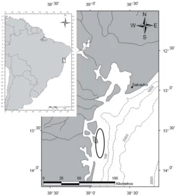
Four samples were collected from four different patches of Trichodesmium ssp.
blooms near the coastal platform off Camamu-Almada, in the Boipeba Island and
Maraú peninsula on the coast of Bahia state (Figure 1). The samples were collected
on 02/01/2007 (#1; 13°78´33´ W, 38°90´50´ S); 02/08/2007 (#2; 13°75´27´ W,
38°90´85´ S); 04/05/2007 (#3; 13°78´72´ W, 38°91´64´ S) and 04/07/2007 (#4;
13°75´99´ W, 38°86´50´ S) and then fixed in a 2% formalin solution to permit the
identification and counting of phytoplankton. The scum was concentrated on a piece
of filter paper and frozen at –18°C for toxin analysis and toxicity assays.
Samples fixed in formalin were observed through a binocular microscope for species
identification. Trichodesmium cell counts were calculated by determining the average
number of cells per trichome (30 trichomes per sample) in a 1 mL Sedgwick-Rafter
and expressing the final cell counts as cell/L. The other organisms were counted in
an IX 50 inverted microscope (Olympus, Japan) using Utermhöll 3 mL settling
chambers at respective magnifications of 200 and 400x (12). All organisms were
identified to the most precise taxonomic level possible (2, 13-15).
The presence of phycotoxins was investigated by high performance liquid
chromatography (HPLC) and bioassays. Each sample was initially weighed (wet
weight) and then dried at 40°C for 24 hours in an oven and subsequently reweighed
(dry weight). The dried samples were extracted into 3 to 5 mL volumes of 80%
methanol. A 500 µL subsample was saved for HPLC analysis as described
subsequently. Before the assays, the extracts were clarified in fiberglass filters and
dried in a rotoevaporator to eliminate the methanol. Again, a subsample was
preserved for the sea urchin test and the remainder partitioned with ethyl acetate.
The two phases were dried and assayed by means of a mouse bioassay.
For the sea urchin larval assay, to test its activity, 10 mL of the extract was put into
15-mL flasks, after correcting the salinity to 30%. In each flask, 200 µL of fertilized
egg suspension, containing a minimum of 300 eggs, was added. Eggs and sperm of
Lytechinus variegatus Lamarck had been obtained by previous spawning induction
with injections of KCl 0.1 M. The test was carried out for 24 hours at controlled
temperature (25°C) and illumination (12:12 light and dark cycle) (16). The test ended
with the addition of formalin solution (4%). Six dilutions of the initial extract from the
dried material were tested (100, 50, 25, 12.5, 6.1 and 3.7%) and compared with the
control containing marine water.
The mouse bioassays were based on Harada et al. (17) and consisted of the
intraperitoneal inoculation of 1 mL of both hydrophilic and lipophilic phases of the
extract. Three Swiss mice, weighing between 17 and 20 grams were used, and
tested for 24 hours with water and food ad libidum. The extracts had been diluted to
obtain a salinity of 0.9%. The injected dose was based on the toxicity scale for
cyanobacteria, as follows: more than 1,000 mg/kg → non-toxic; 1,000 to 500 mg/kg → low toxicity; 500 to 100 mg/kg → high toxicity (18). ELISA assays for microcystin were performed only on the sample collected on 04/07/2007 using a microcystin
HPLC analysis of saxitoxin and congeners, microcystins and cylindrospermopsin
were carried out on the initial 80% methanolic extract using a HPLC system
(Shimadzu, Japan) as described below. Saxitoxin and congeners were analyzed
according to the post-column derivatization method (19). The chromatography was
conducted at 30°C on a Luna® 4.6 x 250 mm column (Phenomenex, USA) filled with
5 µm C8 particles. After the chromatography, the column eluents were derivatized
with buffered 50 nM periodic acid and stabilized with acetic acid using a two-piston
LC 10® pump (Shimadzu, Japan). The peaks were detected on a RF 10Alx®
fluorometer (Shimadzu, Japan). Two runs with different mobile phases were
performed for each sample to separately determine gonyautoxins (GTX1-GTX4),
decarbamoyl gonyautoxins (dcGTX2, dcGTX3) and saxitoxin (STX), neosaxitoxin
(neoSTX) and decarbomoyl neosaxitoxin (dcSTX). The system was calibrated with a
standard purchased from the National Research Council, Canada.
HPLC analysis of cylindrospermopsin was carried out according to methods
described by Li et al. (20), using a Lichrosphere RP 18® (Merck , Germany) (5 μm)
(125 x 4.6 mm) column whereas detection was conducted by means of a diode array
SPD M10A® (Shimadzu, Japan). The process was calibrated with pure
cylindrospermopsin acquired from CyanoHAB, USA. The same column and
equipment were employed to investigate microcystin. The system was calibrated with
pure microcystin-LR (Sigma Chemical Co., USA). The four standards (Dleu-LR-Mic,
LY, LW and LF) were kindly provided by Dr. Yunes from the Federal University of Rio
Grande, FURG.
RESULTS AND DISCUSSION
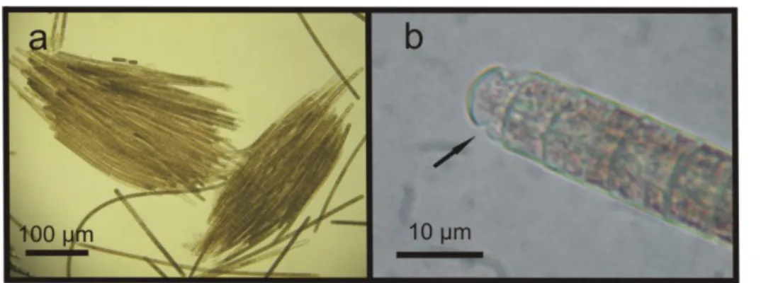
The microscopic analysis in all samples confirmed that the blooming organism,
forming the patches, was the cyanobacterium T. erythraeum, with cell counts as high
as 577.20 x 10⁶ cell/L (found in sample #2 from 02/08/2007). The conspicuous
brownish patches presented different shapes and sizes, ranging from a few meters to
several kilometers. The lowest count, 8.32 x 10⁶ cell/L, was found in sample #4 from
04/07/2007. The solitary or colony-forming parallel straight T. erythraeum trichomes,
with an attenuated end cell, presented cells measuring from 7.5 to 15 µm in width
and 5 µm in height. The trichome length ranged from 246 to 417 µm with a cell
number ranging from 49 to 83. The number of trichomes per colony varied from 29 to
Figure 2. Trichodesmium erythraeum, from sample #2, showing (a) parallel trichomes and, in detail, (b) showing the caliptra (arrow).
Despite the presence of other organisms – which included diatoms; dinoflagellates
such as Prorocentrum rhathymum Loeblich, Shirley & Schmidt and P. minimum
(Pavillard) Schiller; tintinids (samples #1, 3, 4) and copepods (#2) – their numbers
were very low when compared to Trichodesmium (Table 1).
Table 1. List of phytoplankton taxa and their abundances in the four samples analyzed from the Camamu-Almada region
Samples
Phytoplankton taxa 1 2 3 4
Cyanophyta
Trichodesmium erythraeum (x 106) 11.24 577.20 19.00 8.32
Bacillariophyta
Hemiaulus hauckii 3700
Mastogloia minuta 10764
Navicula sp. 26572
Nitzschia sp. 333 1333 221662
Plagiotropis sp. 12109
Pseudo-nitzschia sp. 5382
Thalassionema sp. 333 12109
Gomphonema sp. 333
Dinophyta
Prorocentrum minimum 3333
Prorocentrum rhathymum 333 4667
Extracts from all samples showed antimitotic activity observed in sea urchin larvae,
even with the more diluted extracts. These results agree with previous studies that
found the presence of substances toxic to invertebrates in extracts of T. erythaeum
from the São Paulo coast (10). Guo and Tester (6) showed that while intact, cells
were not toxic to the copepod Acartia tonsa Dana, but their hydrosoluble extracts
revealed toxicity, indicating the presence of endotoxins that may be released into the
water. Hawser et al. (21) found substances that were only toxic to microcrustaceans
in T. thiebautii extracts, but not in those from T. erythraeum. Despite this finding, it
appears that such toxicity does not have a direct impact on mussel aquaculture as in
other algae, such as dinoflagellates (22). Rather, these authors found that mussels
can be affected by the low nutritional value of these cyanobacteria.
The mouse bioassay did not show acute toxicity. All results from both hydrophilic and
lipophilic extracts were negative in the 24-hour test. Some adjustments were
necessary to decrease the salinity of extracts by dilution with water, since differences
in salinity could mask the results. Although we have found the presence of toxins, as
described below, the doses were not sufficient to produce death among tested mice
in the manner described in freshwater cyanobacteria toxicity assays (23).
Saxitoxin analogues were measured by HPLC analysis in all extracts, expressed as
parts per million (Table 2). At this concentration, the extract did not kill mice under the
test conditions. In all nine analogues analyzed, we found only decarbamoyl saxitoxin
(dcSTX) and gonyautoxin 3 (GTX 3), which were expressed as a saxitoxin equivalent
(19).
Table 2. Toxin concentrations of scum extracts from the Camamu-Almada region
Sample Saxitoxin analogues
STX eq. µg/g
Microcystins µg/g
#1 1.95 89.6
#2 3.40 82.7
#3 9.45 302.0
#4 2.28 9.7
Cylindrospermopsin was not found in the analysis. Table 2 shows the results
all samples by HPLC analysis and were confirmed by ELISA in the sample from
04/07/2007.
The results show that all samples were toxic. The role of toxic substances in
Trichodesmium spp. is related mainly to allelopathic functions, which may be very
important since these organisms live in environments with low nutrient availability,
indicating that competition for this resource may be high (6). Therefore, allopathic
substances could be of evolutionary importance. In the same manner, toxic
compounds might limit or restrict the grazing on Trichodesmium by microcrustaceans
or even by fish. On the other hand, compounds other than the toxins analyzed, such
as MIC and STX analogues, can have similar effects since several components can
be produced by the same organisms (6, 10, 21, 24).
The presence of microcystin has been reported in samples from a T. erythraeum
bloom from Canary Island region (25). Sudek et al. (24) reported the presence of a
cyclic peptide, called trichamine, with biological activity. Both, saxitoxin and
microcystins are produced by freshwater cyanobacteria including Microcystis,
Planktothrix, Oscillatoria, Nostoc, Anabaena, Anabaenopsis and Hapalosiphon.
These are also found in transitional environments including estuaries, whereas
recent studies have shown the presence of microcystin in tiny (picoplankton)
Synechococcus spp. in marine environments (26, 27). Therefore, it is possible that
the microcystin found in the present study is not necessarily related to
Trichodesmium erithraeum but rather to co-occurring organisms, or others such as
picoplanktonic cyanobacteria. The same is valid for saxitoxin. In extracts from
Australian Trichodesmium, Negri et al. (22) did not observe the presence of saxitoxin.
But these toxins were observed in a Trichodesmium erythraeum bloom off the Rio
Grande do Sul coast, Brazil, in March 2004 (28). In this event, SXT, GTX1, GTX3
and GTX4 were identified and toxicity was confirmed by the mouse bioassay. In
marine environments, several dinoflagellates produce these toxins, as do some
bacteria (29). In our case, further assays will be necessary to investigate the true
source of the toxins. Samples of T. erythraeum from off the Santa Catarina coast did
not present either mice toxicity or microcystin (4). It is possible, therefore, that other
co-occurring organisms may also produce the toxic compounds found in these tests.
Prorocentrum rhathymun and P. minimum, for example, are dinoflagellates that can
In humans, both saxitoxin and microcystin toxins can become problematic. Drinking
water containing microcystins and other cyanotoxins causes shortages in water
supplies and can lead to several health problems (23). In Brazil, the presence of such
compounds in potable and recreational water is regulated by specific laws (31).
However, microcystin does not cause harm through direct contact. Thus, although
the toxins are present in these microorganisms, we do not consider these blooms a
health problem. There is no report of Trichodesmium toxin magnification through
trophic chains by mussels, filter feeders or fishes, in contrast with dinoflagellates (22).
Blooms reaching mussel culture areas in Santa Catarina have not caused any
observable damage.
Despite their broad geographic occurrence along the Brazilian coast, up to now, the
only reported harmful effect on humans, published by Satô et al. in 1963 (5), was
human illness along the coast of Pernambuco (more precisely, at Tamandaré, a city
by the sea). Since that episode, no other harmful event has been registered in Brazil.
For example, in 2004, a large portion of Cassino beach, in Rio Grande do Sul, was
covered by a T. erythraem bloom but no injurious effects were registered (28). The
same was true for Santa Catarina, Paraná and Bahia states. The etiology of the
Tamandaré event needs to be investigated more thoroughly. In the study that reports
the illness associated with the incident, no attempts were made to elucidate the toxin
origin or to perform toxicity tests. Since then no other paper has been published
relating human illness to the presence of Trichodesmium spp. blooms.
In general, no direct actions have been taken following the discovery of
Trichodesmium spp. blooms in any place. In spite of this, CETESB, the
environmental agency of São Paulo state, usually advises people not to use beaches
during an algal bloom. This may be a valid precautionary procedure since it is known
that some cyanobacteria can cause dermatitis, and that dinoflagellates may produce
toxins carried by aerosols. At this point, we cannot definitively associate harmful
effects with Trichodesmium spp. blooms.
REFERENCES
1. Janson S, Siddiqui PIA, Walsby AE, Romans KM, Carpenter EI, Bergmen B.
Cytomorphological characterization of the planktonic diazotrophic cyanobacteria
Trichodesmium spp. from the Indian Ocean and Caribean and Sargasso Seas. J
2. Desikachary TV. Cyanophyta. New Delhi: Indian Council Agricultural Research;
1959. 686 p.
3. Capone DG, Zehr JP, Paerl HW, Bergman B, Carpenter EJ. Trichodesmium, a
globally significant marine cyanobacterium. Science. 1997;276(5316):1221-9.
4. Rörig L, Yunes JS, Kuroshima K, Schettini CAF, Pezzuto PR, Proença LAO.
Studies on the ecology and toxicity of Trichodesmium spp. blooms in Southern
Brazilian coastal waters. 8th International Conference on Harmful Algae; 1998.
Espanha: Harmful Algae; 1998. 22-5 p.
5. Satô S., Paranaguá MN., Eskinazi E. On the mechanism of red TIDE of
Trichodesmium in Recife north eastern Brazil, with some considerations of the
relation to the human disease Tamandaré Fever. Trabhs Inst Oceanogr (Univ
Recife). 1966;5(6):7-49.
6. Guo C, Tester PA. Toxic effect of the bloom-forming Trichodesmium sp.
(Cyanophyta) to the copepod Acartia tonsa. Nat Toxins. 1994;2(4):222-7.
7. Brandini FP. Composição e distribuição do fitoplâncton na região sudeste do
Brasil e suas relações com as massas de água (Operação sudeste julho/agosto
1982). Ciência e Cultura. 1988;40(4):334-41.
8. Siqueira A, Kolm HE, Brandini FP. Offshore distribution patterns of the
cyanobacterium Trichodesmium erythraeum ehrenberg and associated phyto- and
bacterioplankton in the southern Atlantic Coast (Paraná, Brazil). Braz Arch Bioll
Technol. 2006;49(2):323-37.
9. CETESB (Companhia de Tecnologia e Saneamento Ambiental do Estados de São
Paulo). Florações de microalgas no ambiente marinho [Internet]. São Paulo:
CETESB; 2007. Available from:
<http://www.cetesb.sp.gov.br/Agua/praias/algas.asp>.
10. Fonseca ML, Silva MA, Freitas JC. Ensaios preliminares da toxicidade de
extratos de uma amostra de Trichodesmium erythraeum do Canal de São Sebastião,
SP. XVII Simpósio de Biologia Marinha - Cebimar/USP; 2002. São Sebastião, SP:
Caderno de Resumos e Palestras do XVII Simpósio de Biologia Marinha -
Cebimar/USP; 2002.
11. Gianesella-Galvão SMF, Costa MPF, Kurtner MB. Bloom of Oscillatoria
(Trichodesmium) Erythraea (Ehr.) kutz in coastal waters of the Southweast Atlantic.
12. Utermohl H. Zur vervollkommnung der quantitativen phytoplankton methodik. Mitt
int Verein theor angew Limnol. 1958;9(1):1-38.
13. Cortés-Altamirano R, Sierra-Beltrán AP. Morphology and taxonomy of
Prorocentrum mexicanum and reinstatement of Prorocentrum rhathymum
(Dinophyceae). J Phycol. 2003;39(1):221-5.
14. Cupp ED. Marine plankton diatoms of the west coast of North America. Bull
Scripps Inst Oceanograf. 1943;5(1):1-238.
15. Steidinger KA, Tangen K. Dinoflagellates. In: Tomas CR, editor. Identifying
marine diatoms and dinoflagellates. San Diego: Academic Press, Inc.; 1996. p.
387-584.
16. CETESB (Companhia de Tecnologia e Saneamento Ambiental do Estado de São
Paulo). Água do mar – teste de toxicidade crônica de curta duração com Lytechinus
variegatus Lamarck, 1816 (Echinodermata: Echinoidea). São Paulo: CETESB; 2002.
22 p.
17. Harada K, Kondo F, Lawton L. Laboratory analysis of cyanotoxins. In: Chorus I.,
Barthram J, editors. Toxic cyanobacteria in water: a guide to their public health
consequences, monitoring and management. New York: World Health Organization,
E&F.N. Spon; 1999. 368-405 p.
18. Lawton L, Beattie KA, Hawser SP, Campbell DL, Codd GA. Evaluation of assay
methods for the determination of cyanobacterial hepatotoxicity. In: Codd GA,
Jefferies TM, Keevil CW, Potter E, editors. Detection methods for cyanobacterial
toxins. Cambridge: The Royal Society of Chemistry; 1994. 111-6 p.
19. Oshima Y. Postcolumn derivatization liquid chromatographic method for paralytic
shellfish toxins. J AOAC Int. 1995;78:528-32
20. Li R, Carmichael WW, Brittain S, Eaglesham GK, Shaw GR, Liu Y, Watanabe
MM. First report of the cyanotoxins cylindrospermopsin and deoxicylindrospermopsin
from Raphidiopsis curvata (Cyanobacteria). J Phycol. 2002;37(6):1121-6.
21. Hawser SP, O'neil JM, Roman MR, Codd GA. Toxicity of blooms of the
cyanobacterium Trichodesmium to zooplankton. J Appl Phycol. 1992;4(1):79-86.
22. Negri AP, Bunter O, Jones B, Llewllyn L. Effects of the bloom-forming alga
Trichodesmium erythraeum on the pearl oyster Pinctada maxima. Aquaculture. 2004;
23. Chorus I, Bartram J. Toxic cyanobacteria in water: a guide to their public health
consequences, monitoring and management. New York: World Health Organization,
E&F.N. Spon; 1999. 416 p.
24. Sudek S, Haygood MG, Youssef DT, Schmidt EW. Structure of trichamide, a
cyclic peptide from the bloom-forming cyanobacterium Trichodesmium erythraeum,
predicted from the genome sequence. Appl Environ Microbiol. 2006;72(6):4382-7.
25. Ramos AG, Martel A, Codd GA, Soler E, Coca J, Redondo A, Morrison LF,
Metcalf JS, Ojeda A, Suárez S, Petit M. Bloom of the marine diazotrophic
cyanobacterium Trichodesmium erythraeum in the northwest African upwelling.
Marine Ecology, 2005;301:303-5.
26. Carmichael WW, Li R. Cyanobacteria toxins in the Salton Sea. Saline Systems.
2006;2:5.
27. Domingos P, Rubim TK, Molica RJR, Azevedo SMFO, Carmichael WW. First
report of microcystin production by picoplanktonic cyanobacteria isolated from a
northeast Brazilian drinking water supply. Environ Toxicol. 1999;14(1):31-5.
28. Silva LM. Ocorrência de cianobactérias no estuário e costa adjacente à Lagoa
dos Patos, Rio Grande, RS: avaliação preliminar dos riscos a balneabilidade
[master’s thesis]. [Rio Grande do Sul (RS)]: Universidade Federal do Rio Grande,
FURG; 2005. 129 p.
29. Kodama M, Ogata T, Sato S, Sakamoto S. Possible association of marine
bacteria with paralytic shellfish toxicity of bivalves. Mar Ecol Prog Ser.
1990;61:203-6.
30. Moestrup Ø, editor. IOC taxonomic reference list of toxic plankton algae.
Intergovernmental Oceanographic Commission of UNESCO [internet]. Available
from: <http://www.bi.ku.dk/ioc/introduction.asp>.
31. Brasil. Ministério da Saúde. Portaria n. 518, de 25 de março de 2004. Brasília: