Research Signpost
37/661 (2), Fort P.O., Trivandrum-695 023, Kerala, India
Vanadium Biochemistry
MONOMERIC VERSUS DECAMERIC VANADATE IN
THE ELUCIDATION OF MUSCLE CONTRACTION
REGULATION: A KINETIC, SPECTROSCOPIC AND
STRUCTURAL OVERVIEW
Teresa Tiago1, Carlos Gutiérrez-Merino2 and Manuel Aureliano1
1) Centre for Marine Sciences (CCMAR) and Faculdade de Ciências e Tecnologia, Universidade do Algarve, Campus de Gambelas, 8005-139 Faro, Portugal; 2) Grupo de Bioenergética en Neuronas y Miocitos, Departamento de Bioquímica y Biología Molecular, Facultad de Ciencias, Universidad de Extremadura, Av. Elvas s/n, 06071 Badajoz, Spain
Abstract
Vanadium (V) was rediscovered for biology as a “muscle inhibitor factor” when it was found in commercial ATP prepared from equine muscle almost thirty years ago. Since then it has been used as a molecular probe of the mechanisms of several enzyme reactions involving hydrolysis of phosphate ester bonds. Besides acting as a phosphate analogue, vanadate has also the potential to exhibit biological activities through oligomeric vanadate species. Among the vanadate oligomers, decavanadate is one of the most potent inhibitors and has revealed an excellent kinetic and spectroscopic probe. This is particularly relevant for myosin, the major muscle ATPase which along with actin is able to convert the chemical energy of ATP hydrolysis into mechanical work. Apparently, vanadate is able to populate different conformational states of the myosin ATPase cycle depending on its oligomerization state. While monomeric vanadate (VO43-) mimics the transition state for the
γ
-phosphatehydrolysis blocking myosin in a pre-power stroke state, decameric vanadate (V10O286-) induces the formation of the
intermediate myosin·MgATP·V10 complex blocking the actomyosin cycle in a pre-hydrolysis state. These recent findings, that are now reviewed, point out to the importance of taking into account vanadate species variety in studies describing the interaction of vanadate with biological systems and incite the use of decavanadate as a biochemical tool to the elucidation of muscle contraction regulation.
.
*Correspondence/Reprint request: Teresa Tiago, Faculdade de Ciências
e Tecnologia, Universidade do Algarve, Campus de Gambelas, 8005-139 Faro, Portugal. e-mail: [email protected]
1. INTRODUCTION
Skeletal muscle cells and vanadium are strongly connected to each other, since Josephson and Cantley [1] identified an impurity in commercial ATP prepared from equine muscle as orthovanadate (V), a potent inhibitor of the sodium pump. Although thirty years have passed since the discovery of this naturally occurring inhibitor, the function of this element in muscle cells is still unidentified. This is not surprising if one consider the exceptional complex chemistry of vanadium in solution especially in what concerns the propensity of vanadium species to condense at higher concentrations or acidic pH’s, forming vanadate oligomers, or to interact with countless compounds of biological interest [2,3].
Numerous researchers working in different areas ranging from life sciences to chemical engineering, using vanadate in their studies, have to deal with the tendency of vanadate species to oligomerize. At neutral pH and through the range of concentrations most frequently employed in biochemical studies, the predominant vanadate species are the monomer (H2VO4-), dimer (H3V2O7-) and tetramer (V4O124-) [4,5]. Given the rapid
anion and consequently an equilibrium mixture must be examined [6], which may complicate studies aimed to determine the effect of a specific anion. At alkaline pH, the monomeric species is more prevalent and therefore researchers frequently prepare “activated” solutions of monomeric vanadate by adjusting the pH of a stock solution of sodium orthovanadate above 10 and heating near 100ºC until the solution is colourless [7]. However, for studies at physiological pH these highly basic vanadate solutions require neutralisation with HCl raising the potential for serious contamination with substantial amounts of decameric species (V10O286-). Since methods for preparing
vanadate solutions are not frequently cited it is uncertain if studies using vanadate solutions also include meta- or decameric species as contaminants.
Our laboratory has dedicated over the last 20 years many efforts to learn more about the contribution of oligomeric vanadate species to vanadium effects on muscle contraction/relaxation, namely on sarcoplasmic reticulum Ca2+-ATPase transmembrane transport system and on myosin and actomyosin ATPase activities. Among the vanadate oligomers, decameric species has revealed the most interesting in biochemical terms. Besides having high potency in a number of systems, the lower lability of this large polyoxoanion allows it to be use as an excellent kinetic and spectroscopic probe. The effects of decavanadate on Ca-pump have been recognized for some time and reviewed elsewhere [chapter 7] and therefore are out of the scope of this review. The focal point of this chapter concerns the use of decameric vanadate as a structural-functional tool for further understanding the mechanism of actomyosin action based on studies combining kinetic, spectroscopic and molecular modelling methodologies. In the next sections our recent findings on the structural/functional interaction of vanadate oligomers with the actomyosin system are summarized and the decameric vanadate binding mode and inhibition mechanism compared to the well established monomeric vanadate reactivity towards myosin.
2. MYOSIN AND ACTOMYOSIN ATPase ACTIVITY INHIBITION BY VANADATE
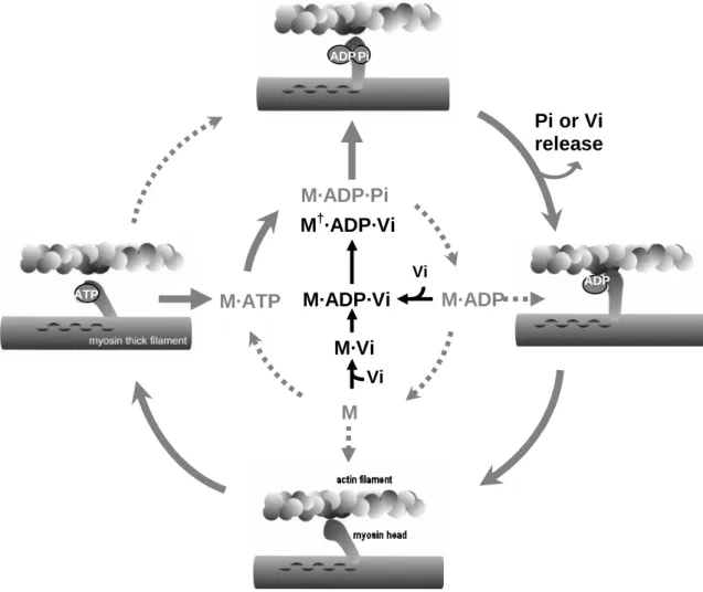
Most of the biological importance of vanadium is associated with the pentavalent form (vanadate) due to the similarities between the phosphate and vanadate chemistries in solution. The enzyme regulatory properties of vanadate depend on its esterification with phosphate or hydroxyl groups of biological molecules [8]. Thus, enzymes involved in phosphohydrolase and phosphotransferase reactions can accept vanadium as an analogue of orthophosphate, forming an inhibitory complex between the enzyme and vanadate. This is particularly relevant for myosin, the major ATPase of muscle which along with actin is able to convert the chemical energy of ATP hydrolysis into mechanical work during the process of muscle contraction [9]. The head segment of myosin, called subfragment-1 (S1), contains the binding sites for ATP and actin, and is responsible for the ATPase activity [10]. During the contractile cycle, binding of ATP reduces the affinity of myosin for actin, and the subsequent hydrolysis of ATP results in a ternary complex between myosin, ADP and inorganic phosphate (Pi). The rate-limiting step of the ATP hydrolysis is the release of Pi from myosin, which is accelerated by the rebinding of actin (Fig. 1). Goodno [11] has originally demonstrated that, in the absence of actin, vanadate inhibits myosin ATPase activity by forming a very stable complex with MgADP that mimics either the transition state of hydrolysis or the ADP·Pi intermediate state. This inactive complex has a stoichiometry of one ADP and one vanadate (Vi) per myosin active site and a half-life of approximately 3 days at
25 ºC. This complex arrests the contractile cycle at a critical point and therefore has provided an opportunity to study this important state.
The behaviour of the myosin·MgADP·Vi complex was also studied in the presence of actin [12,13]. However, just as actin increases the rate of release of Pi from the myosin·MgADP·Pi complex, it also manifestly increases (by 105) the rate of Vi dissociation from the myosin·MgADP·Vi complex [14]. Therefore, there is a considerably difference in the ability of vanadate to inhibit the myosin and the actomyosin ATPase activity. While in the absence of actin Vi inhibits myosin ATPase activity in the submicromolar concentration range, in the presence of actin much higher concentrations (> 900 microM) are needed [13]. Clearly, the need for such high concentrations strongly suggested that this effect could be due to oligomeric vanadate species that are favoured at higher vanadate concentrations and likely to occur through a different mechanism. This possibility was further investigated starting by clarifying which vanadate oligomers interact more potently with myosin and how is this interaction modulated by the myosin natural ligands such as ATP and actin.
Figure 1. Relevant steps of the actomyosin ATP hydrolysis cycle schematically represented (grey) as
perturbed by orthovanadate binding (black). M, myosin; Vi, orthovanadate. The dominant process of ATP hydrolysis observed in vitro is indicated by the filled arrows. Formation of the M·ADP·Vi complex is fast
and reversible but isomerization is essentially irreversible forming a stable ternary complex M†·ADP·Vi
as described by Goodno [11]. AP M·ADP·Pi M M·ADP M·ATP M·ADP·Vi M·Vi Pi or Vi release Vi Vi M†·ADP·Vi Pi ATP ADP ADP ATP
3. VANADATE OLIGOMERS INTERACTION WITH MYOSIN: 51V-NMR SPECTROSCOPY ANALYSIS
Vanadate compounds are excellent probes of their surroundings due to the rapid quadrupolar relaxation in solution, large magnetic moment and high natural abundance (99.76%) of the 51V nucleus. Resolvable 51V NMR spectra can be obtained in a matter of minutes and the chemical shifts can be use to monitor changes in the vanadium (V) coordination sphere as they are very sensitive to changes in the electronic nature of the vanadium atom [15-17]. The distribution of the different vanadate oligomers present in the 51V NMR spectra can reflect changes in ionic strength, concentration, pH and temperature [18-20]. Therefore, 51V NMR spectroscopy besides being a convenient method to monitor the speciation of oxovanadates has proved to be highly informative in biological systems as experiments can be designed to evaluate specific interactions of the different vanadate oligomers with compounds of the biological system [21-26].
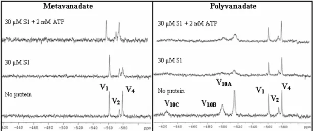
The 51V NMR approach was applied to myosin and subfragment-1 and demonstrated that, in the absence of ATP, of the various forms of vanadate in solution essentially only the tetrameric form binds to the protein [27,28]. Ringel and collaborators showed soon after that the presence of myosin or S1 in a metavanadate solution produced a significant broadening of the signal of each form of vanadate (di-, tetra- and hexa-vanadates) being interpreted as indicative that all of them bind to the protein [29]. Nevertheless, since these oxovanadates are in rapid equilibrium [5], the broadening of the signals is dictated by the total population of free exchanging oxovanadates and may imply that only one of the labile species is binding to the protein. In subsequent studies with meta- and decavanadate solutions it was obtained a relative order of line broadening upon myosin addition of V10 > V4 > V1 where no changes were
observed for monomeric vanadate [30,31].
Decavanadate, contrarily to the labile oxovanadates (V1, V2, V4 and V5) has an
equilibration time with the other species sufficiently long that the disappearance of the NMR signals can be attributed to protein binding [5,6]. Taking advantage of this lower lability, the binding mode of decavanadate to myosin was further explore by analyzing the competition with the other oligomers for the binding sites on myosin under the influence of natural ligands, such as actin and ATP [32]. For that purpose changes in the
51
V NMR signals were monitored upon protein addition to a metavanadate solution (2 mM) containing approximately 766 µM V1, 146 µM V2, and 236 µM V4 (Fig. 2A) and
to a mixed solution containing 2 mM-metavanadate and 5 mM-decavanadate (polyvanadate solution), containing approximately 908 µM V1, 300 µM V2, 219 µM V4
and 461 µM V10 (Fig. 2B). These studies pointed out to a competition between
decameric and tetrameric vanadate species for the same myosin binding sites. The most reasonable explanation for these results is that V10 and V4, as polyanionic species, bind
to a cationic portion of myosin S1 with electrostatic forces involved as it was confirmed by the results, namely: (i) V10 has a greater affinity for the myosin binding sites than V4,
in the same way that, V4 has greater affinity than the lower vanadate oligomers; (ii) V10
interaction is favored for higher ionic strengths (not showed). In addition, these studies confirmed that the interaction of the oligomers with myosin S1 is modulated by the enzyme natural ligands, i.e., is affected in the presence of the nucleotide and appears to be favored in the presence of F-actin filament.
Figure 2. 51V NMR spectra of metavanadate (2.0 mM) and polyvanadate (2.0 nominal metavanadate + 5.0 mM nominal decavanadate) solutions in the absence and presence of 30 µM S1 and 2 mM ATP. The
spectra were collected in a medium containing 25 mM KCl, 25 mM Hepes (pH 7.0) at 25 ºC. V1, V2 and
V4 are referring to monomeric, dimeric and cyclic tetrameric forms of vanadate, respectively. V10A, V10B,
and V10C correspond to the three chemical environmental different vanadate atoms in the decameric
vanadate structure.
4. COMPOSITION OF DECAVANADATE SOLUTIONS AND STABILITY OF DECAMERIC SPECIES
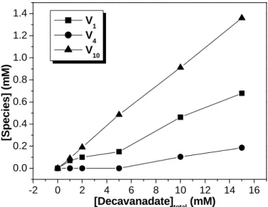
Preparation of vanadate solutions containing decameric species have been developed to study the interaction and/or effects promoted by these species in the biological systems [21,33-39]. Nevertheless, the denominated “decavanadate” solutions containing presumably only the decamer are often contaminated with other species. In fact, the composition of the decavanadate solutions will depend on the time after which the decavanadate stock solution is diluted in the reaction medium and on the conditions of the medium itself (pH, temperature and ionic strength). As appraised by 51V NMR spectroscopy, a decavanadate stock solution of 100 mM total vanadium concentration at pH 4.0 will contain 10 mM decameric vanadate species and, therefore, with no contamination of any other species. Upon dilution into the reaction medium, at physiological pH and room temperature, the decavanadate solution will contain besides the decamer, a contamination of about 5-10% of other vanadate species [40]. This percentage is independent of the total vanadate concentration for a fixed time (approximately 30 min) and therefore V10 shows a linear correlation with the increase in
total vanadate concentration (Fig. 3). Since V10 is in equilibrium with V1, the
appearance of other vanadate oligomers is dependent on V1 concentration, i.e., above
200 µM V1 starts to oligomerize giving rise to di- and tetrameric vanadate species.
Therefore, when a stock solution of decavanadate is diluted into the reaction medium, for instance, at a concentration of 1 mM (total vanadium), 51V-NMR spectra shows an
additional signal from V1 (∼100 µM), that is about 1:1 stoichiometry of decameric and
monomeric vanadate species.
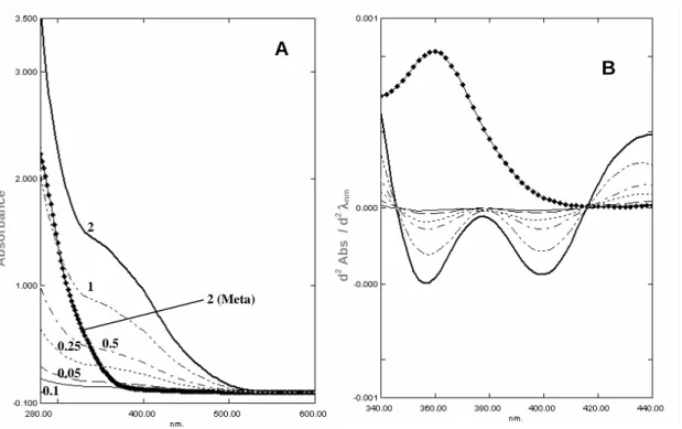
Although NMR spectroscopy is an essential technique for the analysis of vanadate oligomers composition, the poor sensitivity as well as the long time it takes to acquire a spectra, unable its use to follow the dissociation kinetics of decameric species. However, the disappearance of the decamer can be easily followed by UV/Vis spectroscopy. Lacking d-electrons, vanadium(V) shows no transitions in the visible
-2 0 2 4 6 8 10 12 14 16 0.0 0.2 0.4 0.6 0.8 1.0 1.2 1.4 [S p e c ie s ] (m M ) [Decavanadate] total (mM) V1 V 4 V10
region of the electromagnetic spectrum and the yellow color observed for decavanadate solutions is due to an intense absorption tailing in from the ultraviolet [2]. The broad absorption band observed for decameric species (Fig. 4A) is well resolved by the second derivative of the UV/Vis spectra (Fig. 4B) presenting the shape of two valleys with minimum centered at 358 and 400 nm [41]. These two bands are characteristic of decameric species as they are lacking in the 2nd derivative spectra of metavanadate solutions containing mono-, di- and tetrameric species [40]. Moreover, a linear correlation is obtained between the intensity of the two bands and the concentration of decavanadate solution (from 0.05 to 2 mM total vanadium) (Fig. 5). Therefore, decameric species as low as 5 µM (50 µM total vanadium concentration) can be detected by UV/Vis spectroscopy and the dissociation kinetics followed [40]. Decavanadate decomposition follows a first order kinetic process, assessed by measurements of the absorption band at 400 nm as a function of time, with half-life times that are exceedingly dependent on the conditions of the medium including pH, temperature, ionic strength, buffer and other assay components. In addition, decameric vanadate may become inaccessible to decomposition due to their stabilization upon binding to target proteins. An increase of the half-life time from 5 to 17 and 27 hours was found in the presence of sarcoplasmic reticulum vesicles and actin, respectively, at room temperature and pH 7.0 [42]. This stabilization effect is not extendable to all proteins known to interact with decavanadate. For the particular case of myosin, the half-life time of decameric vanadate was found to be the same (~158 min) in the absence or presence of myosin in a medium containing 25 mM KCl, 25 mM Hepes (pH 7.0) at 25 ºC.
Figure 3. Vanadium (V) species composition of decavanadate solutions upon dilution in a reaction
medium containing 25 mM KCl, 25 mM Hepes (pH 7.0) at 25 ºC. A series of 51V NMR spectra of the
decavanadate solutions were recorded at concentrations up to 15 mM (total vanadium concentration). The concentration of each vanadium (V) species was calculated from the fractions of the total integrated areas
observed in the spectra. Notice that V10 shows a linear correlation with the increase in total vanadate
concentration.
Decameric species is thermodynamically unstable but the lability of this oxoanion allows it to be used in studies of limited duration. By selecting the proper conditions, it is possible to determine the effects of decameric species in the activity of enzymes, at physiological pH and room temperature, provided that the putative effects
of the monomeric species are deduced. For this reason, it is desirable to precisely characterize the composition and the interaction of the decavanadate solutions with the system before attempting to understand the promoted effects. In chapter 9 of this book, aspects related with decameric vanadate in vivo effects are being reviewed.
Figure 4. UV/Vis absorption spectra (panel A) and resultant second derivative (panel B) of decavanadate
(0.05; 0.1; 0.25; 0.5; 1 and 2 mM total vanadium) and metavanadate (2 mM) upon dilution of the concentrated stock solutions in 25 mM KCl, 25 mM Hepes (pH 7.0) at 25 ºC. The broad absorption band observed for decameric species is well resolved by the second derivative of the UV/Vis spectra presenting the shape of two valleys with minimum centered at 358 and 400 nm.
5. INHIBITION OF MYOSIN ATPase ACTIVITY AND MODULATION OF ACTIN-MYOSIN INTERACTION BY DECAVANADATE
Among the free or complexed forms of vanadate, decameric species has an outstanding potency in a number of systems. Numerous enzymes were shown to be inhibited by this oxoanion including several kinases, phosphorylases, the glycolytic enzyme aldolase as well as enzymes of inositol phosphate metabolism [reviewed in ref
0.05 0.1 0.25 0.5 1 2 2 (Meta) d 2 A b s / d 2 λλλλ n m A b s o rb a n c e A B 0.0 0.5 1.0 1.5 2.0 -0.5 0.0 0.5 1.0 1.5 2.0 2.5 3.0 3.5 4.0 2 n d d e ri v a ti v e b a n d i n te n s it y ( u .a .) [Decavanadate] (mM) 358 nm 400 nm
Figure 5. Intensity (arbitrary units) of the 2nd
derivative bands (shown in Fig. 4B) as a function of decavanadate concentration (total vanadium concentration). A linear correlation is obtained between the intensity of the two bands and the concentration of decavanadate solution.
6,8]. Rabbit skeletal muscle adenylate kinase was the first enzyme reported to be inhibited by decavanadate (Ki = 1 µM) [43]. Apparently the susceptibility to V10 is due
to the tetraphosphate domain over the AMP/ATP binding site on the enzyme. In fact, decavanadate is known to bind at the polyphosphate binding sites of enzymes or receptors either in the substrate domain or in an allosteric effector site and it does not tend to follow a simple competitive model. For example, hexokinase was reported to be inhibited (Ki = 62 µM) by decavanadate in a noncompetitive way regarding both ATP and glucose [44]. Phosphofructokinase inhibition (Ki = 45 nM) is antagonized by positive allosteric effectors and is synergistic with ATP, suggesting that decavanadate may interact with both the substrate and effector binding sites [44]. Cyclic AMP-dependent protein kinase was found to be non-competitively inhibited regarding ATP (Ki = 0.8 mM) and competitively inhibited regarding the substrate, kemptide (Ki = 1.4 mM), by decavanadate [45]. Ribonuclease A inhibition by decavanadate suggests a model with more subtlety than a simple competition with poly(C) for the active site [46].
Further evidence for the interaction of decavanadate at polyphosphate binding sites is the inhibition of ATPases although little is known about the mechanism of inhibition of this type of enzymes. ATPases hydrolyse phosphate-anhydride bonds and, therefore, inhibitory studies assume that vanadate acts as a phosphate analogue inhibiting the enzyme as a transition state analogue for the phosphoryl group transfer. However, decavanadate is structurally different from orthovanadate and cannot certainly mimic orthophosphate in its interaction with enzymes. For example, it has been reported that the K channel interacts specifically with decavanadate and not with orthovanadate [47]. Binding studies with the sarcoplasmic reticulum Ca2+-ATPase showed that whereas orthovanadate is bound to the phosphorylation site, decavanadate interacts with the nucleotide site [39]. For the mismatch DNA-binding protein - MutS, inhibition by orthovanadate takes place by a similar mechanism to that described for other ATPases while decavanadate appears to produce a steric impediment of the protein ATP/ADP exchange by interacting with the complex MutS-ADP-Mg [48]. More recently, we have recognized a similar pattern of inhibition with respect to the myosin ATPase activity, i.e., ortho- and decavanadate appear to act through distinct inhibition mechanisms.
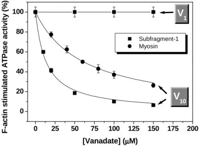
Enzymatic kinetic studies indicated that, unlike orthovanadate, decameric species is able to strongly inhibit the actin-stimulated ATPase activity of myosin or S1 with an IC50 in the micromolar range (Fig. 6). These results attested the initial
hypothesis that the inhibition of the actin-stimulated myosin ATPase activity by milimolar vanadate concentrations is, in reality, due to the presence of vanadate oligomers and not to the monomeric species. The chance that this inhibition could be a result of a direct competition between actin and decavanadate was excluded because the increase of actin concentration fixing myosin concentration did not increase the IC50 for
V10 but at most slightly decrease it, being 20-fold higher in its absence [49]. A more
detailed kinetic analysis revealed that the inhibition is also non-competitive to ATP yielding an inhibition constant (Ki = 0.27 ± 0.05 µM) that is in the range of Ki values previously reported for enzymes inhibited by decavanadate (see above).
The modulation of the actin-myosin interaction by decavanadate was further examined using the light-scattering technique [49]. These studies indicated a positive correlation between the extent of V10 inhibition of the actomyosin ATPase activity and
the time needed for the re-association of actin to myosin, after ATP addition. On the other hand, decavanadate binding did not dissociate either the ATP-free rigor actin-myosin complex or the actin-actin-myosin-MgADP complex. In other words, while decavanadate does not promote dissociation of myosin from actin, it does prevent its
formation until ATP level in the solution is sufficiently low for the re-association process to occur. Moreover, fluorescent studies confirmed that the inhibition is the result of decavanadate binding to a high affinity site (Kd = 0.16 -0.27 µM) near the myosin catalytic centre and that this affinity is modulated by the conformational changes that takes place during the catalytic cycle, as indicated by the 2-3 fold increase of the dissociation constant produced in the presence of ADP·Vi and ADP·AlF4, which
induce the conformation of the intermediate metastable state, generated during the contractile cycle [49]. Altogether, these results suggested that the inhibition of the actin-stimulated myosin ATPase activity by decavanadate was the consequence of slowing down the catalytic cycle by binding at a regulatory site different from - but near - the nucleotide site of the catalytic centre, and likely to be located close to the functional protein domains altered upon F-actin filament binding to S1.
Figure 6. Inhibition of the actin-stimulated ATPase activity of myosin (circles) or myosin Subfragment-1
(squares) by vanadate (given as total vanadium concentration). In the presence of actin, decameric vanadate, unlike the monomer, is able to inhibit the myosin ATPase activity in the micromolar range. The
assays were performed using a coupled enzyme system as described elsewhere [49] with 50 µg/ml
enzyme and 5 µM F-actin, at 25ºC, in a medium containing 10 mM Tris-HCl, pH 7.0, 2.5 mM MgCl2 and
2 mM ATP. The lines are the nonlinear least squares fit of the data to a Michaelis-Menten-like equation
with the following parameter values: Imax = 100 and IC50 = 6.11 ±0.74 µM V10 (or 61.1 ±7.4 µM total
vanadium) and IC50 = 1.36 ±0.14 µM V10 for myosin and S1, respectively [40].
6. DECAVANADATE AS A PHOTOCLEAVAGE AGENT OF MYOSIN
Unlike phosphate, vanadate is able to induce photolytic cleavage of vanadate-protein complexes under UV irradiation. The identification of the cleaved polypeptide may provide remarkable structural information about vanadate binding sites on enzymes extending the use of vanadate as a chemical probe. Dynein [50,51] and myosin [29,52-58] were the first enzymes shown to be photomodified or photocleaved by irradiation of vanadate complexes. Several other enzymes have followed including adenylate kinase [59], tubulin [60], aldolase [61], sarcoplasmic reticulum Ca-ATPase [36,37], phosphofructokinase [62] or glycogen phosphorylase [63]. Although the vanadate mediated photocleavage phenomenon has been reported for at least 10 different enzymes, myosin is one of the few systems in which the mechanism of the reactions involved are well characterize. This is due in part to the difficulty in sequencing the
0 25 50 75 100 125 150 175 200 0 20 40 60 80 100 Subfragment-1 Myosin
V
10V
1 F -a c ti n s ti m u la te d A T P a s e a c ti v it y ( % ) [Vanadate] (µµµµM)amino termini resulting from cleavage. The photocleavage of skeletal muscle myosin at the active site turned out to be an unusual case as the oxidative steps can be performed sequentially and the stable intermediates characterized [64]. Irradiation of the myosin·MgADP·Vi complex starts to covalently modify the peptide backbone by promoting the oxidation of the hydroxymethyl side chain of Ser-180 to a serine aldehyde. This photomodified myosin reforms a new MgADP·Vi complex and upon a second irradiation, the myosin heavy chain is specifically cleaved at the active site. A key step in this process involves addition of molecular oxygen to a stabilized seryl radical generated by a light-catalyzed vanadate reduction to VIV (vanadyl ion). Detailed mechanistic studies have found out that the cleaved peptide is in the glicine-rich phosphate binding loop (P-loop) homologous to the nucleotide binding site of several enzymes including ATP-binding cassette (ABC) transporters or other ATPases. This sequence has the general formula G-X1-X2-X3-X4-G-K-G/T in which X2 corresponds to
the aminoacid cleaved: for example, an alanine in dynein and β-ATP synthase, a proline in adenylate kinase, an arginine in α-ATP synthase and a serine in the particular case of myosin [50,53,54,59,65]. Curiously, the cleavage is determined by the location of the aminoacid and not by its nature.
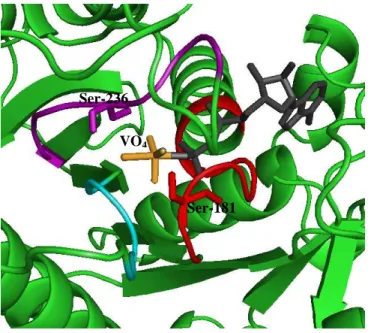
In the absence of bound nucleotides, vanadate was found to be able to cleave the myosin heavy chain (95 kDa) only when the concentration was above 0.2 mM [29]. Since 51V NMR studies had shown before that tetrameric vanadate binds most strongly to myosin and is known to be present at vanadate concentrations higher than 0.2 mM, this species was suggested to be responsible for the photocleavage reaction. In the presence of 0.2 mM vanadate concentration, the extent of cleavage was small occurring in two distinct sites located at 23 and 31 kDa from the N-teminal. With the increase of vanadate concentration, the extent of cleavage increased and an additional product with 74 kDa appeared in the electrophoretic pattern. While the presence of ATP specifically inhibits the vanadate-associated cleavage of myosin heavy chain at the 23 and 31 kDa sites, actin specifically inhibits the cleavage at the 74 kDa site [29,58]. The exact positions of the 23 and 31 kDa cleavage sites were assigned to Ser-180 [53] and Ser-243 [64] respectively, both located on highly conserved regions at the myosin active site of rabbit skeletal muscle myosin. According to the X-ray structure of the truncated head of Dictyostelium myosin complexed with MgADP, the side chains of the analogues of
Figure 7. Ribbon representation of
the γ-phosphate pocket of the
truncated head of Dictyostelium
myosin complexed with MgADP·VO4.
Protein coordinates were taken from PDB with the accession number 1VOM [66]. The heavy chain is
displayed in green except the
conserved motives forming the Pi-tube which are colored in red, magenta and cyan for the P-loop, switch I and
switch II domains, respectively.
MgADP, VO4, Ser-181 and Ser-236
are depicted in gray, yellow, red and
magenta, respectively, as stick
representation. Note that both Ser-181 and Ser-236 are in position to form hydrogen bonds with the oxygens of the vanadate molecule.
Ser-181 Ser-236
these two residues (Ser-181 and Ser-236) form hydrogen bonds with the oxygens of the vanadate molecule (Fig. 7) [66]. The aminoacids involved in the third cleavage site (74 kDa), although not yet identified were demonstrated to be placed at the 50/20 kDa proteolitic fragments junction where the Loop-2 (residues 627-646 of rabbit skeletal muscle myosin) is encountered. This lysine-rich polyphosphate binding site was proved to participate in the myosin-actin interaction.
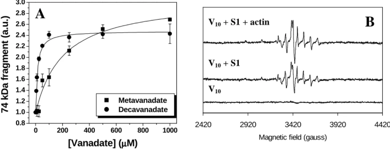
More recently, it has been demonstrated that besides monomeric and tetrameric vanadates, also the decamer is photoactive being rapidly decomposed, through the photoreduction of vanadate atoms to vanadyl, during the photocleavage process (Fig. 8). While for decavanadate cleavage initiates at vanadate concentrations as low as 5 µM (~ 0.5 µM V10 species), for metavanadate cleavage is almost negligible at concentrations
lower than 100 µM and only above 200 µM (when tetrameric species start to appear) the extent of cleavage is comparable to the decavanadate-promoted photocleavage [32]. Additionally, the identical cleavage patterns found for meta- and decavanadate solutions in the absence or presence of the myosin natural ligands, ATP and actin [32], suggest that V4 and V10 share the same myosin binding sites (in agreement with previous NMR
studies), namely one of higher affinity (23 kDa) at the consensus ATP binding site and another one of lower affinity (74 kDa) at the actin-binding interface.
Figure 8. Vanadate-dependent cleavage of myosin Subfragment-1 (4.5 µM) upon UV irradiation in a
medium containing 25 mM KCl, 2.5 mM MgCl2, 25 mM Hepes (pH 7.0) at 25 ºC. A: Development of the
74 kDa cleavage product upon increasing vanadate concentration [32]. B: The products of decavanadate reduction in the presence of myosin subfragment-1 and actin give rise to EPR signals typical of vanadyl
(VIV) ions [40].
7. VANADIUM(V) AS A POWERFUL TOOL IN THE ELUCIDATION OF MYOSIN ATPASE ACTIVITY
The ability to visualize a transition state analogue is a powerful tool for understanding the dynamics of enzyme mechanisms as they can help mapping the enzyme active site and reveal conformational changes that may take place in the course of the catalytic cycle. The vanadate ion (VO43-) is able to act as an analogue for the
conformation of the phosphate group at the transition state expected for phosphoryl transfer or hydrolysis, in part because it has very similar size and charge to inorganic phosphate, but also because it tends to adopt pentacoordinate complexes exhibiting trigonal bipyramidal geometry [67]. In the myosin·MgADP·VO4 complex, vanadate
exhibits this geometry forming one apical bond to the β-phosphate of ADP but with no covalent linkages to the enzyme (see Fig. 7). This structure is a realistic analogue of the
0 200 400 600 800 1000 0.8 1.0 1.2 1.4 1.6 1.8 2.0 2.2 2.4 2.6 2.8 3.0 7 4 k D a f ra g m e n t (a .u .) [Vanadate] (µµµµM) Metavanadate Decavanadate 2420 2920 3420 3920 4420
Magnetic field (gauss)
V10 V10 + S1 V10 + S1 + actin
A
metaphosphate-like transition state for phosphoryl transfer [66,68]. Vanadate is frequently replaced by aluminium and berilium fluoride as phosphate analogues on account of vanadate tendency to polymerize and due to its photoreactivity. Although the ions AlF4- and BeF3- approximate the shape and charge of phosphate, they do not
function as true analogues of the transition state for phosphate ester bond cleavage [66-68]. While beryllium fluoride moiety in the myosin active site is tetrahedral, representing the ground state prior hydrolysis, aluminium fluoride is approximately square bipyramidal [69]. The X-ray resolution of the MgADP·VO4 structure in the
catalytic domain of myosin Dictyostelium disciudeum (S1dC) revealed how the protein ligands and water structure surrounding the γ-phosphate pocket were oriented to stabilize a water molecule in an appropriate position for in-line nucleophilic attack on the γ-phosphorus of ATP [66]. Besides providing insights into the structural basis of ATP hydrolysis, the ADP·vanadate structure has also captured a key conformational change playing an important role in the generation of movement. It has been observed that in the structure of the ADP·vanadate complex there is reduce access of the solvent to the γ-phosphate pocket. This is caused by a closure of the Pi-tube located at the apex of a large cleft that divides the 50 kDa fragment into upper and lower domains at the rear opening of the active site [66]. However, in the structure of the S1dC complexed with MgADP·BeF or MgATP the γ-phosphate is partially visible because there is a clear opening of the Pi-tube into the large 50 kDa cleft [69]. The former conformational change has been suggested to be required for hydrolysis and attainment of the ADP·Pi state meaning that the phosphate group will be trapped in the Pi-tube until a second conformational rearrangement occurs by the rebinding of myosin to actin [70]. It is thus comprehensible that considerable biochemical and biophysical effort has been expended on the S1·MgADP·VO4 complex as part of the greater goal of understanding the
molecular basis of muscle contraction.
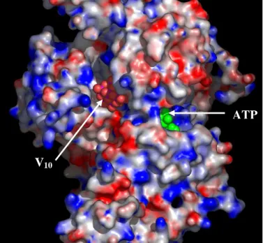
Apparently, decavanadate cluster is too large to form an enzyme-vanadate complex capable of providing insight into phosphoryl transfer reactions. Nevertheless, if decavanadate is able to trap the contractile cycle at a critical point, that is necessarily different from the one trapped by orthovanadate, then, it could provide a good opportunity to further comprehend the modulation of the actin-myosin interaction. Indeed, based on molecular docking simulations of the oligomer on three different structures of the myosin motor domain of Dictyostelium discoideum representing distinct states of the ATPase cycle, decavanadate was suggested to prevent a crucial conformational change for the hydrolysis of the ATP-γ-phosphate [71]. The theoretical results indicated a clear preference of V10 to bind at the rear opening of the Pi-tube (the
so called “back-door” [72,73]) but only on the “open” structures where there is access to the phosphate binding-loop (P-loop) such as the free or the ATP-bound conformations. As expected, in the S1dC·MgADP·VO4 X-ray structure, the back-door does not
represent an energetically favourable solution since the 50 kDa cleft is closed blocking the Pi-tube and concomitantly the access of decavanadate to the P-loop. The proposed binding location explains how the 10 Å decavanadate molecule gains access to Ser-181, a residue located on the P-loop, previously shown to be cleaved upon irradiation in the presence of the oligomer, without having to squeeze through the narrow entrance of the Pi-tube at the nucleotide binding site. Furthermore, it provides a simple mechanism for the experimentally observed non-competitive inhibition pattern of V10 towards both
ATP and actin as it does not interfere with the nucleotide binding site or the actin binding surface.
The enzymes inhibited by decavanadate appear to be all prearranged to bind phosphoryl groups, but decavanadate inhibition is by no means a general property of all
such enzymes and, therefore, the structural and conformational characteristics of the enzymes are certainly important. For instance, the walker A motive (corresponding to the P-loop in myosin) of ABC ATPases has been shown to be a highly adapted anion-binding domain that can bind decavanadate with high affinity [48]. Unlike orthovanadate, decavanadate appears to interact outside the walker A loop but needs to be stabilized by certain residues nearby. As a result, only some members of the ABC superfamily are able to interact with the oligomer. In what concerns myosin, the specific phosphate-binding domains in the vicinity of the back-door provide electrostatic interactions which favor an approach of this polyanionic species to the rear opening of the Pi-tube (Fig. 9). Once “accommodated” to the 50 kDa cleft and stabilized by certain residues nearby, the large size of decavanadate is likely to interfere with movements associated with closure of the cleft, a critical conformational change necessary to carry out the ATP-γ-phosphate hydrolysis. Therefore, by forming the intermediate myosin·MgATP·V10 complex, decavanadate will act as a “back-door” stop blocking the
actomyosin cycle, most likely, in the pre-hydrolysis state.
8. CONCLUDING REMARKS
Interest in the interaction of vanadium oxoanions with biological systems has increased gradually since it was demonstrated to have a variety of physiological effects, acting either as a phosphate analogue in the monomeric form (H2VO4-) or through
oligomeric vanadate species. In muscles, where it was rediscovered for biological studies, several enzymes are inhibited by this oxoanion, namely myosin, a highly specialized protein which along with actin is able to convert the chemical energy of ATP hydrolysis into mechanical movement. Monomeric vanadate affects the ATPase activity of myosin through the formation of a complex with MgADP that mimics the conformation of the myosin head in the transition state for ATP hydrolysis. As would be expected for such a transition state mimic, vanadate has been widely used as a very useful tool to elucidate myosin catalytic mechanisms and substrate binding modes. Although the mechanism of inhibition of the monomeric species towards myosin has been relatively well characterized, very little has been described about the contribution
ATP
V10
Figure 9. Map of the surface
electrostatic potential of myosin subfragment-1, with the resulting
docked conformation of
decavanadate (red spheres) on the
“back-door” region. The ATP
molecule depicted in green has been introduced to show the location of the active site with respect to the decavanadate molecule. Positive,
neutral and negative surface
potential are coloured in red, white and blue, respectively. Note how the decavanadate molecule with an
ellipsoidal shape is perfectly
adjusted to the 50 kDa cleft at the rear of the nucleotide site.
of vanadate oligomers in the biochemical/structural properties of myosin. The latter point is of relevance since it could provide structural/functional tools for further understanding the mechanism of myosin action and it could address one of the most interesting questions of the field: the role of vanadate in the in vivo regulation of myosin and other molecular motors.
Very often studies describing interactions of vanadate with proteins consider only monomeric vanadate as the active species, disregarding the possible contribution of other vanadate oligomers which are favoured at higher vanadate concentrations or through a slight acidification of the medium. In the present review we become aware of the fact that observation of inhibition of an enzyme system cannot be taken as evidence that monomeric vanadate is the inhibiting species and that individual vanadate species can differently affect an enzyme mechanism. In what concerns myosin,vanadate is able to populate different conformational states of the myosin ATPase cycle depending on its oligomerization state. While monomeric vanadate (VO43-) mimics the transition state for
the γ-phosphate hydrolysis blocking myosin in a pre-power-stroke state, decameric vanadate (V10O286-) induces the formation of the intermediate myosin·MgATP·V10
complex blocking the contractile cycle presumably in the pre-hydrolysis state. The former complex is destabilized in the presence of actin, comparable to the in vivo conditions, whereas the latter is not. Therefore, the mode of action of vanadate on the inhibition of the actomyosin ATPase activity depends, at least in part, on the charge and size of the vanadate species, being favoured for those with higher oligomerization state, such as the decameric species.
Unlike the simple and labile vanadate oligomers including the monomer (V1),
dimer (V2), tetramer (V4) and pentamer (V5), which interchange with each other on the
millisecond to second time scale in neutral and alkaline aqueous solutions, decavanadate interconversion with the other vanadate oligomers occurs on a considerably longer time scale. Moreover, even though at physiological pH decavanadate is not the most thermodynamically stable, hydrolysis into other vanadate oligomers is very slow and therefore it is sufficiently long lived so its effects can be determined in the biological systems under study. This constitutes a great advantage in regard to the other vanadate oligomers in which the effects of each species must be examined in an equilibrium mixture and correlated with changes in the concentration of the different vanadate ions. In addition to its lower lability, the physicochemical properties of decavanadate allows it to be analysed through several techniques including 51V NMR and UV-Vis spectroscopy, as well as to be used as a probe through photolytic cleavage reactions. Regardless of the controversy around the possibility of existence of the decameric species at physiological conditions, which is out of the scope of this review, decavanadate remains as an excellent biochemical tool in such important aspects as: 1) to gain a deeper knowledge about the modulation of actin-myosin interactions in the process of muscle contraction; 2) to the description of the location and function of the phosphate binding sites on myosin; 3) to further understand the basic structural requirements for high affinity protein binding sites to decavanadate; and 4) to a more general knowledge about polyanion-enzyme interaction.
ACKNOWLEDGMENTS
This work has been supported by POCTI program financed through FEDER for the research project 38191/QUI/2001 (to M.A.). The authors also thank to Joint Spanish-Portuguese Grant HP2004-0080, Spanish-Spanish-Portuguese CRUP project E-105/06 (to C.G.-M. and C.G.-M.A.), and to Grant 3PR05A078 of the Junta de Extremadura (to C.G.-C.G.-M.). Dr.
T. Tiago is the recipient of a post-doctoral fellowship (SFRH/BPD/20777/2004) from the Portuguese Foundation for Science and Technology (FCT).
REFERENCES
[1] Josephson, L., and Cantley, L.C. Jr. 1977, Biochemistry, 16, 4572. [2] Chasteen, N. D. 1983, Struct. Bond., 53,105.
[3] Nriagu, J.O. 1998, Vanadium in the environment (Vol. 1 and 2), J.O. Nriagu (Ed.), John Wiley & Sons, New York,.
[4] Petterson, L., Andersson, I., and Hedman, B. 1985, Chemica Scripta, 25, 309. [5] Crans, D.C., Rithner, C.D., and Theisen, L.A. 1990, J. Am. Chem. Soc., 112, 2901. [6] Crans, D.C. 1994, Comments Inorg. Chem., 16, 35.
[7] Gordon, J.A. 1991, Methods Enzymol., 201, 477.
[8] Stankiewicz, P.J., Tracey, A.S., and Crans, D.C. 1995, Metal ions in Biological Systems: Vanadium and its Role in Life, H. Sigel and A. Sigel (Eds.), Marcel Dekker, New York, 287.
[9] Lymn, R.W., and Taylor, E.W. 1971, Biochemistry, 10, 4617.
[10] Toyoshima, Y.Y., Kron, S.J., McNally, E.M., Niebling, K.R., Toyoshima, C., and Spudich, J.A. 1987, Nature (Lond.), 328, 536.
[11] Goodno, C.C. 1979, Proc. Natl. Acad. Sci. U.S.A., 76, 2620. [12] Goodno, C.C. 1982, Methods Enzymol., 85, 116.
[13] Goodno, C.C., and Taylor, E.W. 1982, Proc. Natl. Acad. Sci. U.S.A. 79, 21. [14] Smith, S.J., and Eisenberg, E. 1990, Eur. J. Biochem., 193, 69.
[15] Gresser, M.J. and Tracey, A.S. 1986, J. Am. Chem. Soc., 108, 1935. [16] Heath, E. and Howarth, O.W. 1981, J. Chem. Soc. Dalton Trans., 1105. [17] Howarth, O.W. 1990, Prog. NMR Spectrosc., 22, 453.
[18] Crans, D.C. 1994, Comments Inorg. Chem., 16, 1. [19] Rehder, D. 1982, Bull Magnetic Resonance, 4, 33.
[20] Baruah, B., Roden, J.M., Sedgwick, M., Correa, N.M., Crans, D.C., and Levinger, N.E. 2006, J. Am. Chem. Soc., 128, 12758.
[21] Csermely, P., Martonosi, A., Levy, G.C., and Ejchart, A.J. 1985. Biochem. J., 230, 807.
[22] Borah, B., Chen, C.W., Egan, W., Miller, M., Wlodawer, A., and Cohen, J.S. 1985, Biochemistry, 24, 2058.
[23] Butler, A. and Eckert, H. 1989, J. Am. Chem. Soc., 111, 2802. [24] Crans, D.C. and Schelble, S.M. 1990, Biochemistry, 29, 6698.
[25] Wittenkeller, L., Abraha, A., Ramasamy, R., Mota de Freitas, D., Theisen, L. A. and Crans, D. C. 1991, J. Am. Chem. Soc., 113, 7872.
[26] Liu, S., Gresser, M.J., and Tracey, A.S. 1992, Biochemistry, 31, 2677. [27] Cremo, C.R., Grammer, J.C., and Yount, R.G. 1988, Biophys. J., 53, 236a. [28] Cremo, C.R. and Wilcott, J. 1990, Biophys. J., 57, 329a.
[29] Ringel, I., Peyser, Y.M., and Muhlrad, A. 1990, Biochemistry, 29, 9091. [30] Aureliano, M. 2000, J. Inorg. Biochem., 80, 141.
[31] Tiago, T., Aureliano M., Duarte, R.O., and Moura, J.J.G. 2002, Inorg. Chim. Acta., 339, 317.
[32] Tiago, T., Aureliano, M., and Moura J.J.G. 2004, J. Inorg. Biochem., 98, 1902. [33] Varga, S., Csermely, P., and Martonosi, A. 1985, Eur. J. Biochem., 148, 119. [34] Csermely, P., Varga, S., and Martonosi, A. 1985, Eur. J. Biochem., 150, 455. [35] Coan, C., Scales, D.J., and Murphy, A.J. 1986, J. Biol. Chem., 261,10394.
[36] Vegh, M., Molnar, E., and Martonosi, A. 1990, Biochim. Biophys. Acta, 1023, 168.
[37] Molnar, E., Varga, S., and Martonosi, A. 1991, Biochim. Biophys. Acta, 1068, 17. [38] Aureliano, M., and Madeira, V.M.C. 1994, Biochim. Biophys. Acta, 1221, 259. [39] Hua, S., Inesi, G., and Toyoshima, C. 2000, J. Biol. Chem., 275, 30546.
[40] Tiago, T. 2005, PhD thesis, University of Algarve.
[41] Aureliano, M. and Madeira, V.M.C. 1998, Vanadium in the environment. Part 1: Chemistry and Biochemistry, J.O. Nriagu (Ed.), John Wiley & Sons, New York, 333.
[42] Ramos, S., Manuel, M., Tiago, T., Duarte, R., Martins, J., Gutierrez-Merino, C., Moura, J.J., and Aureliano, M. 2006, J. Inorg. Biochem., 100, 1734.
[43] DeMaster, E.G., and Mitchell, A. 1973, Biochemistry, 12, 3616.
[44] Boyd, D.W., Kustin, K., and Niwa, M. 1985, Biochim. Biophys. Acta, 827, 472. [45] Pluskey, S., Mahroof-Tahir, M., Crans, D.C., and Lawrence, D.S. 1997, Biochem.
J., 321, 333.
[46] Messmore, J.M., and Raines, R.T. 2000, Arch. Biochem. Biophys., 381, 25. [47] Proks, P., Ashfield, R., and Ashcroft, F.M. 1999, J. Biol. Chem., 274, 25393. [48] Pezza, R.J., Villarreal, M.A., Montich, G.G., and Argarana, C.E. 2002, Nucleic
Acids Res., 30, 4700.
[49] Tiago, T., Aureliano, M., and Gutierrez-Merino, C. 2004, Biochemistry, 43, 5551. [50] Lee-Eiford, A., Ow, R.A., and Gibbons, I.R. 1986, J. Biol. Chem., 261, 2337. [51] Gibbons, I.R., and Mocz, G. 1991, Methods Enzymol., 196, 428.
[52] Grammer, J.C., Cremo, C.R., and Yount, R.G. 1988, Biochemistry, 27, 8408. [53] Cremo, C.R., Grammer, J.C., and Yount, R.G. 1988, Biochemistry, 27, 8415. [54] Cremo, C.R., Grammer, J.C, and Yount, R.G. 1989, J. Biol. Chem., 264, 6608. [55] Cremo, C.R., Long, G.T., and Grammer, J.C. 1990, Biochemistry, 29, 7982. [56] Cremo, C.R., Grammer, J.C., and Yount, R.G. 1991, Methods Enzymol., 196, 442. [57] Mocz, G. 1989, Eur. J. Biochem., 179, 373.
[58] Muhlrad, A., Peyser, Y.M., and Ringel, I. 1991, Biochemistry, 30, 958.
[59] Cremo, C.R., Loo, J.A., Edmonds, C.G., and Hatlelid, K.M. 1992, Biochemistry, 31, 491.
[60] Correia, J.J., Lipscomb, L.D., Dabrowiak, J.C., Isern, N., and Zubieta, J. 1994, Arch. Biochem. Biophys., 309, 94.
[61] Crans, D.C., Sudhakar, K., Zamborelli, T.J. 1992, Biochemistry, 31, 6812.
[62] Signorini, M., and Bergamini, C.M. 1990, Biochem. Biophys. Res. Commun., 172, 919.
[63] Bergamini, C.M., and Signorini, M. 1991, Biochem. Int., 25, 143.
[64] Grammer, J.C., Loo, J.A., Edmonds, C.G., Cremo, C.R., and Yount, R.G. 1996, Biochemistry, 35, 15582.
[65] Saraste, M., Sibbald, P.R., and Wittinghofer, A. 1990, Trends Biochem. Sci., 15, 430.
[66] Smith, C.A., and Rayment, I. 1996, Biochemistry, 35, 5404. [67] Davies, D.R., and Hol, W.G. 2004, FEBS Lett., 577, 315. [68] Rayment, I. 1996, J. Biol. Chem., 271, 15850.
[69] Fisher, A.J., Smith, C.A., Thoden, J.B., Smith, R., Sutoh, K., Holden, H.M., and Rayment, I. 1995, Biochemistry, 34, 8960.
[70] Fisher, A.J., Smith, C.A., Thoden, J., Smith, R., Sutoh, K., Holden, H.M., and Rayment, I. 1995. Biophys. J., 68, 19S.
[71] Tiago, T., Martel, P., Gutiérrez-Merino, C., and Aureliano, M. 2007, BBA-Proteins and Proteomics (in press).
[72] Yount, R.G., Lawson, D., and Rayment, I. 1995, Biophys. J., 68, 44S.