MELLATIE R. FINISIE, ATCHE JOSUÉ, VALFREDO T. FÁVERE and MAURO C. M. LARANJEIRA
Departamento de Química, Universidade Federal de Santa Catarina, Campus Universitário, Trindade – 88040-900 Florianópolis-SC, Brazil. Manuscript received on December 20, 2000; accepted for publication on August 29, 2001;
presented byFernando Galembeck
ABSTRACT
Bioceramic composites were obtained from chitosan and hydroxyapatite pastes synthesized at physiological temperature according to two different syntheses approaches. Usual analytical techniques (X-ray diffrac-tion analysis, Fourier transformed infrared spectroscopy, Thermo gravimetric analysis, Scanning electron microscopy, X-ray dispersive energy analysis and Porosimetry) were employed to characterize the resulting material. The aim of this investigation was to study the bioceramic properties of the pastes with non-decaying behavior from chitosan-hydroxyapatite composites. Chitosan, which also forms a water-insoluble gel in the presence of calcium ions, and has been reported to have pharmacologically beneficial effects on osteoconductivity, was added to the solid phase of the hydroxyapatite powder. The properties exhibited by the chitosan-hydroxyapatite composites were characteristic of bioceramics applied as bone substitutes. Hydroxyapatite contents ranging from 85 to 98% (w/w) resulted in suitable bioceramic composites for bone regeneration, since they showed a non-decaying behavior, good mechanical properties and suitable pore sizes. Key words:bioceramic, chitosan, hydroxyapatite, composites, bone regeneration.
INTRODUCTION
Calcium phosphate based bioceramics have recently received special attention as bone replacement ma-terials, as they behave similarly to the mineral con-stituent of bones (Martin and Brown 1995, Felício-Fernandes and Laranjeira 2000, Pereira et al. 1999, Kawachi et al. 2000, Shareef et al. 1993, Sivakumar et al. 1996).
Among their most attractive properties, these materials have shown no local or system toxicity, no response to strange bodies or inflammation, and apparent ability to attach themselves to host tissues (Kawachi et al. 2000). The mechanism of tissue attachment is directly related to the type of tissue
Correspondence to: Mauro C.M. Laranjeira E-mail: [email protected]
response at the implant interface (Hench 1991). Im-plants always elicit a response from living tissues (Hench 1991), since replacement materials in living tissues are never inert.
Pores larger than 100µm are ideal for
bioce-ramics (Kawachi et al. 2000), as they maintain vas-cularity and long-term viability (Hench 1991). In order to form macro porous biomaterials (pore size ranging from 50 to 250µm) a polymer or organic
substance is mixed to a powder material. Alter-natively, it can be wetted with hydrogen peroxide that decomposes releasing oxygen gas to form pores (Kawachi et al. 2000). In the present work, the use of aluminum powder in alkaline media to form hy-drogen gas is discussed.
be used depending whether a resorbable or bioac-tive material is desired (Hench 1991). The stability of calcium phosphate phases depends considerably upon temperature and moisture, either during pro-cessing or use.
HAp [Ca10(PO4)6(OH)2] is the main inorganic constituent of human and animal bones and teeth. It has a Ca/P ratio of 1.67 and water contents of 1.79 wt.%.
The resorption and biodegradation of phos-phate ceramics are caused by (1) physiochemical dissolution; (2) physical disintegration into small particles due to preferential chemical attack of grain boundaries; and (3) biological factors, such as phagocytes, which decreases local pH (Hench 1991).
The synthesis of calcium phosphate has been accomplished by different methods, including: pre-cipitation in aqueous solutions, solid-state reactions, hydrothermal methods (Kawachi et al. 2000), (Hench 1991), sol-gel process and, more recently, micro emulsion (Kawachi et al. 2000). The phos-phates can be transformed into biocompatible and osteoconductor ceramics, capable of inducing bone growth both on the surface and through the pores of the material (Kawachi et al. 2000, Hench 1991).
Chitosan, a poly-2-amino-2-deoxy-β
-(1,4)-D-glucopyranose, is derived from chitin, poly-2-aceta-mide-2-deoxy-β-(1,4)-D-glucopyranose (Knaul et
al. 1999). Chitin is one of the most abundant nat-ural polysaccharides, primarily obtained as a sub-product of seafood. Chitosan has been used as a floc-culent and adsorbent in wastewater treatment. Re-cently it has been applied in the biomedical and phar-maceutical areas, mainly because of its biodegrad-ability, low toxicity, and good biocompatibility (Kawachi et al. 2000, Tas 2000).
The HAp-chitosan combination can be used to prepare composites with controlled bioactivity (biodegradability). Chitosan is insoluble in water and, consequently, in physiological environments. HAp can act as a reticular modifier agent (due to the presence of calcium and phosphorous) and its use
as bone former (Felício-Fernandes and Laranjeira 2000), has also been appraised. Its superior osteo-conductive properties render HAp a vast potential to be used in implants as bone substitute (Sivakumar et al. 1996).
In the present paper, we report the preparation and characterization of bioceramic composites in the form of pastes from hydroxyapatite and chitosan. This work also examines the probable interaction between amine group of chitosan and phosphate of hydroxyapatite. The chemical combination of chi-tosan and hydroxyapatite by the employed methods yielded a new material, which could be used as al-ternative bioceramic in the bone regeneration.
EXPERIMENTAL
Materials and methods
the storage temperatures, may drastically affect the phase purity and high-temperature stability of the produced HAp powders, as well as the kinetics of the precipitation processes.
TABLE I
Compositions of the prepared HAp-Chitosan pastes
Samples % HAp % Chitosan
I 55.6 44.4
II 71.4 28.6
III 86.2 13.8
IV 93.1 6.90
V 96.2 3.80
VI 98.0 2.00
NaCl (99.5%), NaHCO3(99.5%), KCl (99.0%), Na2HPO4·2H2O (99.5%), MgCl2·6H2O (99.0%), CaCl2·H2O (99.0%), Na2SO4, (CH2OH)3CNH2 (99.5%), and HCl (37 vol%,) were used in the prepa-ration of the SBF (Tas 2000).
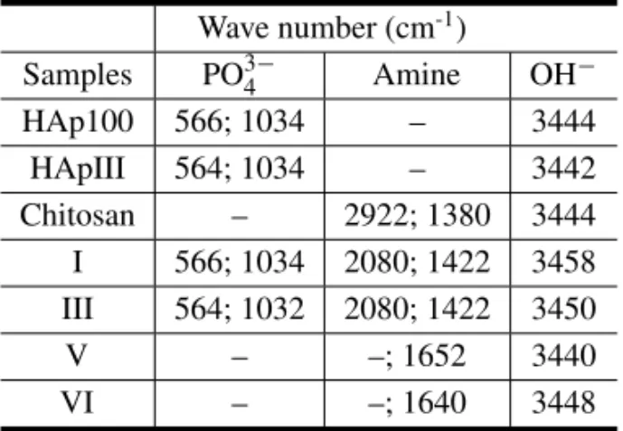
TABLE II
The shifts observed at the FTIR spectra of the characteristic bands from the functional groups
Wave number (cm-1) Samples PO3−
4 Amine OH−
HAp100 566; 1034 – 3444
HApIII 564; 1034 – 3442
Chitosan – 2922; 1380 3444
I 566; 1034 2080; 1422 3458 III 564; 1032 2080; 1422 3450
V – –; 1652 3440
VI – –; 1640 3448
The pastilles were then freeze-dried and char-acterized by conventional analytical techniques. In order to enhance the porosity, HAp powder was mixed with aluminum powder (0.3−1.1%), and
then transformed into paste. Due to the toxicity of aluminum, all the metal was eliminated by dissolu-tion in 2 mol/L NaOH soludissolu-tion, after immersing the pastilles in the alkaline solution. This resulted in the formation of sodium aluminate in solution and hydrogen gas, which led to the forming of pore at the order larger than 100µm. The sodium aluminate
was removed by extensive washing with deionized water. The resulting pastilles after immersed for 7 days in synthetic body fluid (SBF) at 37◦C (Tas 2000) were also characterized by conventional ana-lytical techniques.
All samples were freezes-dried and character-ized by the following techniques: Infrared absorp-tion spectroscopy using KBr pellets (FTIR); X rays powder diffraction (XRD); Scanning electronic mi-croscopy (SEM), where the pastille surfaces were covered with a thin layer of gold; Analysis of X rays dispersive energy (EDX); Porosimetry by mercury intrusion; and TG analysis of powder of composites (TGA).
RESULTS AND DISCUSSION
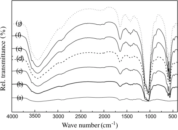
Fig. 1 – FTIR spectra from: (a) Chitosan, (b) HAp100, (c) HApIII, (d) composite I, (e) composite III, (f) composite V, and (g) composite VI.
groups (protonated amine and phosphate) to lower wave number (2080 and 1422 cm-1for composites I and III, and 1652, 1640 cm-1for compositesV andVI respectively) was observed in Figure 1d–1g for the functional group of chitosan, and may be attributed to interactions involving these groups. The shift of the characteristic bands of phosphate group was only observed on the FTIR spectrum of composite VI in Figure 1g.
The XRD patterns shown in Figure 2 reveal that the method used to synthesize HAp100 resulted in better crystallinity (Figure 2c.), compared to thein situapproach (HApIII in Figure 2b). In addition, powder samples from bioceramic composites (Fig-ure 2a and 2d) were similar to those representatives of HAp100 (Figure 2c).
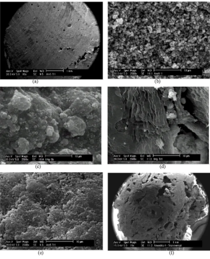
A morphological study (Figure 3) was also car-ried out and showed that composites prepared from HAp100 (Figure 3a, 3b, 3d, 3e) had quite porous sur-faces, exceeding the porosity present on those from HApIII as showed in Figure 3c. This was probably due to the uniform grain distribution observed from HAp100. Additional, using aluminum (0.2−0.5 wt.%) results in pores in excess of 100µm in
aver-age diameter (Figure 3f).
TG plots are shown in Figure 4. It can be seen that no phase transformation took place upon heating HAp100 (Figure 4e) or HApIII (Figure 4d). There is no notable occurrence of weight loss in both HAp products. The weight loss observed from chitosan at 281.01◦C and 295.89◦C (Figure 4a), probably cor-responded to the decomposition and elimination of the polymeric constituent. Bioceramic composites showed in Figure 4b and 4c weight loss at higher temperature (298.27◦C and 307.08◦C respectively). The weight loss observed at 281.01◦C in the thermo-graph of chitosan disappeared in those of the com-posites I and III (Figure 4b and 4c, respectively). This confirmed the hypothesis of interactions be-tween the functional amine groups from chitosan and phosphate groups from HAp. The amplitude of the temperature shift observed was inversely pro-portional to the chitosan contents present in the ceramic sample. The compressive strength of
bio-ceramic pastilles varied from 3−7 MPa, where the compositions with hydroxyapatite contents ranging from 85 to 98 wt.% showed the highest values.
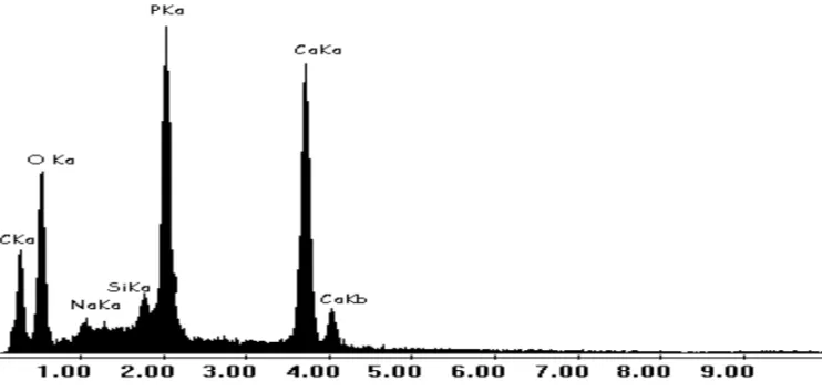
An analysis of the X ray dispersive energy pat-tern shown in Figure 5 revealed the absence of resid-ual aluminum in the bioceramic composite. All alu-minum was eliminated due to its toxicity by disso-lution in 2 mol/L NaOH sodisso-lution.
Both methods used to synthesize HAp were ef-ficient, since the resulting bioceramic composites showed certain porosity and crystallinity. The composites presented pore sizes larger than 100µm,
which is the requirement for the bone ingrowths through the pore channels. The techniques used to prepare bioceramics from calcium phosphate and chitosan biopolymer led to the preparation of ma-terials with the necessary requirements for bone re-generation: inhibition of decay of the pastilles in body fluids, suitable pore sizes and mechanical re-sistance.
We recommend the composites with hydrox-yapatite contents ranging from 85 to 98 wt.% for the production of ceramic pastilles, since decay of the paste was effectively inhibited at this compo-sition range, which also demonstrated the highest mechanical properties. In vivoevaluation of these HAp-Chitosan composites is awaited based on this initialin vitrostudy.
RESUMO
Fig. 3 – SEM micrograph results from: (a) composite V, (b) composite III, (c) composite III (from HApIII), (d) composite I transversal section, (e) composite V transversal section, and (f) composite IV transversal section (with 0.3% Aluminum addition).
íons cálcio, e tem mostrado possuir efeitos farmacologica-mente benéficos na condutividade óssea, foi adicionada à fase sólida do pó de hidroxiapatita. As propriedades
Fig. 4 – Thermographs from sample of: (a) Chitosan, (b) composite I, (c) composite III, (d) HApIII, and (e) HAp100.
Fig. 5 – X ray dispersive energy pattern from sample of: composite IV.
entre 85 a 98% em peso resultou em compósitos bio-cerâmicos adequados para regeneração óssea, visto que apresentaram um comportamento de não-desintegração, boas propriedades mecânicas e tamanhos de poros apro-priados.
Palavras-chave: biocerâmica, quitosana, hidroxiapatita,
compósitos, regeneração óssea.
REFERENCES
Hench LL.1991. Bioceramics: From Concept to Clinic. J Am Ceram Soc 74: 1487-1510.
Kawachi EY, Betran CA, dos Reis RR and Alves OL.
2000. Biocerâmicas: Tendências e Perspectivas de uma Área Interdisciplinar. Quim Nova 23: 518-522.
Knaul JZ, Hudson SM and Creber KAM.1999. Im-proved Mechanical Properties of Chitosan Fibers. J Appl Polym Sci 72: 1721-1732.
Martin RI and Brown PW.1995. Mechanical
proper-ties of Hydroxyapatite formed at Physiological Tem-perature. J Mater Sci: Mater Med 6: 138-143.
Pereira APV, Vasconcelos WL and Oréfice
RL.1999. Novos biomateriais: híbridos orgânico-Inorgânicos bioativos. Polim: Cienc e Tecnol 9: 104-109.
Shareef MY, Messer PF and van Noort R.1993. Fabrication, Characterization and Fracture Study of a Machinable Hydroxyapatite Ceramic. Biomaterials 14: 69-75.
Sivakumar M, Sampath Kumar TS, Shantha KL and
Panduranga Rao K.1996. Development of Hy-droxyapatite Derived from Indian Coral. Biomateri-als 17: 1709-1714.
Tas AC.2000. Synthesis of Biomimetic