!
" ! #
! " $ ! % &
!
"#
+ ! ! , - , . / 0& 1 ! 2 ! "
$ ! % 3 # 4 $5 # 6 / 0& 1 7 ! 2
$8 !9% # ! 4 $#6 0 % % - . - : 4 “The
Molecules of Colour in Art: a photochemical study” + !! - % ! 0 .
/- : & ; ! % 0 ! / 0& 8 % < ! 2 4 6
- : &
+ ! ! ! , - , !! - ! ! - /- : 4 / 0& 1 % / ! 2 $5
# 6 0 - # !. 0 % + , - %
0 !. = / 0& / 2 $5 # 6 0
-- % ; ! 0! .! - > = / 0& !
2 $8 4 $6 0 - - ! - =
-% ! 2 $5 # 6 0 ! % % % - % !
- % = % 2 $5 # 6 0 - ?/ 5 !.
- / < ! = / 0& 2 ! . 0 3 . 0
4 5 6 0 - % ; ! ! ! / 0& 1
2 . 0 / 4 /6 0 - % ; ! / !. &
; % 0 ! !! - ! - ! 0 - 00
% . !. 4 6 Dragon’s blood samples4 - ! % 0
! % 0 : 1 0 - ! % 0
- # ! / , 0 0 - Dracaena draco ! / 0& 1& / ! 0
-Dracaena cinnabari ! & ! + ! ! , - , & @ . 0 - % ; !
! ?& - 0 - A & % 0 ! ,
2 . ! ' 4 ' B +6 +- - ! - ! % 0 % ; ! '
!! ! 0 - ! ! & '6 Mauve dye samples: /
-2 ! 6 0 . - ! . # + C ,
. - 0 - ! & ! + ! ! , - , /&1&
2 6 ! 0 % !! - ! 0 - ! !
& ! ; % 0 ! / 0& - . $ 2 . & ! 0
-? . /- ! -. 0 6 / 0& ? . > 2 ! !! % 6 +- + .
- ! 0 ! - 0 - . ! &
! + ! ! , - , . /- !! % +- - . - : .
+ - < - % 0 , +! % & ! % 0 ! - D 00E
F A ! % 0 !! - &
!!. ; % 0 ! / 2/ G@ GHHIJ(G())K /$ G $GIHKKHG())I6 $
#$!
# 0 " - 9 L ! ! !
% & ! ! > ! %L
= #' $ (' ) !- ! > !
0 L 8 !' *' 7 !
- 9 7 L M & 5
!-! > 9 ! ! "< &
+ '*% & # '"$('&# 0 90 F !
!-% % & ! J K;5 - < 5H5 !0! L! 2 0! L! 6 0
! > 0 > ?/ 5 5 #
% Dracaena draco 7 7 ! J K;5 - < 0! L!
0 0 ! > % Dracaena cinnabari& H)
% % " - 0 ! 0
! ?/ 5 ! 5 J I5 - < 5H5 !0! L! 2 6
! J K;5 - < 5H5 !0! L! 2 0! L! 6 ! J K;5 - < 0! L!
8 Daemonorops spp. Dracaena draco
Dracaena cinnabari & 8 0 !
0 !- 8 ! N N . ! ' B +
2 ! . ! ' B +6&
> ! < O 7 L ! 7
0! L! O F ! % F ! 7 ! 8
7 !L ? K5I 0! L! 8 !- 7 ! 2 6&
0 % % & L % !M ! F% 2L % 6 0
! > !L7 9! &
0 % F PPH IQ) & 0 !
% 0 ?/ 5 5 L % 7 7
L % ! !0 & ! 0
0 F! > "< ! F ! / 85
! / (uma colaboração com o Museu of Fine Arts, Boston).
> !' *' ?/ 5 5 # ! 7
! ! 8 ! < L QP
90 < & F! - 9 ! 0 L ! 0 7
! % ! 0 Q*HI5HJ / , < "<
- 9 & - 9 ! < L0
D ! % ! ! > R !! / , Q*HIE 0 >
,#"(' "
- > - ! ! ! ! 0 - - . + , 4
-! , $ - - . 0 - , % - (
% ; ! - 0 . - . + - - %- ! ! !'$* &
$-! $-! ! ! % + < 4 - - > 0 ! - - 0
% S ! - . 0 % - - .&
'"$(' - '*. &$! + - - - ! 0
! % S ! & J K;5 -. <.5H5 - <.0! .! 2 0! .! 6 +
0 - 0 0 ! 0 - dragon’s blood < 0
-Dracaena draco& ! J K;5 -. <.0! .! + 0 0 - 0
-! 0-! .! Dracaena cinnabari & !! + % - ! - 0 0! .!
, 0. - 0 % ; ! &
$- - + ! 5 - !. 0 - H) ! 0 00
0 - 0 !!. QT- . B + !! (in a collaboration
with Kew Gardens, Kew). - ! < + , 0 ! - !
! %- !. - 0! .! % 7 !
! - - ! 0 - - ! 7
+- - - : - ? % K5I&
$- - % 0 & & + 5 ! ! % +
! 7 % > & /- % 7 . ! + 0
- PPH IQ) = - - % +
0 . ?/ 5 5 % 0 % & $- ! . 0 %
- 0 % +- + !! .
/ < ! (in a collaboration with Museum of Fine Arts, Boston).
$- - > 0 !'$* ! - !. +- - !
- . ! < < 0 ! QP - - + - - J5 5H5 - .!5P5
2 - .! 6 - > 5H5 & - !. 0 - ! +
! 0. - - D % ! E - !. . 0 Q*HI5J . / ,
< - < ! ! & $- - ! ! !. ! %
-! . - 0 - D % ! 0 . R !!
/ , Q*HIE + 0 ! - Q*I((in a collaboration with Science Museum,
.!, # '
"'"&
#
@
5
@ >
?U ! .!
' ? !
) 0 - 0 !! 0! .! - 7 !
5 - !
5 5 - ! >
$ 5 - !
5 $ 5 - ! >
B Q 7 ! 0 0! .! ! % - 7 2 6
B ( 7 ! 0 7 ! % - >
7 2 56
B- 7 ! 0 0! .! -. ! % - - ! 2'6
B 7 ! 0 5 0 - - !
B 7 ! 0 0 - - !
2 6
ε ! . 00
?/ 5 ? %- 0 ! 7 - % -. 3 .
λ < < + ! % - - ! ! 5 !
hν
%-I) . 0 - !
%-I ! !
%-5 - % -. 3 < - % - % -.
/5 !. ! ! 3 .
k 0 !
k 0 . %
k0 0 0!
-.
5 ! 5
? ? %-5 !
5 7 - % -. 3 .
# # ! %
Q? # / # ! %
QP
# QP # ! %
C ! .
? ' ? ! ! ! ' !
? @ ? ! ! ! @
-? @ ? ! %! @
-# C # ! - 00 .
δ - ! - 0
/ / ! !.
Φ @ . ! 0
) %!
Q < %
&
&
-$ $ ! - % 2 6
A5A ! ! 5 ! .
/ -
"
"#
(' "( $ "& 0 1
Q& / ! Q
(& - - - > Q
P& /- -. ! - > (
K& /- - ! - > K
2'3" ( 1 0 (' # 4
Q&Q + H
1.1.1 Dracaenacea 6
1.1.2 Palmae 8
1.1.3 Euphorbiaceae 9
1.1.4 Others 9
Q&( - ! 3 $- ! . Q)
Q&P ! QP
1.3.1 Dragon’s blood resins data library 13
1.3.2 Flavylium markers identification 14
1.3.2.1 Dracaena draco: 7,4’9dihydroxy959methoxyflavylium 16
1.3.2.2 Dracaena cinnabari: 7,4’9dihydroxyflavylium 16
1.3.2.3 Daemonorops draco: 79hydroxy959methoxy969methylflavylium 17
1.3.3 The Economic Botany Collections at the Royal Botanic Gardens (EBC, RBG) – Kew
17
1.3.3.1 Daemonorops draco (synonym, Daemonorops propinqua) and
Daemonorops sp.
18
1.3.3.2 Dracaena cinnabari, Dracaena ombet (synonym D. schizantha) and
Dracaena sp.
20
1.3.3.3 Dracaena draco 24
1.3.4 Flavylium markers characterization 27
1.3.4.1 Flavylium chemical reactions network – the dragon’s blood red
colour
27
1.3.4.1.1 Dracoflavylium 28
1.3.4.1.2 Dracorhodin and 7,4’9dihydroxyflavylium 29
2'3" ( 5 0 & . 65
(&Q O + P(
(&( - ! 3 ! % - ! ! P(
(&P& % - % PP
(&K ! PH
2.4.1 Monochromatic irradiation in homogeneous media 36
2.4.1.1 Indigo in DMF 36
2.4.1.2 Indigo carmine in DMF and water 38
2.4.2 Monochromatic irradiation in heterogeneous media 39
2.4.3 Polychromatic irradiation in heterogeneous media 41
2.4.4 Characterization of the degradation products in Andean millenary textiles 42
(&H ! KK
2'3" ( 6 0 '$* . 74
P&Q + KH
P&(& - ! 3 % 0 ! KJ
P&P ! H)
3.3.1 Syntheses 50
3.3.2 Original samples 53
3.3.2.1 Original mauve textile samples 53
3.3.2.1.1 Group I 9 Perth, Science Museum F5 and F6 55
3.3.2.1.2 Group II 9 ScMF1, F2, F3 and F4 56
3.3.2.2 Original mauve salt samples 57
3.3.2.2.1 Science Museum 1, Chandler Museum and Science
Museum 3
58
3.3.2.2.2 Museum SI Manchester 1, Science Museum 4 and Science
Museum 2
59
3.3.2.2.3 Museum SI Manchester 2 and JCE 1926 60
3.3.3. Accelerated aging study 61
3.3.3.1 Mauve dyed textile reconstruction 61
3.3.3.2 Mauve dyed historic textiles 62
P&K ! I(
(' $#& 87
33 &/ 9 /3 (&! "' # "& :7
&Q ! JK
&( JK
I.2.1 HPLC9DAD 74
I.2.2 LC9MS 76
I.2.3 MS 77
I.2.4 NMR spectroscopy 77
I.2.5 IC9AEC 77
I.2.6 ICP9AES 78
I.2.7 Optical Microscopic 78
I.2.8 Monochromatic irradiation 78
I.2.9 Solar Box Camera 78
I.2.10 UV/Vis spectra 78
&(&QQ ! J*
&P - JT
I.3.1 Dragon’s Blood 79
I.3.1.1 Resin samples 79
I.3.1.2 Collection/sampling of resin samples 87
I.3.1.3 Extraction of the dragon’s blood dye chromophores, purification and characterization of the natural flavylium markers
88
I.3.1.4 PCA analysis 89
I.3.1.5 Characterization of the flavylium compounds network chemical
reactions
89
I.3.2 Indigo 89
1.3.2.1 Actinometry 89
I.3.2.2 Homogeneous media –monochromatic irradiation 91
I.3.2.2.1 Quantum yield 91
I.3.2.3 Heterogeneous media –monochromatic irradiation 92
I.3.2.3.1 Quantum yield 92
I.3.2.4. Heterogeneous media 9 polychromatic irradiation 93
I.3.2.5 Indigo photodegradation HPLC9DAD calibration curves 93
I.3.2.6 Andean indigo dyed fibres extraction 93
I.3.3 Mauve dye 94
I.3.3.1 Synthesis 94
I. 3.3.2 Mauve dye sources 94
I.3.3.3 Extraction and characterization of the mauve dye 98
I.3.3.4 Mordant analysis 100
&K 0 Q)Q
33 &/ 9 (' # , '"' 1;5
&Q # - > Q)(
II.1.1 Dracoflavylium 102
II.1.2 Dracorhodin 103
II.1.3 7,4’9dihydroxyflavylium 104
&( / !. Q)H
&P # + , 0 - ! Q)J
II.3.1 Dracoflavylium 107
II.3.1 A9 concentration in the equilibrium 108
II.3.2 Dracorhodin and 7,4’9dihydroxyflavylium 109
&K 0 Q)T
33 &/ 9 & . '"' 11;
&QI0 and photodegradation quantum yields QQ)
&(Indigo photodegradation calibration curves HPLC9DAD QQQ
&P ?/ 5 - > QQ(
III.3.1 Indigo dye 112
III.3.2 Indigo carmine 112
&K ! ' < < QQP
&H % $ < ! QQP
&I 0 QQK
33 &/ 9 '$* . '"' 114
A&Q . - 3 - 0 - - - QQH
IV.1.1 Formation of Mauveine A 115
IV.1.2 Formation of Mauveine B 116
IV.1.3 Formation of Pseudo9mauveine 117
A&( . > - > QQT
A&P ?/ 5 G 5 - > Q()
IV.3.1 Mauve dyed textiles 120
IV.3.2 Mauve salts 124
IV.3.3 Mauve from other sources: mauve9dyed textiles 125
A&K # - > 2 ! 6 Q(I
IV.4.1 Mauveine B2 126
IV.4.2 Mauveine C 127
IV.4.4 Mauveine C25a 129
IV.4.5 Mauveine C25b 130
A&H /5 - > 0 - 0 . < ! QPQ
A&IA < - % - % -. 0 0 ! QPQ
A&J ? % 5 !. - QPP
/ - -& $( #
2'3" ( 1
% Q&Q5 / ! 5 < -+ . 0 < ! ! (
% Q&( 5 1 ! , - P
% Q&P3a) 0 Dracaena draco =b) 0 Daemonorops micrantha
2 00&6 ' !
I
% Q&K 3Dracaena cinnabari Balf I
% Q&H 3Dracaena serrulata' , I
% Q&I 3Dracaena ombet Kotschy & Peyr J
% Q&J 3Dracaena draco L. J
% Q&* 3Dracaena draco & *
% Q&T 3Daemonorops draco sp. T
% Q&Q) 3Croton lechleri T
% Q&QQ5Pterocarpus oficinallis Q)
% Q&Q(5Dracaena cochinchinensis2 &6 Q)
% Q&QP 3 - ! 0 - . QQ
% Q&QK 3 # + , 0 - ! 0 J K;5 -. <.5H5 - <.0! .! Q(
% Q&QH 3 / !. 0Dracaena draco Dracaena cinnabari Daemonorops
draco?/ ! . - %
QI
% Q&QI 3 / !. 0Dracaena draco Dracaena cinnabari Daemonorops
draco?/ ! . ' B + - %
Q*
% Q&QJ 3 ?/ 0 ! 0Dracaena cinnabari Dracaena schizantha !
0 ' B !!
(P
% Q&Q* 3 ?/ 0 ! 0 + ! ! !! Dracaena draco0
-D % $ E 0 $ 0 5 ' B !!
(H
% Q&QT 5 A5A 0 0! .! + - ? : (*
% Q&() 5 ! 0 + - ? 0 0! .! - 7 ! (*
% Q&(Q 5 A5A 0 - + - ? : (T
2'3" ( 5
% (&Q 3 / 0 % 0 ! ! PP
% (&( 5 % PK
% (&P 3 % - PH
% (&K 3 A5 0 % PI
% (&H 3 > . ?/ 5 0 % PPH
- %
P*
% (&J 5 > . ?/ 5 0 % - % - ! K(
2'3" ( 6
% P&Q 3 0 - #5 - .! - > ! . &
-& ? Q*TP & # >, Q*TI
KT
% P&( 3 . -5 - .
-QTTK
KT
% P&P 5 !!. - ! 0 .
-- ! ! !
H(
% P&K 3 . < ! ! 0 !! HK
% P&H 3 . - +! I ! HH
% P&I 3? ! ! ! & H*
% P&J 3 . < ! 0 0 K*- 0 IQ
33 &/ 9 /3 (&! "' # "&
% &Q 3 6 !! 0 - -= 6 !! 0 - = 6
< 0 - + - 0 ? 2 ?56 ? 2 6
*J
% &( 3Dracaena Daemonorops ! **
% &P 3 ! % % ! 0 6 0 6 PPH TP
% &K 3 % %! ! - ! <& TP
% &H 3 / - ! 0 0 < + - ? G ? ? TT
33 &/ 9 (' # , '"'
% &Q 5 J K;5 -. <.5H5 - <.0! .! -. % ! - Q)(
% &( 5 J5-. <.5H5 - <.5I5 -.!0! .! Q)P
% &P 5 J K;5 -. <.0! .! & Q)H
% &K 5 / !. 0Daemonorops spp. ! Q)I
% &H 5 / !. 0Dracaena spp. ! Q)I
% &I 5 ? : 0 - J K;5 -. <.5H5 - <.0! .! 0 Q *&* Q)*
33 &/ 9 & '"'
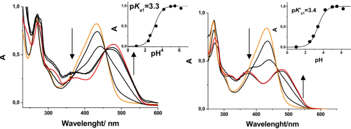
% &Q 3 ?/ 5 - % 0 % . QQ(
% &(3 ?/ 5 - % 0 % QQP
% &P 5 ?/ 5 - % 0 % < ! ! QQK
33 &/ 0 '$* . '"'
% A&Q 3 0 QQH
% A&P 3 0 5 QQJ
% A&K 3 . < ! ?/ 5 - % Q()
% A&H 5 ?/ 5 ! - % 2$ 6 Q ! ! Q(Q
% A&I 5 ?/ 5 ! - % 2$ 6 0 I Q((
% A&J 5 ?/ 5 $ 0 - ( ! ! Q(P
% A&*0 ! ?/ 5 - % Q(K
% A&T 5 5 . < ! ?/ 5 - % 0 - Q(H
% A&Q) 5 '( Q(I
% A&QQ 5 Q(J
% A&Q(5 / 5 Q(*
% A&QP 3 (H Q(T
% A&QK 3 (H QP)
/ - ', #
2'3" ( 1
$ ! Q&Q 5 - ! ! 0 - ! % ; ! QQ
$ ! Q&( 3 ?/ 0 Daemonorops draco Dracaena draco Dracaena cinnabari
0! .! , & QH
$ ! Q&P 3Daemonorops ! 0 ' B !. . ?/ 5 QT
$ ! Q&K 5Dracaena cinnabari ! 0 ' B !. . ?/ 5 (Q
$ ! Q&H 3Dracaena draco ! 0 ' B !. . ?/ 5 (K
2'3" ( 5
$ ! (&Q 5 < < 00 0 % %
$V(TP B PI
$ ! (&( 5 @ . ! 0 IΦ 0 % $V(TP B PJ
$ ! (&P 3 0 . ?/ 5 + - - - % 0 %
+ + - PPH PT
$ ! (&K 5 @ . ! 0 IΦ 0 % 7 % !
+ $V(TP B K)
$ ! (&H 3 ! 0 - ! -
-0 $ < ! . ?/ 5 KP
2'3" ( 6
$ ! P&Q 3 . - 0 . + - 00 0 ! ! H)
$ ! P&( 3 ! % 0 - - - - . - >
. HP
$ ! P&P 5 ! % 0 - - - 0 - . < !
! HK
$ ! P&K 5 ! % 0 - - - 0 - !
! 5 HJ
33 &/ 9 /3 (&! "' # "&
$ ! &Q 3 ! % 0 . !. JH
$ ! &( 5 . ! !. . ?/ 5 *)
$ ! &P 3 ? ! ! TH
$ ! &K 3 < - . !, < ! TT
$ ! &H 3 - - . < ! Q))
33 &/ 9 (' # , '"'
$ ! &Q 3Q? QP 5# 0 J K;5 -. <.5H5 - <.0! .! Q)P
$ ! &( 3Q? QP 5# 0 J5-. <.5H5 - <.5I5 -.!0! .! Q)K
$ ! &P 3Q? QP # 0 J K;5 -. <.0! .! Q)H
33 &/ 9 & '"'
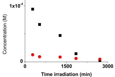
$ ! &Q 3I0 0 - PPH IQ) QQ)
$ ! &( 3 % Φ 0 - PPH IQ) QQ)
$ ! &P 3 % Φ 0 - PPH IQ) QQQ
$ ! &K 3 ?/ 5 ! 0 % QQQ
33 &/ 0 '$* . '"'
$ ! A&Q 3 0 - % ! - K . . - QQ*
$ ! A&( 3 0 B( ( J ?( K . 0 . - QQ*
$ ! A&P 5 > ! 0 QQT
$ ! A&K 5Q?5 QP 5# 0 - ! '( Q(I
$ ! A&H 5 Q?5 QP 5# 0 - ! Q(J
$ ! A&I 5Q?5 QP 5# 0 - ! 5 Q(*
$ ! A&J 5Q?5 QP 5# 0 - ! (H Q(T
$ ! A&* 5Q?5 QP 5# 0 - ! (H QP)
$ ! A&T 5 !. 0 - 5 . < ! ! QPQ
Before the discovery of mauve dye and later thousands of synthetic dyes that resemble a
colourful rainbow [1] in the nineteenth century, the colours used to dye a textile since pre
historic times were obtained from natural sources – vegetable or animal [2,3]. For the blue
and purple colours, indigo derivatives were mainly used. For red dyed textiles,
anthraquinone based dyes could be found and the yellows were obtained from an
enormous variety of local plant species, mostly from the flavonoid family [2,3]. From these
organic dyes, dragon’s blood and indigo were chosen for the red and blue colour study,
respectively. The synthetic mauve dye, a landmark in the history of chemistry and dye
industry, was also studied.
A characterization at the molecular level of dragon’s blood, indigo and mauve dye is the
main aim of this thesis. A more in depth understanding will in turn enable a better
conservation and access to our cultural heritage, namely to ancient textiles. This PhD
thesis was carried out in the framework of the project Molecules of Colour in Art: a
Photochemical Study, where the molecular studies evolved along three main axes with: i)
structural characterization of the most relevant chromophores in each dye; ii)
photophysical characterization and iii) photochemical characterization. In this thesis, the
structural characterization of dragon’s blood, indigo and mauve dye, as well as the
photodegradation of indigo will be presented.
In each chapter an introduction to each organic dye is presented, followed by its molecular
characterization and photodegradation for the indigo dye. A brief summary of the principal
conclusions is also presented.
Organic dyes are usually a complex mixture of different chromophores that bring colour to
the textile. In natural dyes, such as dragon’s blood, the presence of several chromophores
with different relative proportions according to the dyeing source, the local, date and
season of the sampling amongst others [4,6], can give a kind of fingerprint very useful in
the identification of dyes in works of art, such as textiles [2,7]. With this information
amongst others factors, it will be possible, for example, to establish conception dates,
assign production centres, identify textile trade routes, etc [8]. Moreover, successful
involved are well characterized structurally. In synthetic dyes, like the mauve dye, a
complex pattern of chromophores can also be found, when the synthetic procedure gives
several coloured products. Nevertheless, a simpler pattern can be found in natural and
synthetic dyes as the case of the indigo dye where usually only the indigotin chromophore
is retained on the fibres during the dyeing bath [2]. For this reason the photodegradation of
indigo in homogeneous and heterogeneous media was carried out.
Organic dyes being coloured molecules will absorb in the UV Vis region and therefore
photochemical reactions that will dictate the stability of the molecule can occur. When
these molecules absorb radiation, the resulting extra energy produces an excited state
molecule, which can be considered an entire new molecule since several properties as its
polarity, acidity, oxidation and reduction properties, just to name a few, are entirely
different from the ground state molecule [9 12]. The lifetime of an excited molecule is
usually very small, in the order of nanoseconds or even less. It can return to the ground
state with emission of fluorescence (i.e. emission of photons), internal conversion (i.e.
direct return to the ground state without emission of fluorescence), intersystem crossing
(possibly followed by emission of phosphorescence), intramolecular charge transfer;
proton transfer; photochemical reactions, amongst others (see figure 1.1), which compete
with each other while the molecule is in the excited state.
Figure 1.1 Possible de excitation pathways of excited molecules, adapted from [9].
ν
ν
ν
ν
fluorescence emission
intersystem crossing
internal conversion
intramolecular charge transfer
conformational change
electron transfer proton
transfer energy
transfer excimer /
exciplex formation photochemical
transformation
When the excited molecule loses energy through chemical reactions we are in the
photochemistry field but when the excited molecule is converted to the ground state, with
the excess of energy being released as radiative or non radiative energy we are in the
photophysics domain [11,12]. In the excited state, bonds can be broken and new ones
formed; if the processes involving bond breaking and formation are reversible, they will be
considered as non radiative photophysical deactivations (figure 1.2). If they are
irreversible, they will be studied as a photochemical reaction and an important parameter
for its characterization is defined as the quantum yield of reaction (see below).
Figure 1.2: Adapted Jablonski scheme [9], where the kf (fluorescence deactivation) and kp
(phosphorescence deactivation) are the rate constants for the radiative processes and the
k’isc (intersystem crossing deactivation), kic (internal conversion deactivation) and kisc
(intersystem crossing deactivation) are the non radiative processes. The S0 and S1 are the
singlet and excited ground state respectively and T1 is the first triplet state.
For a full photophysical characterization, absorption, fluorescence and phosphorescence
spectra can be obtained; triplet triplet absorption analysis is also performed. Moreover,
fluorescence lifetimes and quantum yields can be determined and the rate constants
responsible for the excited state deactivation can be calculated.
The photophysical characterization will enable a better understanding of the
photochemistry as well as more precise lifetime of the molecule. The study of the influence
of oxygen in the photochemistry and reactivity of these dyes will, in turn, enable a better
understanding of which are the photophysical parameters that play a relevant role in the
photodegradation mechanism. Furthermore, it will enable the prediction of the colour
changes over time.
One important parameter, as mentioned before, in the characterization of photochemical
reactions is the photodegradation quantum yield of a reaction that can be defined as:
Φ = Amount of reactant consumed or product formed per unit of time Amount of photon absorbed
The quantum yield obtained should take into account the role of oxygen in the
photodegradation reaction as photooxidation can contribute for the global mechanism of
fading. Therefore, the photodegradation quantum yield in the presence and absence of
oxygen should be calculated.
Moreover, the main photoproducts should also be identified as well as the intermediates
formed after monochromatic irradiation, in order to obtain the principal degradation
mechanisms occurred during the light induced fading.
A systematic approach to the study of the photophysics and photochemistry of these
colourants cannot be found in the literature. Particular importance has been given to the
molecular characterization of the cited molecules rather than on a full understanding of the
photodegradation mechanisms. It is important to remember that the most known organic
dyes had to overcome the barrier of light and washing fading during hundreds of years.
Therefore, their degradation is somehow a slow process and for that reason the
photophysical and photochemical characterization of these organic dyes is a complex and
time consuming task. Nevertheless, it is important to know their molecular mechanisms of
degradation, in order to be able to plan strategies to prevent their fading and improve their
durability.
With the results presented in this thesis, new knowledge into historic dyes at their
molecular level is provided and, from now on, it will be easier to perform a detailed
photophysical and photochemical study of these organic dyes as well as applying the
Dragon’s blood is a natural resin [13] with a rich deep red colour obtained essentially from
three different families of plants, namely Dracaenaceae, Palmae and Euphorbiaceae [14
17]. It may sound like an exotic ingredient of a witch's brew or a magic potion. Indeed,
legends refer that dragon’s blood was the result of a fight between a dragon and an
elephant until death, where a dragon tree sprung up from the congealed blood dropped by
both animals, giving “magical” properties to the red resin [14 17]. In the Greek mythology it
was mentioned that Hercules killed Geriones (the three headed monster) from the Eriteya
Island, and from his blood a tree was also born with red fruits which produced the dragon’s
blood resin [18]. Curiously, the red resin has been applied for centuries in traditional
medicine with different clinical and ethnomedical uses where pharmacological assays
have frequently corroborated its medical applications (and therefore the “magical”
properties of dragon’s blood) against cancer, ulcers and wounds, amongst others [19 28].
Apart from this, dragon’s blood has also been used in the past with several artistic
purposes [14 17,29].
Attempts to unveil the trade history of dragon’s blood resins, the use of its sources and
even its chemical composition have been difficult due to botanical misunderstanding and
the diversity of dragon trees [14 17,30 32]. It is believed that in the past, one of the
principal sources of dragon’s blood was Dracaena cinnabari Balf. (Dracaenaceae) from
Socotra Island [14 16]. Today the main dragon’s blood resin commercially available is
obtained from the species Daemonorops draco (Willd.) Blume (synonym Daemonorops
propinqua Becc.) from Thailand, Sumatra and Borneo [14 16,30,33]. However, a great
diversity of dragon’s blood resins has been used since ancient times until today, due to the
different dyeing sources of the resin available in each geographic area [14 16,27 28].
The resin can be collected from natural exudates that occur in injured areas in the stem
and branches of the tree (Dracaenaceae andEuphorbiaceae families, figure 1.3a) or can
be obtained from the fruits which are covered with small scales through where the resin
exudes forming a brittle red resinous layer outside the fruits (Palmae family, figure 1.3b)
Figure 1.3– a) Resin from Dracaena draco tree; b) Resin from Daemonorops micrantha
(Griff.) Becc. [13].
!
In the family, which comprises between 60 and 100 species, only 5
species have the growth habit of a tree with an umbrella type crown producing dragon’s
blood resin [34,35,38 41]. These trees are the (figure 1.4)
which is endemic of Socotra Island [38]; ! (figure 1.5) from the
south western Arabia [34,35]; the " # $ % # (synonym
! & figure 1.6) from North East tropical Africa and western Arabian
peninsula [40]; ' (figure 1.7) from the Macaronesian Islands as Madeira,
Azores, Cape Verde and Canarias [34,35] or from Morocco being reported there
' ( )* ( + [41], and finally the recently identified
) , $ (similar to Dracaena draco L.) from the Gran
Canaria (Canary Islands)[39].
Figure 1.4 – Dracaena cinnabari Balf, Socotra [42].
Figure 1.5 – Dracaena serrulata Baker [43].
(
-Figure 1.6 – Dracaena ombet Kotschy & Peyr [44].
Figure 1.7 – Dracaena draco L., Lisbon.
It is believed that these dragon trees with a bizarre prehistoric appearance share a
common origin due to the similar morphological features and the existence of comparable
fossils (Dracaenites brongniartii Saporta, Dracaenites narbonenis Saporta and
Dracaenites sepultus Saporta) from Tertiary deposits (65 million to 1.8 million years ago)
in Southern Europe [38,39]. During the Tertiary period, the drastic climate changes at the
end of the Oligocene brought about the almost extinction of the subtropical vegetation
throughout the South of Europe and North Africa, from the Atlantic to the Indian Oceans
and lead to a biogeographic dissociation between the living dragon’s tree of East and West
Africa. Only a few specimens of the ancient flora survived on both sides of the African
continent where the environmental conditions were more stable due to the tempering
effect of the sea [39]. Far from one another, these remaining colonies continued to develop
independently, giving rise to the actual five species of dragon’s tree of the Dracaena
genus. As long isolation exists, each species will tend to become slightly different from
others [37,39]. Nevertheless, similarities can be drawn between the five species
mentioned.
Important colonies of this ancient relic flora are the Dracaena cinnabari dragon’s tree of
Socotra Island with an age between 200 and 300 years old [38]. However, studies point
out that this Dracaena cinnabari woodland will reach the stage of intensive disintegration
within 30 77 years due to climate changes [38,45]. Other very famous exemplar was the
Great Dragon Tree of Orotova (Tenerife) believed to have an estimated age of 6000 years
old. The dragon tree fell in 1817 due to a hurricane. Today, the oldest Dracaena draco tree
in Icod (Tenerife) has an estimated age of 700 years old [13 16] (figure 1.8). Being
monocots, its age is very difficult to estimate due to the lack of annual growth rings.
Nevertheless, the dragon’s tree age can be estimated with different indirect methods being
the most usual the number’s determination of the sausage shaped section that normally
display between 11 and 16 years for Dracaena draco and 14 30 years for Dracaena
cinnabari [38].
Figure 1.8 – Dracaena draco L., Icod [46]
" # $ !
In the family, the species that produce dragon’s blood resin belong to genus rattan
Daemonorops which comprises about 115 species [36,37,47]. The Daemonorops genus is
divided into two sections, the Cymbospatha and the Piptospatha being the most important
product of the last one, the dragon’s blood powder. The dragon’s blood species are
confined to Malaysia, Thailand and West Indonesia, where the red resin is an item of trade
between Borneo, Sumatra, the Malay Peninsula and even China [16]. As in the
Dracaenaceae family, they are a natural unit taxa where some species have slightly
diverged because of isolation or adaptation [37 39]. As mentioned before, the most
commonly used Daemonorops sp. dragon’s blood species is Daemonorops draco (Willd.)
Blume (synonym Daemonorops propinqua Becc. Species, figure 1.9), but up to 10 species
are known, namely the 0 from Peninsula Malaysia
to North Sumatera, 12 3 4 ! from Peninsula
Thailand to West Malesia, Sumatera, 5 from Sumatera
(Kep. Mentawai), % 5 6 from Sumatera,
3 from Borneo, 7 8 . from
Peninsula Malaysia and Borneo, 75 9 2 , 8 6 from
Jawa, from Peninsula Thailand to West Malesia [36,37], and
the recently discovered 5 6 from Sumatera as well the
Figure 1.9 – Daemonorops draco sp. [48].
# & $ !
In the # family the production of dragon’s blood belongs to genus Croton (figure 1.10) widespread in tropical regions of the Old and New World with circa 1300
species [27 28].
Figure 1.10 – Croton lechleri, [49].
Several species of Croton, namely from South American (Ecuador, Peru,
Colombia and Bolivia), " + from South American tropics
(Peru), 6 $ from Mexico and Central America&
from Brazil and Paraguai, ,: ) from Peru,
+ , ' from Sri Lanka, and
+ have been used in the production of the red resin [27, 28].
'
Other known sources of dragon’s blood are the % 3 ;
(synonymous of Pterocarpus draco L., Leguminosae family, figure 1.11) from West Indian,
Caribean, coastal areas of Central and northern South America [15,16,50] and the shrub
7' 8 (Dracaenaceae family, figure 1.12) from
China, S.W. Guangxi to S. Yunnan, to Indochina believed by some authors to be the
original source of the dragon’s blood used for thousands of years in traditional Chinese
Figure 1.11Pterocarpus oficinallis [52]
Figure 1.12 Shrub Dracaena cochinchinensis (Lour.) [53]
However, it is known that for Chinese herbal medicine Daemonorops was also imported
into China from South East Asia, and others sources as % 7, ! 8 5 2( (tropical and subtropical Asia to North Australia including Taiwan, Guangdong and
Yunnan) and % 2 . (South Hainan to Indochina) [52]
were also used.
<(
The dragon’s blood resins are a complex mixture of several compounds. Recent
phytochemical studies on the genera Dracaena and Daemonorops identified several
compounds as saponins [19 21,55], chalcones [23], flavonoids [23,56 58], sterols [59], and
flavans [58,59], amongst others. These compounds are colourless or display a yellowish
colour. However, for the Daemonorops sp. resins, besides the presence of chalcones,
flavans and flavonoids, red flavylium pigments as dracorhodin and dracorubin were also
identified [60,62 69]. It was in 1936 that Brockmann and Haase published the first attempt
to identify the red colourants of a powdered commercial dragon's blood resin probably
from a Daemonorops sp. source in which they isolated one of the red compounds and
named it as dracorubin [62]. A second colourant, dracorhodin, was identified in 1943 by
Brockmann and Junge that have also synthesized the molecule and concluded it was a
natural 2 phenyl 1 benzopyrylium (a flavylium salt) from the anthocyanins family [64]. A
more straightforward synthesis, and consequent confirmation of the structure, was
published by Robertson and Whalley, in 1950 [65]. The structure of dracorubin was
in 1975, by Whalley[66]. In the meantime, two other natural red flavylium colorants from
dragon's blood were characterized and named as nordracorhodin and nordracorubin
[60,67] (table 1.1).
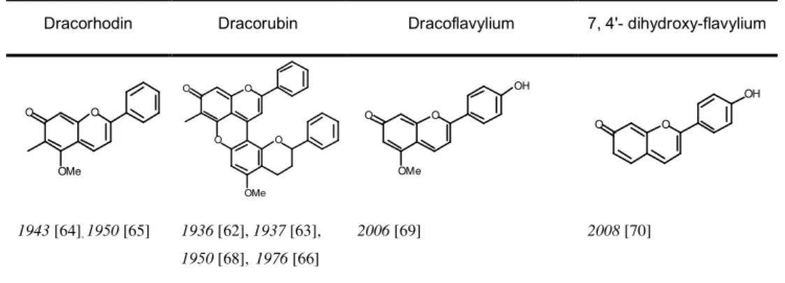
Table 1.1 Chemical structures responsible for the red colour in dragon’s blood resins.
The structures correspond to the quinoid bases ()).
Dracorhodin Dracorubin Dracoflavylium 7, 4' dihydroxy flavylium
O O
OMe
O O
O O
OMe
O O
OMe
OH
O O
OH
1943 [64],1950 [65] 1936 [62], 1937 [63],
1950 [68],1976 [66]
2006 [69] 2008 [70]
The presence of natural flavylium compounds in dragon’s blood resin from Daemonorops
sp. has been mislaid during the last decades [19 22,26,30 32]. However, those natural
flavylium compounds contribute significantly to the final deep red colour of dragon’s blood
resin.
Synthetic flavylium salts, natural flavylium and anthocyanins have in common the 2
phenyl 1 benzopyrylium chromophore unit. In terms of molecular structure, flavylium salts
were the first to be discovered, Bülow [71] 1901, followed by the natural anthocyanins,
Willstätter [72], and finally by natural flavylium compounds [73].
Anthocyanins are characterized by the existence of an O glucoside in position 3
(monoglucoside). A sugar can also be present in position 5 (diglucosides) or less
frequently in position 7 [74], figure 1.13. On the other hand, in anthocyanidins hydroxyl
groups take the positions of the glucosides, leading to unstable structures in solution. On
the contrary, the so called desoxyanthocyanidins, that corresponds to “anthocyanidins”
lacking the hydroxyl in position 3 (but bearing a hydroxyl in position 5), are quite stable in
solution.
Figure 1.13 – In the basic chemical structure of anthocyanins, an hydroxyl group is
present in positions 4’ and 7, and a sugar in position 3 (monoglycosides) or 3 and 5
(diglycosides)
O
OGl HO
OH R'3
OGl
R'5
3 5
7 A
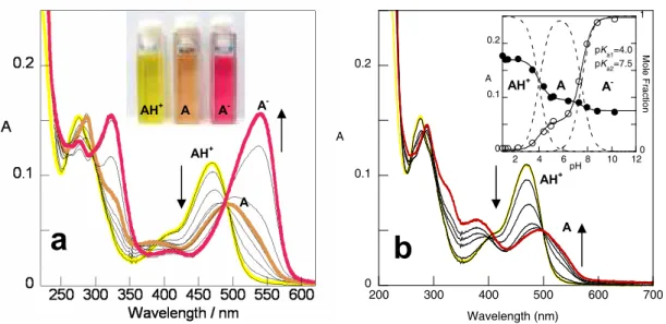
In the seventies of the last century, it was firmly established by Dubois and Brouillard
(anthocyanins) [75] and McClelland (synthetic flavylium salts) [76] that both families of
compounds undergo multiple structural alterations in aqueous solution, following the same
basic mechanism[77] (see figure 1.14).
The flavylium cation ()4=) is the dominant species in very acidic solutions, but with
increasing of pH a series of more or less reversible reactions occur: 1) deprotonation
leading to the quinoid base ()), 2) hydration of the flavilyum cation giving rise to the
colourless hemiacetal ( ), 3) tautomerisation reaction, responsible for ring opening, to give
the pale yellow Z chalcone form ( ), and finally, 4) cis trans isomerisation to form the
pale yellow chalcone ( ). Furthermore, at higher pH, and depending on the number of
hydroxyl groups, further deprotonated species are found, such as and ) . The
relevant contribution for the colour is given by )4= and the quinoid bases.
Figure 1.14 – Network of chemical reactions for 7,4’ dihydroxy 5 methoxyflavylium in
solution [69] (see section 1.3.4.1).
In this work, the discovery of natural flavylium compounds in resins [69,70], namely the
compounds 7,4’ dihydroxy 5 methoxyflavylium and 7,4’ dihydroxyflavylium from Dracaena
draco and from Dracaena cinnabari, respectively, is reported for the first time. A fingerprint
study of these red chromophores in dragon’s blood resins from Dracaena and
O O O -A -O O OH A O HO OH + AH+ O HO OH HO B OH HO OH O Cc OH HO Ct O OH OH -O O OH OH -O O O -Ct
-Ct2
-K a1 K a2 K h Kt K i K
Ct1 KCt2
+ H+
+ H+
+ H+
+ H+ + H+
OCH3 OCH
3 OCH3
OCH3 OCH3
OCH3
Daemonorops trees was performed using high performance liquid chromatography with
diode array detector (HPLC DAD) and principal component analysis (PCA).
These natural flavylium markers do not fit the commonly accepted definition of
anthocyanidin or 3 deoxyanthocyanidin [78] as some authors recently have proposed for
analogous structures from Arrabidaea chica[79], as a methoxy group was found in position
5. Their structure and chemical behaviour are closer to the so called synthetic flavylium
salts, as will be described bellow.
)
Due to the great diversity of dragon’s blood resins and botanical misunderstandings
amongst others in the literature, previously to the resin red compounds characterization,
an HPLC DAD data base was built (see appendix I experimental section, section I.3
p.79). Moreover, a method based on flavylium markers was developed for the resins
identification and applied to the XIX century dragon’s blood collection of the Economic
Botany Collections at the Royal Botanic Gardens, Kew (EBC, K). Afterwards the dragon’s
blood flavylium characterization was performed.
From the circa 30 species referred in the section 1.1, only three species (Dracaena draco,
Dracaena cinnabari and Daemonorops draco), which were probably the most important
species used in Europe, were selected for the construction of the dragon’s blood HPLC
DAD library (see appendix I experimental section, table I.2, p. 80, for library HPLC DAD
dragon’s blood samples). The aim of the HPLC library was the distinction of dragon’s
blood species and subsequent identification of unknown resins. An initial HPLC DAD
screening revealed the presence of different flavylium compounds responsible for the red
colour of the resins. The use of the flavylium compounds as potential species markers for
dragon’s blood resins was for the first time investigated.
Circa 50 samples from Dracaena draco, Dracaena cinnabari and Daemonorops draco
(mostly collected in botanical gardens) were analyzed by HPLC DAD. The results obtained
were subsequently applied to 37 samples of dragon’s blood from EBC, K (labelled as
Daemonorops draco, Daemonorops sp., Dracaena cinnabari, Dracaena draco, Dracaena
schizantha and Dracaenasp.). The EBC, K contains perhaps the largest and most reliably
identified assemblage of dragon’s blood resins dating from the 19th century which were
donated by Sir Isaac Bailey Balfour or the Pharmaceutical Society of Great Britain,
* %
Samples from known provenance, with the species correctly identified by experts, were
used to build up the HPLC DAD database (i.e. for the Dracaenaceae, 33 samples of
Dracaena draco and 9 samples of Dracaena cinnabari). It was also possible to
characterize roughly the tree age (for more details see appendix I – experimental section,
table I.2, p. 80). The situation was different for the Daemonorops draco resin, where only 2
commercial samples were acquired. However, this limitation was overcome with the
analysis of the dragon’s blood EBC, K collection (see section 1.3.3.1).
In all the samples analysed, the red colour resulted from the contribution of single flavylium
chromophores, as for instance, dracorhodin, and condensed flavylium molecules, such as
dracorubin [23,24,62 69] (see table 1.1). More importantly, it was observed (table 1.2) that
Dracaena draco, Dracaena cinnabari and Daemonorops draco presented each one a
characteristic flavylium: 7,4’ dihydroxy 5 methoxyflavylium (dracoflavylium), 7,4'
dihydroxyflavylium and 7 hydroxy 5 methoxy 6 methylflavylium (dracorhodin),
respectively. As can be observed in table 1.1, these compounds have the 2 phenyl 1
benzopyrylium core in common, but a different substitution pattern, consequently each
exhibit characteristic UV Vis spectra and retention times (table 1.2). This, in turn, enables
a straightforward identification of these flavylium chromophores by HPLC DAD, which
leads to the identification of the dragon’s blood source (table 1.2). Besides these markers,
the chromatograms acquired at the wavelengths for red detection (462 nm) for each resin
type are also characteristic for these species and, after PCA, could also be used as a
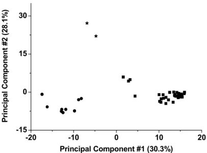
fingerprint (see table 1.2 and figure 1.15).
The PCA principal components represented in Figure 1.15 can be analysed according to
the corresponding loadings (each sample PCA score is the inner product between the
sample chromatogram and the loading corresponding to a given principal component). It
was observed that the loading for the first score, PC#1, contains strong positive peaks at
20.5, 27.4 and 27.9 min and strong negative peaks at 18.0, 18.7 and 23.3 min. The former
correspond to Dracaena draco elution peaks while the latter correspond to Dracaena
cinnabari peaks. Therefore, the first PCA component, PC#1, is able to discriminate
between these two Dracaena species (positive scores for Dracaena draco and negative
scores for Dracaena cinnabari in the first component axis). The third score, PC#3, exhibits
two strong positive peaks at 21.1 and 21.7 min. These peaks correspond to peaks
observed in Daemonorops dracochromatograms (see table 1.2). No other relevant peaks
were observed in the third loading which means that this component captures only the
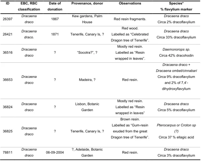
Table 1.2 – HPLC chromatogram profiles, retention times and absorption maxima for
Daemonorops draco, Dracaena draco, Dracaena cinnabari resins and respective flavylium
markers.
Species Chromatogram Flavylium marker Spectrum’ Flavylium marker
Dracaena cinnabari Balf. f. (Source: Firmihin and Hamadero in
Socotra Island; mean age circa 250 years old) -& &> &. ) ( ( . 6 & (1) 7,4’ dihydroxyflavylium
tr = 18.03±0.15
min
λmax = 462 nm
- --& &. & ) ( (
? @ &
Dracaena
draco L.
(source: Natural Park
of Madeira; age: circa 200
years old)
-& &> &. ) ( ( / . 6 & (2) Dracoflavylium 7,4’ dihydroxy 5 methoxyflavylium
tr =
20.51±0.12min
λmax = 476 nm -
--& &. & ) ( (
? @ &
Daemonorops
draco (Wild.)
Blume (source: Zecchi; age unknown) -) ( ( > 6 & (3) Dracorhodin 7,6 dihydroxy 5 methoxyflavylium
tr =
21.76±0.07min
λmax = 438 nm -
--& &/ & ) ( (
-%
6
A
7
>
B
8
% 6 A 7 B8
Figure 1.15 – PCA analysis of Dracaena draco (squares), Dracaena cinnabari (circles) and
Daemonorops draco (stars) with HPLC data library chromatograms acquired at 462 nm..
+ , - -.- %
The HPLC DAD library for Dracaena draco was created with resins freshly gathered from
Madeira, Lisbon and Cape Verde. In all the samples analysed the 7,4’ dihydroxy 5
methoxyflavylium (dracoflavylium) was always present. Samples enclosed by the dotted
line are centenary trees c. 200 years old, where dracoflavylium content was the major red
compound (c. 32%) (figure 1.15). In the other samples this flavylium was present as a
minor product of the total amount of the red colourants of resin, ranging from 1 10%
(relative area). Although relative concentration of dracoflavylium is variable, this flavylium
was only found in Dracaena draco resins.
+ , - %
In the chromatograms used to build up the HPLC DAD library for Dracaena cinnabari, the
quantity of 7,4’ dihydroxyflavylium varied from 5% to 15% of the relative area for the total
amount of the red colourants of resin. The colour of this resin is due to a complex mixture
of red compounds (table 1.2), with 7,4’ dihydroxyflavylium being one of the major
+- -.- -/- %
In the Daemonorops draco resins, 7 hydroxy 5 methoxy 6 methylflavylium (dracorhodin)
was the major red compound. The occurrence of dracorhodin in Daemonorops sp. resins
has been described already in the literature [23,24,62 65].
( # 0 ) 0 1 2 3
After obtaining the results described above, identification of dragon’s blood resin sources
based on flavylium markers was applied to the largely 19th century collection of dragon’s
blood at EBC, K. These items comprise not only resins from the already described
Daemonorops draco, Dracaena draco and Dracaena cinnabari, but also unnamed
Daemonorops and Dracaena sp. resins and one resin from Zanzibar tentatively labelled
Dracaena schizantha (a synonym of Dracaena ombet). The EBC, K collection is very
heterogeneous with very different grades of resin purity. Just over 50% of the resin
collection labelled as Daemonorops draco (or its synonym Daemonorops propinqua),
Dracaena cinnabari and Dracaena draco species were examined.
The results obtained were analyzed and compared with the HPLC DAD data library, using
the flavylium markers and the PCA of the full chromatograms acquired at 462 nm. Both
approaches gave identical results and successfully discriminated Daemonorops draco,
Dracaena cinnabari and Dracaena draco (see figure 1.16). On the other hand, it was not
possible to distinguish unambiguously the EBC, K sample labelled Dracaena schizantha (a
synonym of Dracaena ombet) from the circa 30 Dracaena cinnabari samples analysed
(HPLC DAD library and EBC, K collection). All the samples, had the same flavylium
marker; however, due to the small number of samples labelled Dracaena schizantha, 1
from EBC, K and 1 from the living collections (Horticulture & Public Education, Kew HPE,
K) a more conclusive result using PCA could not be drawn.
PCA shows that all the EBC, K samples are in accordance with the built up data library
(figure 1.16), therefore, being possible to establish and verify the species source. Only two
samples lay outside the three established areas: the samples EBC, K 36653 (labelled
Dracaena draco), which is probably a mixture of resins, and EBC, K 36825 (labelled
Dracaena draco), which is not actually based on flavylium compounds. In the next sections
the results obtained will be described in more detail and, whenever possible, they will be
-%
6
A
7
>
B
8
% 6 A 7 B8
Figure 1.16 PCA analysis of Dracaena draco (squares), Dracaena cinnabari (circles) and
Daemonorops draco (stars); HPLC chromatograms acquired at 462 nm for data library
samples (open symbols) and EBC, K samples (solid symbols). The areas assigned
represent three major distinct zones enabling the species identification of the samples
analyzed.
4 !
5
In the EBC, K samples labelled as Daemonorops draco or D. propinqua, 7 hydroxy 5
methoxy 6 methylflavylium (dracorhodin) was identified as the major compound. This
observation is in line with the recent publication which announced the two taxa as
synonyms [37], Daemonorops draco being the accepted name [33]. Furthermore, the
chromatograms obtained for these samples are very similar, as confirmed by PCA analysis
(see appendix II – Dragon’s blood data, section II.2, p. 105, for PCA graphics).
The HPLC DAD data showed that of the 9 analyzed samples labelled Daemonorops
draco, Daemonorops propinqua or Daemonorops sp., only 6 samples had a correct
attribution (see table 1.3). In 4 samples (35489, 35495, 35500, 35526), the relative
percentage of 7,6 dihydroxy 5 methoxyflavylium (dracorhodin) was circa 65% of the total
area of red chromophores as found in the commercial Daemonorops sp. resins. In
samples that were processed (35490 and 35505), the relative percentage of 7 hydroxy 5
( < Specimens of Daemonorops samples from EBC, K analysed by HPLC DAD
and percentage of the flavylium markers identified.
1 & " % @ &
C( @
B @# 6 6 ! D
35487 Daemonorops
draco ?
India,
India Museum Mixture of resin, bark and powder.
Dracaena cinnabari
Circa 6% 7,4’ dihydroxyflavylium
35489 Daemonorops
draco 1851
Singapore A.S. Hill & Son
Mostly resin.
Labelled as “Calamus draco, in lumps, colour of powder, brick red. Contains about 12 per cent of insoluble matter”. Paler and duller than other samples.
Daemonorops draco
Circa 65% dracorhodin
35490 Daemonorops
draco 1851
India, Calcutta, Royal Commonwealth
Exhibition
Mostly resin.
Labelled as “Reed dragon’s blood”
Daemonorops draco
Circa 55% dracorhodin
35495 Daemonorops
draco ?
?, British Museum (Natural History)
Mixture of resin and powder. The sample appears to be small pieces from
a reed resin.
Daemonorops draco
Circa 65% dracorhodin
35499 Daemonorops
draco ? Sumatra, ? Resin attached to fruit scales.
Daemonorops sp. (?)
Circa 65% unknown compound (tr=21.03 min,
λmax=453 nm)
35500 Daemonorops
propinqua 1896 Sumatra, ? Resin attached to fruit scales.
Daemonorops draco
Circa 65% dracorhodin
35526 Daemonorops
propinqua 1890
?, A Hill & Son
Mixture of resin, powder and contaminants. It looks paler and much less resinous than some other samples
of lump dragon’s blood.
Daemonorops draco
Circa 65% dracorhodin
35527 Daemonorops
propinqua ?
?,
Savory & Co Mixture of resin and powder. Lump dragon’s blood.
Dracaena cinnabari
Circa16%7,4’ dihydroxyflavylium
35505 Daemonorops
sp. 1851
Sumatra, Royal Commonwealth
Exhibition
Mostly resin. Similar to lump dragon’s blood.
Daemonorops draco
Circa 55% dracorhodin
DThe relative peak areas were calculated with the chromatographic program ChromQuest
4.1 at the maximum wavelength absorption for each flavylium marker selected: 438 nm for
dracorhodin (Daemonorops sp.), 462 nm for 7,4’ dihydroxyflavylium (Dracaena cinnabari
and Dracaena schizantha) and 476 nm for 7,4’ dihydroxy 5 methoxyflavylium (Dracaena
In two further samples, 35487 and 35527, labelled Daemonorops draco and Daemonorops
propinqua respectively, the presence of 7,4’ dihydroxyflavylium and absence of
dracorhodin prompt the conclusion that these resins were from Dracaena cinnabari. Both
these samples appeared more glossy or resinous than other lump resins classed with this
genus. Also, at least one of these 2 samples was sourced in India (see table 1.3).Finally,
sample 35499 (labelled as Daemonorops draco) revealed the presence of an unknown red
chromophore in its composition (see table 1.3) and therefore no conclusion concerning its
provenance could be drawn. It seems that the names of samples of lump dragon’s blood
resins currently housed under the name Daemonorops in the EBC, K may not be accurate,
and analysis of further samples using these techniques would be profitable. Some of the
naming errors may have arisen due to past curation of the samples where Latin names
have been assumed from the vernacular. These errors are more likely to occur from
samples sourced in India as both Daemonorops draco and Dracaena cinnabari were
traded there [16].
5 !
5
All 15 EBC, K Dracaena cinnabari samples showed the presence of 7,4’
dihydroxyflavylium (5 20%), in agreement with the species attribution; these samples were
acquired most directly from Socotra, where the species is endemic and some from market
( Dracaena cinnabari samples from EBC, K analysed by HPLC DAD.
1 & 5
% @ & C( @
B @# 6 6 ! D
36489 Dracaena
cinnabari ?
?, labelled Socotra dragon’s
blood from Allen & Co. Mixture of resin, bark and powder
Dracaena cinnabari
Circa 7% 7,4’ dihydroxyflavylium
36542 Dracaena
cinnabari 07 1881 ?, donated by Prof IB Balfour
Mixture of resin, bark and powder. Labelled as “Socotra Dragon’s blood”.
Dracaena cinnabari
Circa 8% 7,4’ dihydroxyflavylium
36543 Dracaena
cinnabari 1875 ?, donated by Dr Vaughan
Resin.
Labelled as “Socotra Dragon’s blood”.
Dracaena cinnabari
Circa 5% 7,4’ dihydroxyflavylium
36545 Dracaena
cinnabari 10 1899
?, purchased by Mather at Ripley Roberts Drug sale, 3
Mincing lane
Resin.
Labelled as “Extra fine Zanzibar leas”.
Dracaena cinnabari
Circa 5% 7,4’ dihydroxyflavylium
36557 Dracaena
cinnabari 10 1899 Zanzibar, purchased by Mather
Heterogeneous resin. Labelled as “Socotra Dragon’s blood”.
Dracaena cinnabari
Circa 15% 7,4’ dihydroxyflavylium
36563 Dracaena
cinnabari ? ?
Powder.
Labelled as “Socotra Dragon’s blood”.
Dracaena cinnabari
Circa 15% 7,4’ dihydroxyflavylium
36580 Dracaena
cinnabari 10 1899
?, Kurachi, purchased by Mather
Resin.
Labelled as “Fine marbles of dragon’s blood”
Dracaena cinnabari
Circa 5% 7,4’ dihydroxyflavylium
36599 Dracaena
cinnabari 1881 Socotra, ?
Mixture of pigment and resin. Labelled as “dam el akhuwen”
Dracaena cinnabari
Circa 17% 7,4’ dihydroxyflavylium
36611 Dracaena
cinnabari 01 1880 ?, presented by Dr JB Balfour
Tears of resin.
Labelled as “Socotra Dragon’s blood”.
Dracaena cinnabari
Circa 15% 7,4’ dihydroxyflavylium
36622 Dracaena
cinnabari ? ?
Tears of resin.
Labelled as “Socotra Dragon’s blood”.
Dracaena cinnabari
Circa 5% 7,4’ dihydroxyflavylium
36773 Dracaena
cinnabari 1880
Socotra, donated by Prof IB Balfour
Tears of resin. Labelled as “Edah Amsellah”
Dracaena cinnabari
Circa 22% 7,4’ dihydroxyflavylium
36808 Dracaena
cinnabari ?
Socotra,
donated by Prof IB Balfour Resin wrapped in bark.
Dracaena cinnabari
Circa 20% 7,4’ dihydroxyflavylium
36809 Dracaena
cinnabari 1880
Socotra. donated by Prof IB Balfour
Mixture of resin, pigment and bark. Labelled as “Edah Muck Dehar”
“prepared from the boiled dust”
Dracaena cinnabari
Circa 19% 7,4’ dihydroxyflavylium
36823 Dracaena
cinnabari 07 1881
Socotra. donated by Prof IB Balfour
Powder. Labelled as “Edah Dukkah” “consisting of small fragments broken
tears of Dragons blood”
Dracaena cinnabari
79745 Dracaena
cinnabari ? Socotra, ? Tears of resin.
Dracaena cinnabari
Circa 5% 7,4’ dihydroxyflavylium
36816 Dracaena
schizantha
23 04
1871 Zanzibar; ? Resin.
Dracaena cinnabari?
Circa 15% 7,4’ dihydroxyflavylium
Dracaena
schizantha 2007 Kew gardens, Palm House Resin wrapped in bark
Dracaena schizantha (D. ombet)
Circa 2% 7,4’ dihydroxyflavylium
36819 Dracaena sp ? Socotra, ? Mixture of resin, pigment and bark.
Dracaena cinnabari
Circa 5% 7,4’ dihydroxyflavylium
36820 Dracaena sp ? ? Mixture of resin, pigment and bark.
Dracaena cinnabari
Circa 12% 7,4’ dihydroxyflavylium
36821 Dracaena sp 1906 Zanzibar, London Drug market Mixture of resin, pigment and bark.
Dracaena cinnabari
Circa 13% 7,4’ dihydroxyflavylium
36822 Dracaena sp ? ?, donated by East India
Company Mixture of resin, pigment and bark.
Dracaena cinnabari
Circa 5% 7,4’ dihydroxyflavylium
75793 Dracaena sp ? Socotra, Mixture of resin, pigment and bark.
Dracaena cinnabari
Circa 15% 7,4’ dihydroxyflavylium DThe relative peak areas were calculated as described for table 1.3
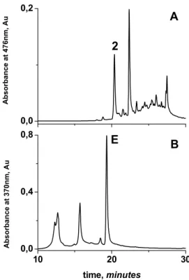
The 36816 EBC, K sample, tentatively labelled as Dracaena schizantha from Zanzibar
and donated in 1871, also revealed the presence of 7,4’ dihydroxyflavylium, in 15% of the
total of the red chromophores, and a similar chromatographic elution profile to Dracaena
cinnabari resins (see figure 1.17A and 1.17B). Moreover, when the 36816 sample was
compared with resin from a living Dracaena ombet (synonym Dracaena schizantha) tree,
from Ethiopia [80], HPE, K collected in 2007, there was no match between the two (see
figure 1.17B and 1.17C). In the living Dracaena ombet tree, the relative percentage area of
7,4’ dihydroxyflavylium (circa 2%) was less than the second flavylium eluted (circa 6%),
contrary to the 36816 EBC, K sample and all the Dracaena cinnabari samples analyzed.
Furthermore, the compounds eluted between 22 and 28 minutes in the sample from the
living Dracaena ombet had different concentrations compared to sample EBC, K 36816
(see figure 1.17B and 1.17C). This is reflected in the PCA analysis that clearly shows that
the sample collected from the living Dracaena ombet is different from the Dracaena
cinnabari samples, whilst sample 36816 EBC, K Dracaena schizantha is similar to the
p. 105 for PCA graphics). It seems that the original label for specimen 36816 only referred
to dragon’s blood and not to the botanical name Dracaena schizantha which was
tentatively, but wrongly attributed some time in the history of its curation. As the analysis
shows, this specimen should have been labelled Dracaena cinnabari which also makes
sense historically as, at this time, there was an established trade route between Socotra
and Zanzibar [16]. Then, D. cinnabari was the most popularly traded dragon’s blood. There
is another sample of D. cinnabari from Zanzibar (EBC, K 36557) to support its presence in
trade there. As no species of Dracaena grow naturally in Zanzibar, the only other likely
source is D.ombet from N.E. African mainland but evidence suggests this species was
very little traded [81].
& &
6 & &
& & &
-)
(
(
.
6
&
$
)
Figure 1.17 – HPLC profile of Dracaena cinnabari and Dracaena schizantha samples from EBC, K collection. A) HPLC chromatogram profile of 36809 EBC, K Dracaena cinnabari sample (1880); B) HPLC chromatogram profile of sample of 36816 EBC, K labelled Dracaena schizantha (1871); C) HPLC chromatogram profile of a resin sample collected in 2007 from a living 28 year old Dracaena ombet (synonym D. schizantha) tree from RBG, Kew. The two first chromatograms are also similar to Dracaena cinnabari resins collected in 2007, see table 1.2, p. 15.
In all the unknown Dracaena sp. samples (36819, 36820, 36821, 36822, 75793) the 7,4’
dihydroxyflavylium was detected and a similar chromatographic elution profile to Dracaena
cinnabari resins, confirmed by PCA, was found; this points to Dracaena cinnabari as the
source (see appendix II – Dragon’s blood data, section II.2, p. 105, for PCA graphics). In
the case of 36822, a sample labelled Dracaena sp. from the East India Company,
![Figure 1.1 Possible de excitation pathways of excited molecules, adapted from [9].](https://thumb-eu.123doks.com/thumbv2/123dok_br/16622967.740261/18.892.220.682.717.1049/figure-possible-excitation-pathways-excited-molecules-adapted-from.webp)
![Figure 1.14 – Network of chemical reactions for 7,4’ dihydroxy 5 methoxyflavylium in solution [69] (see section 1.3.4.1)](https://thumb-eu.123doks.com/thumbv2/123dok_br/16622967.740261/28.892.304.652.473.849/figure-network-chemical-reactions-dihydroxy-methoxyflavylium-solution-section.webp)