Universidade de Lisboa Faculdade de Ciências Departamento de Física
Adipose Stromal Vascular Fraction Cell Sheets as Vascular Strategies for
Tissue Engineering and Regenerative Medicine Applications
Marina Filipa da Silva Costa
Dissertação orientada por:
Professor Alexandre Andrade Professor Rui L. Reis
Mestrado Integrado em Engenharia Biomédica e Biofísica Perfil de Engenharia Clínica e Instrumentação Médica
Universidade de Lisboa Faculdade de Ciências Departamento de Física
Adipose Stromal Vascular Fraction Cell Sheets as Vascular Strategies for
Tissue Engineering and Regenerative Medicine Applications
Marina Filipa da Silva Costa
Dissertação orientada por:
Professor Alexandre Andrade, Instituto de Biofísica e Engenharia Biomédica, Faculdade de Ciências da Universidade de Lisboa
Professor Rui L. Reis, 3Bs Research Group, Caldas das Taipas, Guimarães
Mestrado Integrado em Engenharia Biomédica e Biofísica Perfil de Engenharia Clínica e Instrumentação Médica
Acknowlegdements
In a short essay, I would like to thank all the people who made this internship possible and unforgettable. Thanks to these people, I discover a true passion for the Tissue Engineering and Regenerative Medicine areas and I feel that, in fact, I evolved and developed so much skills and qualities not only professionally but also personally. None of this would be possible without the support of my supervisors, co-supervisor, family, friends and colleagues in the 3B’s research group.
The first people I'll mention are those who contributed the most to help me acquiring all the knowledge I needed for this Internship and the ones who made it happen: my supervisors; Prof. Rui L. Reis and Prof. Alexandre Andrade and my co-supervisor Rogério P. Pirraco, Post-doc Researcher,. I just feel so lucky for having the pleasure to work with all of you, more closely with Rogério P. Pirraco, who was tireless and always offered to help me at all times. I can never thank you enough for all teachings, friendship and for having inspired me to continue this scientific journey.
I would also like to thank all of the people of the 3B’s Research Group especially the ones that made this work possible, besides my supervisors: Mariana T. Cerqueira, Post-doc Researcher, Tírcia C. Santos, Post-doc Researcher, Belém Sampaio-Marques, Post-doc Researcher, Paula L. Ludovico, Principal Investigator, and Alexandra P. Marques, Principal Investigator, who made my work much easier both for the help they gave me and for the good lab environment in which I was integrated.
I would like to thank also my closest friends who have always been by my side throughout these 2 years. For all the laughs, for all the love and dedication to our friendship thank you so much Ana Rita Barbosa, Ana Luísa Ramos, Gabriela Diogo, Teresa Oliveira, Cláudia Lima, Melissa Sirage and Teresa Oliveira.
Last, but not less important my family, who gave me so much support both financially and emotionally deserves a special place on these acknowledgements for all the patience, love and care, for being always my angels.
List of Figures
I.
INTRODUCTION
Figure 1- Traditional cell-based approach in TE. Adapted from:
http://www.wikilectures.eu/index.php/Tissue_engineering_principle ...2 Figure 2 - Formation of blood vessels is regulated by three fundamental processes: vasculogenesis (during the embryo development), angiogenesis and arteriogenesis. Adapted from [21]. ………...4 Figure 3 - Isolation and potential applications of human adipose tissue derived stem cells. Adapted from [39]. ………6 Figure 4 - Cells recovery. Cells can be recovered as single cell suspensions by using proteolytic enzymes that degrade the cell-cell and cell-matrix interactions (upper right) or as cells sheets prepared using a temperature-responsive polymer covalently grafted to a culture dish surface (lower right). ………..8 Figure 5 - HIF pathway for both normoxic and hypoxic conditions. Adapted from:
http://being-bioreactive.com/2014/06/27/focus-on-the-hypoxia-pathway/ ……….12
III. CELL SHEETS OF ADIPOSE TISSUE STROMAL VASCULAR
FRACTION
AS
VASCULARIZATION
UNITS
FOR
TISSUE
ENGINEERING AND REGENERATIVE MEDICINE
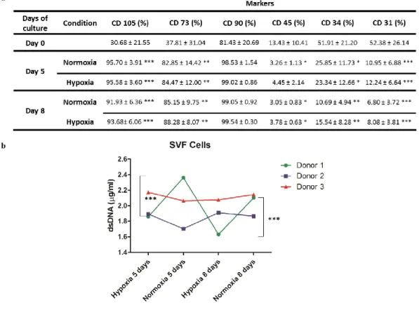
Figure 1 – a. Flow cytometry results obtained after using a gating strategy with DRAQ5 right after isolation (0 days) and for the time points 5 and 8 days. Mesenchymal (CD 105, CD 90, CD 73) and endothelial (CD 31) and hematopoietic (CD 45, CD 34) markers were used. b. DNA quantification on lysed SVF cells for 5 and 8 days in hypoxic and normoxic conditions. DNA results showed significant differences for a p-value < 0.05 for donor 1 between hypoxic and normoxic conditions both at 5 (***) and 8 days (***). Statistically, both flow cytometry and DNA quantification were analyzed using one-way ANOVA and Tukey’s post-tests. ………...37
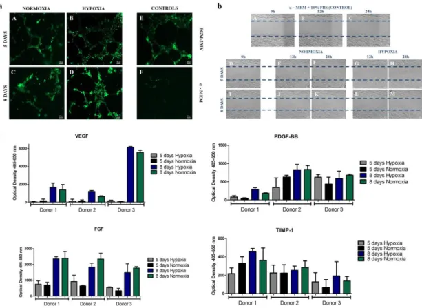
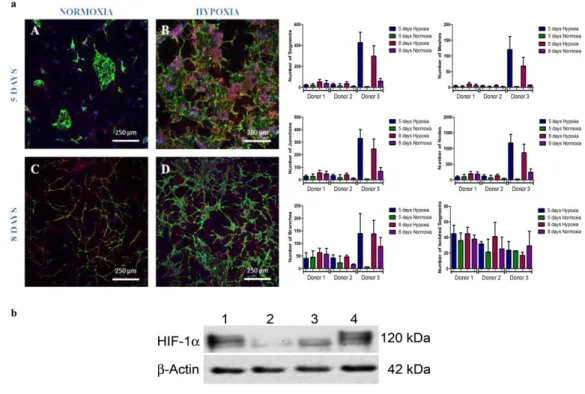
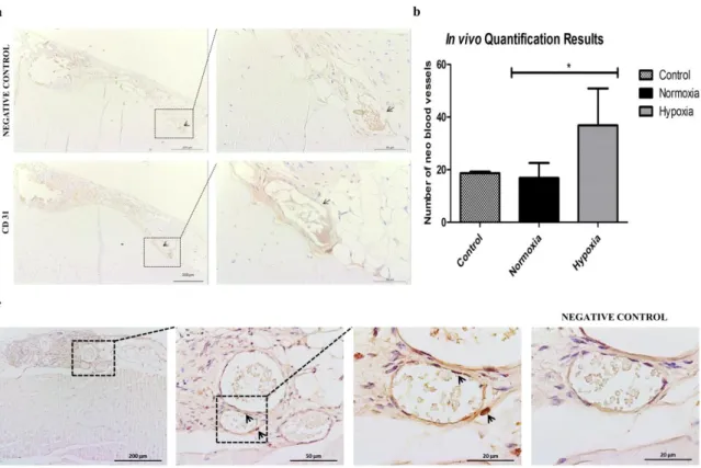
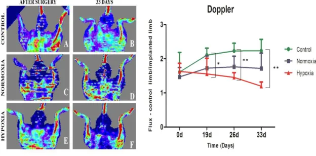
Figure 2 – a. Matrigel assay of HDMECs stained with Calcein-AM using conditioned media withdrawn from donor 3 SVF cell culture in normoxic condition after 5 A) and 8 days C); and from hypoxic cultures after 5 B) and 8 days D). Controls were made with VEGF-containing endothelial cell growth medium which, as expected, stimulated the in vitro formation of capillary-like structures for HDMECS E) and with α-MEM that didn’t promote the same. b. Scratch assay performed with conditioned media from normoxic and hypoxic SVF cultures. Controls were made with α-MEM supplemented with 10% FBS at the beginning of the experiment a) and after 12h and 24h b) and c). Cell migration was assessed at 0h D) and I), 12h and 24h for media collected from 5 days normoxia D), E), F) and hypoxia G), H) samples and 8 days normoxia J), K) and hypoxia L) and M) samples. c. Quantification of growth factors release to the medium by SVF cells assessed by ELISA, including vascular endothelial growth factor (VEGF), platelet-derived growth factor (PDGF-BB), fibroblast growth factor (FGF) and tissue inhibitor of metalloproteinases (TIMP1). Statistical analysis was performed using Kruskall-Wallis test and Dunn’s Multiple Comparison post-test. ………...38 Figure 3 – a. Representative images after immunocytochemistry using endothelial marker CD 31 and CD 146 that stains pericytes in normoxic conditions after 5 A) and 8 days of culture, C) and in hypoxic after 5 B) and 8 days of culture D). Graphic representation of the quantification performed for all biological samples regarding the number of segments, isolated segments, meshes, branches, junctions and nodes, displayed as a mean ± standard deviation. Statistical analysis was performed using Kruskall-Wallis test and Dunn’s Multiple Comparison post-test. b. Western Blot analysis of the nuclear extracts of SVF cells for HIF-1α and for the loading control, actin. Hypoxia is presented at 1), hypoxia supplemented with HIF-1α inhibitor at 2), normoxia at 3) and cobalt chloride II supplementation at 4). ………..40 Figure 4 - SVF cell sheet implantation was performed following hind limb ischemia in a murine model. a. Representative images of histological sections after immunohistochemistry with CD 31 marker. Black arrows indicate the neo blood vessels. b. Quantification of neo blood vessels manually counted by an operator blind to the conditions and based on CD 31 positive cells. The graph presents a mean ± standard deviation of all animals per condition. For statistical analysis one-way ANOVA and Tukey’s post-tests were used. c. Detection of transplanted human cells as SVF CS by chromogenic in situ hybridization 33 days after implantation. Black arrows point to them. In purple are represented cellular nuclei. ………..41 Figure 5 – Laser Doppler results collected up to 33 days. Blood perfusion assessment shows that the animals that received the hypoxic cell sheet had an enhanced blood flux after 33 days. However, statistical analysis shows no statistical differences between normoxic and hypoxic conditions but when comparing hypoxia with the control for a p-value < 0.05, difference was
significant for 19 days (*), 26 days (**) and 33 days (**) with Kruskall-Wallis test, Dunn’s post-test. ………...42
IV. ANNEX
Figure 21 - Gating strategy for nucleated cells in the SVF population, by using DRAQ5. It allowed a clear distinction between our cells of interest and undesired debris. ………..47 Figure 32 - Proliferation of SVF cells conditioned by the HIF-1α inhibitor and cobalt chloride II assessed at day 5 of SVF culture. No statistical differences were found between the two conditions. However, the inhibitor and cobalt chloride II might be causing some cytotoxicity over the cells. ………...48 Figure 43 - Immunocytochemistry against CD 31 and CD146 and quantification of capillary-like structures assessing HIF-1α inhibitor and cobalt chloride II effects over the cells. The lower (by HIF-1α inhibition) and higher (through cobalt chloride supplementation) complexities may
be associated with the HIF-1 α signalling pathway.
………49
Figure 4 - In vivo tests were performed following a hind limb ischemia murine model. After 33 days, the animals were sacrificed and the explants fixed for immunohistochemistry. The image depicts the place of implantation after sacrifice. Inside the boxes are visible the regenerated vessels. ……….50
Abbreviations
α- MEM - Minimum Essential Medium with Alpha Modifications
ANG – Angiopoietin ABTS -
2,2'-azino-bis(3-ethylbenzothiazoline-6-sulphonic acid) APS - Ammonium Persulfate
ARNT - Aryl Hydrocarbon Receptor Nuclear Translocator
BMSCs – Bone Marrow Stem Cells BSA – Bovine Serum Albumin CS – Cell Sheet(s)
CSE – Cell Sheet Engineering DNA - Deoxyribonucleic acid dsDNA – Double-stranded DNA ECs – Endothelial Cell(s)
EPC(s) – Endothelial Progenitor Cell(s) ELISA – Enzyme-linked Immunosorbent Assay
ECM – Extracellular Matrix FGF - Fibroblast Growth factor FBS – Fetal Bovine Serum
hASCs – Human Adipose Stem Cells
HIF – Hypoxia inducible factor MSC – Mesenchymal Stem Cells ODD – Oxygen-Dependent Degradation Domain
PBS – Phosphate Buffered Saline PDGF-BB – Platelet-derived Growth Factor subunit B
PNIPAM - poly(N-isopropylacrylamide) RM – Regenerative Medicine
SVF – Stromal Vascular Fraction TEMED - Tetramethylethylenediamine TE – Tissue Engineering
TERM – Tissue Engineering and Regenerative Medicine
TIMP1 – Tissue Inhibitor of Metalloproteinases
Te - Tris-EDTA (Ethylenediamine Tetraacetic Acid; buffered solution) TGF – Transforming Growth Factor TNF alpha – Tumor Necrosis Factor Alpha VEGF – Vascular Endothelial Growth Factor
ABSTRACT
Tissue Engineering (TE), a subfield of Regenerative Medicine, arises as an attempt to repair diseases or defects by using cells, tissues and biomaterials isolated or in combination, to restore or improve biological functions. Cell sheet (CS) engineering is a TE cell-based technique that uses stimuli-responsive culture surfaces to fabricate robust and intact cell monolayers that can be implanted without requiring the support of a scaffold. The creation of viable thick CS constructs is limited by the lack of suitable vascularization strategies prior to implantation. The herein described strategy results from the optimization of different aspects of cell culture to obtain cell sheets capable of developing a capillary-like network in vitro without adding extrinsic growth factors. These cell sheets are derived from the isolation of the stromal vascular fraction (SVF) of adipose tissue, which is composed of different cell populations including stromal, endothelial and hematopoietic cells, holding thus an intrinsic angiogenic potential. The culture was performed with basal medium in normoxic (pO2 = 21%) and hypoxic (pO2 = 5%) conditions for up to 8 days. Cell proliferation results showed no significant differences between all conditions. Immunocytochemistry performed with the endothelial marker CD 31 and CD 146 that stains pericytes demonstrated the organization of SVF cells in capillary-like structures for both conditions, although the level of branching was much more complex in hypoxia. It was hypothesized that the secretion of growth factors by cells was involved in this effect. A matrigel assay using conditioned media from the different conditions showed increased formation of capillary-like structures by endothelial cells cultured in hypoxic conditioned media. The same conditioned media increased the migration of human adipose stem cells (hASCs) in a scratch assay, when compared with media collected from normoxia. The production of growth factors assessed through ELISA suggested higher secretion of VEGF for hypoxia although statistical analysis showed no significant differences by comparison with normoxia. Western blot for HIF-1α expression highlighted more prominent bands for hypoxic conditions. In vivo implantation of cell sheets cultured for 8 days in normoxia and hypoxia following a hind limb ischemia model showed improved neo-angiogenesis as evidenced by immunohistochemistry and laser Doppler. These results highlight the outstanding potential of combining hypoxic conditions and SVF cells to create cell sheets that can be used as functional vascularization units for TE and RM.
RESUMO
A Engenharia de Tecidos (TE), uma subárea da Medicina Regenerativa (RM), surge como uma tentativa de reparar doenças ou defeitos usando células, tecidos e biomateriais, isoladamente ou em combinação, para restaurar ou melhorar funções biológicas. A Engenharia de Cell Sheet (CS) é uma técnica de TE baseada em células que usa superfícies de cultura sensíveis a estímulos de temperatura para fabricar monocamadas celulares intactas e robustas que podem ser implantadas sem requerer o apoio de um scaffold, evitando assim algumas das várias restrições às quais eles estão associados. A criação de estruturas de CS viáveis e espessas, necessárias para regenerar tecidos mais robustos como o osso, é limitada pela falta de estratégias de vascularização eficazes, antes da implantação. A falta de vascularização adequada resulta, muitas vezes, em necrose celular e rejeição da estrutura sendo, por isso, uma das maiores preocupações na área da TE. Na comunidade científica, diversas estratégias foram propostas neste sentido, muitas delas envolvendo o uso de células endoteliais que, embora bastante promissoras, enfrentam múltiplos desafios como sendo as fontes de células e a estabilidade dos novos vasos sanguíneos formados. A estratégia aqui descrita resulta de uma optimização dos diferentes aspectos relativos à cultura celular para obter CS capazes de desenvolver, in vitro, uma rede de estruturas semelhantes a capilares sem adição de factores de crescimento extrínsecos. Estas CS são derivadas do isolamento da fracção vascular estromal (SVF) do tecido adiposo após dissociação enzimática por acção da colagenase. Este tecido tem vindo a destacar-se na comunidade científica pelas suas características: é abundante, prescindível e pode ser facilmente obtido. Para além disso, a SVF é considerada como sendo detentora de um potencial angiogénico intrínseco, em virtude do tipo de células que a compõem. As células da SVF isoladas a partir de tecido proveniente de lipoaspirações foram distribuídas por placas estéreis de 24 poços numa densidade de 2x10⁵ células nucleadas/poço e cultivadas em condições de normóxia (21%) e hipóxia (5% de oxigénio) com meio basal durante 8 dias. Foram efectuados diversos testes in vitro após terem sido atingidos os time points de 5 e 8 dias, nomeadamente citometria de fluxo (também efectuada imediatamente após o isolamento), quantificação de ADN, ensaios de matrigel e scratch, ELISA, imunocitoquímica e western blot. Para além disso, foram também efectuados ensaios in vivo após indução de isquemia nos membros posteriores que foram avaliados por imunohistoquímica, hibridização in situ e laser
Doppler.
A análise dos marcadores de superfície das células da SVF por citometria de fluxo evidenciou que a expressão dos marcadores endotelial CD 31 e hematopoiético CD 34 decresce ao longo do tempo, o que seria expectável uma vez que a cultura foi efectuada em meio basal, sem adição de factores de crescimento extrínsecos. Na literatura, a SVF do tecido adiposo é descrita como
sendo composta por diversas populações de células, incluindo células estromais, endoteliais e hematopoiéticas, facto que foi comprovado nesta análise. Os resultados relativos à proliferação celular de células da SVF mostram que não há diferenças significativas entre as condições de normóxia e hipóxia sugerindo que a proliferação destas células não é afetada significativamente pelas concentrações de oxigénio. A imunocitoquímica realizada para os marcadores CD 31 e para o CD 146, o qual marca péricitos, demonstram a organização de células da SVF em estruturas tipo capilares para ambas as condições, embora o nível de ramificação tenha sido mais complexo em hipóxia para 8 dias. A comprovar este facto, a quantificação das estruturas tipo capilares no Angiogenesis Analyzer, um plug-in do programa Image J, destacou que as formações seriam mais maduras e organizadas nessas mesmas condições. Esse facto tornou-se mais evidente num dos dadores devido ao decréscimo no número de segmentos e malhas formados pelas células ao longo do tempo, sendo que o nível de ramificação se manteve praticamente inalterado. Isso traduziu-se em segmentos mais alongados e interconectados entre si. Foi levantada a hipótese de que a secreção de factores de crescimento pelas células em condições de hipóxia poderia estar envolvida neste efeito. Como tal, foram efectuados diversos ensaios utilizando os meios condicionados recolhidos das culturas em diferentes condições. Um ensaio de matrigel utilizando um extracto de membrana basal proteico que favorece a angiogénese, evidenciou um aumento na formação de estruturas tipo capilares pelas células endoteliais cultivadas em meio condicionado hipóxico. Os mesmos meios condicionados aumentaram a migração de células estaminais do tecido adiposo humano (hASCs) num ensaio
scratch, quando comparados com os meios recolhidos de normóxia. O ensaio scratch é um
ensaio baseado na raspagem da zona central de uma monocamada celular para avaliar a migração das células em torno da área erodida, sob diferentes condições. A produção de factores de crescimento avaliada por meio de ELISA sugeriu uma maior secreção do VEGF para condições de hipóxia. Colocou-se a hipótese que este facto poderia estar relacionado com a estabilização das subunidades dos HIFs (hypoxia inducible factors) em condições de concentrações baixas de oxigénio. OS HIFs são uma família de factores de transcrição constituída por três subunidades principais HIF-1, HIF-2 e HIF-3 e os seus respectivos heterodímeros (alfa e beta). As subunidades alfa respondem a variações na concentração de oxigénio e sob condições de oxigénio atmosféricas (normóxia) são constantemente expressas e degradadas no citoplasma, mais concretamente no proteassoma. Em condições de stress de oxigénio estas subunidades migram para o núcleo onde se ligam aos seus heterodímeros, os quais são constitutivamente expressos, e estabilizam. Esta estabilização resulta num aumento da secreção de diversos factores de crescimento responsáveis pela regulação do metabolismo da glucose, eritropoiese e angiogénese como sendo o VEGF e o PDGF. Deste modo, foi efectuada uma análise por western blot para o HIF-1α a qual destacou bandas mais proeminentes para condições de hipóxia. Paralelamente às condições de hipóxia e normóxia foram também
testadas a incubação das células com um inibidor da subunidade HIF-1α e com cloreto de cobalto II, um indutor químico de condições de hipóxia. Como seria expectável, a banda obtida na análise de western blot para o inibidor foi bastante ténue contrariamente à obtida com o mimetizador das condições de hipóxia. Em imunocitoquímica isto reflectiu-se num fraco e proeminente desenvolvimento das estruturas tipo capilares, respectivamente, facto que veio reforçar a hipótese proposta.
Finalmente, depois da manutenção das CS por 8 dias em condições de normóxia e hipóxia foi efectuada a sua implantação in vivo em ratinhos durante 33 dias, após indução de isquemia no membro posterior. A imunohistoquímica dos explantes recuperados executada para os marcadores endotelial CD 31 e perivascular α-SMA destacou uma melhoria na neo-angiogénese em condições de hipóxia por comparação com as de normóxia e controlos. Foi efectuado também um ensaio de hibridização in situ para localizar as células humanas nos explantes e verificou-se que algumas destas células permaneciam na área da implantação em torno dos novos vasos sanguíneos formados. Os resultados obtidos através do laser Doppler demonstram que a perfusão dos vasos sanguíneos aumentou nos animais em que foram transplantadas as cell
sheets. Para além disso, foi observado que nos animais que receberam as cell sheets
provenientes de culturas em hipóxia havia uma tendência para uma recuperação mais rápida do fluxo sanguíneo. Tendo em consideração todos os resultados obtidos é importante salientar o excelente potencial da estratégia aqui descrita que resulta da combinação de diversos conceitos como sendo, condições de hipóxia, células da SVF e Engenharia de Cell Sheet (CS) para criar unidades de vascularização funcionais para TE e RM.
TABLE OF CONTENTS
STRUCTURE
………...…………..1I.
INTRODUCTION
………...…………..11. Tissue Engineering and Regenerative Medicine……….1
2. The problem of Vascularization after implantation……….2
3. Stromal Vascular Fraction derived cells………..5
3.1. The intrinsic angiogenic potential of SVF derived cells……….7
4. Cell Sheet Technology……….7
4.1. The principle for cell sheet detachment………..8
4.2. Applications in the Regenerative Field………...9
5. Hypoxia the “physiological normoxia”………...9
5.1. Stem Cells Niche ………..10
5.2. Hypoxia Inducible Factors: the signaling cascade……….10
5.3. The impact of low oxygen tensions………...12
II.
MATERIAL AND METHODS
………141. CULTURE OF SVF CELLS………...14
1.1. Isolation of human adipose stromal cells………..14
1.2. Conditions of Culture………15
1.3. Cryopreservation………16
2. IN VITRO CHARACTERIZATION TESTS……….16
2.1. Flow Cytometry………...16
2.2. DNA quantification on lysed cells……….17
2.3. Matrigel Assay………...18
2.4. Scratch Assay……….18
2.5. ELISA (Enzyme-Linked Immunosorbent Assay)………..19
2.6. Immunocytochemistry vs Immunohistochemistry……….19
2.7. Immunocytochemistry………20
2.8. Nuclear and cytoplasmic fractionation of the cells for Western Blot………….……...20
2.9. Total Protein Quantification (Bicinchoninic acid assay)………...21
2.10. Western Blot………...22
2.11. Implantation of SVF cell sheets following a hind limb ischemia model……...23
2.12. Immunohistochemistry………...23
2.13. In situ hybridization………...24
2.14. Laser Doppler……….………25
3. QUANTIFICATIONS………...25
3.1. Quantification of capillary-like structures in vitro……….25
3.2. Quantification of blood vessels in vivo………..26
III.
ARTICLE
………27ABSTRACT……….27
1. INTRODUCTION……….28
2. MATERIAL AND METHODS……….29
2.1. Isolation of SVF Cells………29
2.2. Culture Conditions……….30
2.3. Flow Cytometry……….30
2.4. DNA Quantification on lysed cells………...30
2.5. Matrigel assay………31
2.6. In vitro scratch assay……….31
2.7. ELISA………31 2.8. Immunocytochemistry………32 2.9. Western Blot………...32 2.10. In vivo tests………....33 2.11. Immunohistochemistry………..33 2.12. In situ Hybridization………...34
2.13. Quantification of capillary-like structures in vitro……….34
2.14. Quantification of blood vessels in vivo………..35
2.15. Statistical Analysis……….35
3. RESULTS………..36
3.1. Flow Cytometry……….36
3.2. DNA Quantification on lysed cells………...36
3.3. Matrigel assay………37
3.4. In vitro scratch assay……….37
3.5. ELISA ………...38 3.6. Immunocytochemistry………39 3.7. Western Blot………...39 3.8. Immunohistochemistry………...40 3.9. In situ Hybridization……….41 3.10. Laser Doppler………....42 4. DISCUSSION………....43 5. CONCLUSIONS………...46
IV.
ANNEX
1. GATTING STRATEGY PERFORMED FOR FLOW CYTOMETRY…………...……….472. ISOLATION OF HUMAN DERMAL ENDOTHELIAL CELLS………47
3. SUPPLEMENTATIONS WITH COBALT CHLORIDE II AND HIF-1Α INHIBITOR….48 3.1. Proliferation of SVF cells………..48
3.2. Immunocytochemistry………49
4. IN VIVO HIND LIMB ISCHEMIA MODEL………50
1
STRUCTURE
The herein presented thesis is divided in four main chapters: chapter I, the state of the art of the theme; chapter II, the detailed description of all materials and techniques applied throughout the work; chapter III, the scientific paper written during this dissertation project that integrates all the results, discussion and final conclusions which, at the time of the delivery of this thesis was in process of submission, and finally chapter IV, the appendices.
II.I.
INTRODUCTION
1.
Tissue Engineering and Regenerative Medicine
Tissue Engineering (TE) is a forefront area in Health Sciences whose purpose is to rebuild or repair damaged or aged cells, tissues or even entire organs by isolated or combined action of cells, growth factors and biomaterials. It is integrated in the field of Regenerative Medicine (RM) that aims the regeneration of tissues and organs using not only TE but also the potential of genetics and cell therapy. Cell-based approaches such as TE and cell therapy take advantage of the biological properties of mesenchymal stem/stromal cells, which can be found in almost all tissues of the human body, to regenerate tissues or entire organs by transplantation into the injured site. Most of TE strategies require a well-orchestrated sequence of events that range from the extraction of cells, which can be expanded or induced to differentiate upon administration of proper biochemical cues in cell culture surfaces, to its transplantation integrated in 3D scaffolds made of both natural and synthetic biomaterials, in the same patient. In order to be functional after implanted in vivo, these scaffolds should have some basic properties: they have to be biocompatible, bioresorbable with a well-controlled degradation rate and facilitate the diffusion of nutrients and oxygen through a porous surface. Besides, this surface should be compatible with cell attachment and constructs must possess specific mechanical properties taking into account the target-tissue[1]. However, biomaterials-based strategies face several constraints associated mainly with immunogenicity problems and poor degradation rate of the scaffold that can lead to fibrosis and necrosis in the bulk of thicker constructs, aiming to target thicker tissues and/or organs[2].
2
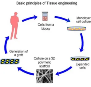
Figure 51- Traditional cell-based approach in TE. Adapted from: http://www.wikilectures.eu/index.php/Tissue_engineering_principle
Another constraint typically associated with cell-based strategies is cell sourcing. Mesenchymal stem cells, also known as multipotent stromal cells, captivated an overwhelming attention in the scientific world as powerful therapeutic tools and inspired several works, a great part of them using MSCs isolated from the bone marrow. The first pioneer works reporting the discovery of a MSC’s niche date back to 1968 when Friedenstein and colleagues discovered that stem cells, derived from the bone marrow (BMSCs), were capable of differentiating into bone [3]. However, the extraction of cells from the bone marrow is often associated with donor-site morbidity, pain and poor yield of cells. Necessity of an alternative spurred scientists to find new MSC sources and currently it is well known that they can be found in almost all tissues of the human body, namely skeletal muscle [4], adipose tissue [5], skin [6], pancreas [7], synovial membrane [8], dental pulp [9], placenta[10], trachea [11], nasal mucosa[12], Wharton’s jelly [13] and also amniotic fluid[14], menstrual blood[15], among others.
There are a lot of studies in clinical trials regarding cell-based strategies involving mainly the use of stem cells isolated from bone marrow, adipose tissue, placenta and umbilical cord blood for several different applications. Amongst this wide range of applications it’s possible to find immunological and blood diseases, cardiac repair and neurological disorders[16].
2.
The problem of Vascularization after implantation
Blood vessels, major components of the circulatory system, are essential for the survival of the tissues and organs by supplying the nutrients and oxygen and collecting the waste products from cellular metabolism, regulating thus diverse functions that allow maintaining homeostasis. Fast
3 neovascularization is a critical aspect for the effective survival of tissue engineered constructs in order to avoid hypoxia and/or anoxia in the inner mass of tissue-engineered constructs, due to a poor diffusion of nutrients and oxygen essential for their successful engraftment. Ultimately, this poor supply of nutrients and oxygen can culminate in unsatisfactory tissue regeneration or even, failure of the construct. Nonetheless, is difficult to build whole prevascularized implants that could mimic tissues and organs thickness without compromising the distance between cells and blood vessels.
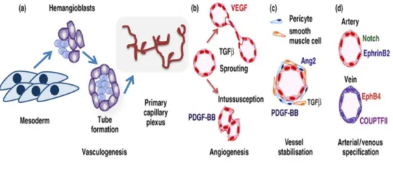
Neovascularization comprises three distinct processes: vasculogenesis, angiogenesis and arteriogenesis. Vasculogenesis corresponds to the process by which de novo vessels are formed during the embryonic phase by endothelial progenitors[17] developing a primitive vascular plexus. The sprouting and stabilization processes that follow vasculogenesis and the reshuffle of the early vasculature into a complex and functional circuit are called angiogenesis and arteriogenesis. For that purpose, the secretion of diverse angiogenic growth factors and recruitment of mural cells like smooth muscle cells and pericytes are required to promote ECs survival, proliferation and differentiation as well as the complexity of the blood vessels[18]. Vascular endothelial growth factor (VEGF), angiopoietin (ANG), platelet-derived growth factor (PDGF), fibroblast growth factor (FGF), TIMP (tissue inhibitor of metalloproteinases), transforming growth factor (TGF-α) and (TGF-β), hepatocyte growth factor (HGF), tumor necrosis factor-α (TNF- α), proteins, cytokines are also known for playing an active role in the process of neovascularization[17]. VEGF is crucial for modulating endothelial progenitor cells (EPCs) behavior [19] and endothelial cells (ECs) survival, inducing a powerful angiogenic response for both neovascularization and pathological conditions[20]. Moreover, stabilization and maturation of primary capillary plexus depends on the coordinated action of PDGF, TIMP (by remodeling of the ECM during the sprouting), TGF-β and FGF (angiogenic promoters released from the ECM by proteinases action)[17].
4
Figure 62 - Formation of blood vessels is regulated by three fundamental processes: vasculogenesis (during the embryo development), angiogenesis and arteriogenesis. Adapted from [21].
Several strategies mainly related with in vitro and in vivo prevascularized engineered constructs, which can involve angiogenic factors or scaffolds, are under investigation, many of them reporting the use of endothelial cells [22–25]. For instance, transplantation of ex vivo expanded human endothelial cells was found to increase the blood vessels and capillary perfusion in a hind limb ischemia induced in mice by Kalka et. al[23]. The same type of cells expanded also ex vivo were used as a therapeutic tool in myocardial ischemia[25]. Co-culturing endothelial cells with bone marrow derived stem cells promoted vascularization after implantation[26,27]. Nevertheless, the previously described strategies reported fairly late blood vessels formation. Scaffolds have been also widely used to support the formation of blood vessels. For instance Santos et. al engineered a cell-based construct using a blend of starch with polycaprolactone and co-cultures of endothelial cells and osteoblasts for bone tissue regeneration. The culture in the first 21 days lead to the formation of microcapillary-like structures, more branched and developed after 35 days[28]. Osteogenic/vasculogenic constructs based on porous hydroxyapatite scaffolds and SVF cells cultured in vitro for 5 days were found to form blood vessels and bone tissue 8 weeks post-transplantation in nude mice[29]. Moreover, spheroid co-cultures of endothelial and osteoprogenitor cells resulted in the formation of prevascularized networks of capillary-like structures 10 days after in vitro culture for bone tissue engineering applications. However, integration with host vasculature after implantation of the construct was found to be poor[30]. The controlled release of angiogenic factors, in the case bFGF, has been achieved by incorporating microspheres composed of poly (lactic-co-glycolic acid) on alginate scaffolds. Capillary density increased since day 10 until 21 days post-transplantation in rat
5 peritoneum[31]. Nano-sustained controlled release of VEGF on polylactic acid microspheres in combination with SVF cells increased also capillary density in nude mice, 2 months after implantation[32]. The potential of SVF cells in a process of tissue expansion in rats was found to be facilitated, possibly due to the secretion of EGF, VEGF and bFG which increased not only blood vessels formation but also cells proliferation[33]. Recently, SVF cells were used as vascularization tools in the generation of a human liver. Co-culturing SVF cells with HepG2 cells resulted in the integration of the first with the host vasculature and in an interactive role with parenchymal cells[34]. Endothelial cells were also used to promote vascularization for regeneration of other tissues such as skin[35] or skeletal muscle[36]. Nevertheless, most of the described strategies have drawbacks such as the late formation of blood vessels in vitro and/or post-implantation, the complexity associated with cultures of more than one cell type isolated from different tissues and formation of fibrous tissue after implantation of engineered constructs. With the exponential growth of the engraftment technology over time there is an increasing need to find more, faster and efficient techniques for neovascularization.
3.
Stromal Vascular Fraction derived cells
Adipose tissue was neglected in previous years in the scientific world because it was seen as an organ exclusively associated with the storage and release of lipids. Nonetheless, their unique properties (accessibility, low-cost and abundance) combined with the fact of being a reservoir of stem and stromal cells made it quite captivating as a cell source [5]. Also, as fat tissue lines and protects several parts of the human body it can be easily identified, namely at the intra-abdominal area and subcutaneously, and extracted through minimally invasive procedures. As stated by the International Society for Cellular Therapy the definition of multipotent mesenchymal stromal cells, typically named mesenchymal stem cells, can be divided in three fundamental topics: the presence or absence of selected surface markers (they must express specific cell surface antigens such as CD73, CD90, and CD105, and lack expression of other markers including CD45, CD34, CD14, CD11b, CD79α, or CD19 and HLA-DR surface molecules) and adhesion and multipotency in vitro (MSCs must be adherent to plastic when kept under standard culture conditions and have the capacity to differentiate in vitro into osteoblasts, adipocytes, and chondroblasts) [37]. Besides these three fundamental topics, mesenchymal stem cells can also be easily identified by their functional properties: the self-renewal and proliferative capacities and their undifferentiated state.
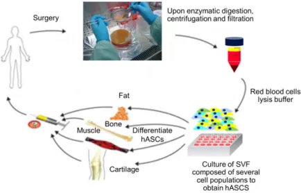
6 The initial works regarding the isolation of stem and stromal cells from the adipose tissue were developed in the 60s by Rodbell et al. in rat fat pads. Briefly, Rodbell et al. chopped the fat and then washed it several times in order to remove unwanted cells. The extracellular matrix (ECM) of the remaining tissue was then digested with collagenase and centrifuged for deposition of stromal vascular fraction (SVF) cells. Then, SVF cells were removed by aspiration and fat cells washed. This process of isolation is very similar to the one that is used now, with human lipoaspirate tissues with the addition of a red blood cells lysis buffer to the equation. Nevertheless, literature already describes non-enzymatic methods for SVF isolation [38].
Figure 73 - Isolation and potential applications of human adipose tissue derived stem cells. Adapted from [39].
Adipose-derived stem and stromal cells are multi and pluripotent cells, respectively, that result from the processing of fat tissue. Cells from the stroma of adipose tissue are quite heterogeneous among themselves since they constitute a cocktail of different populations such as pre-adipocytes, mesenchymal progenitors, fibroblasts, pericytes endothelial and hematopoietic cells [40–42]. This heterogeneous composition makes them ideal candidates for a wide range of applications such as neovascularization.
7
3. The intrinsic angiogenic potential of SVF derived cells
Stromal vascular fraction from adipose tissue has been recently regarded as highly angiogenic and, therefore, quite captivating for improvement of the neovascularization. These cells are known to promote the formation of capillary-like structures both in vitro and after transplantation.
Besides its hematopoietic and endothelial composition others factors have been pointed as conditioning for the angiogenic potential of SVF cells. Koh et al. hypothesized that the network of capillary-like structures they are capable to build is created by the reassembly of endothelial progenitors and mural cells contained in the SVF mixture [43]. Dong et Al. also hypothesized that the recruitment of M2 macrophages may also constitute one important factor influencing this angiogenic potential of SVF [44]. All of the research involving its angiogenic capacity made SVF cells quite attractive not only for in vitro but also for in vivo tests. For instance, the transplantation of the stromal vascular fraction from adipose tissue following a murine hind limb ischemia model have also been shown to promote neovascularization [45,46]. The transplantation was also successful and resulted in the same angiogenic effects in random skin flaps[47]. The great ability of these cells to promote angiogenesis has contributed to physiological and pathological wound healing in mice. The same work also highlighted the potential ability of these cells to enhance reepithelialization[48]. A 3D implant using freshly isolated SVF cells was also built for generation of a functional liver tissue [34]. Nevertheless, more studies need to be developed regarding the process by which SVF cells assemble as capillary-like structures and all variables involved in the process.
4.
Cell Sheet Technology
To overcome some of the problems arising from previous cell-based approaches, namely the low anchorage of cells at the injured site [49] and the limitations related to the use of biomaterials as scaffolds [50], the concept of Cell Sheet Engineering was proposed in 1993, bringing a new approach to tissue engineering: the recovery of a contiguous cell layer as a sheet maintaining the native cell-cell and cell-ECM interactions[51].
8
4.1 The principle for cell sheet detachment
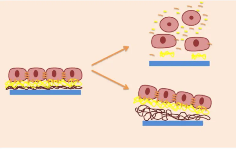
The basic idea of Prof. Okano’s group was to develop a scaffold-free approach by coating culture surfaces through covalent grafting with a temperature-responsive polymer - poly(N-isopropyl acrylamide) (PNIPAM) that transits from a dehydrated to a hydrated state at temperatures below 32º C. Thus, PNIPAM is regarded as being hydrophobic at temperatures above 32º C and hydrophilic below these. The group successfully cultured bovine endothelial cells and rat hepatocytes at 37º C until achieving confluence and then, by decreasing the temperature until approximately 20ºC, the interactions with the ECM were diminished. The critical value of temperature allowed a spontaneous cell detachment [51].
Figure 84 - Cells recovery. Cells can be recovered as single cell suspensions by using proteolytic enzymes that degrade the cell-cell and cell-matrix interactions (upper right) or as cells sheets prepared using a
temperature-responsive polymer covalently grafted to a culture dish surface (lower right).
This came to revolutionize the field, as cell sheet engineering (CSE) avoids the use of proteolytic enzymes such as trypsin and dispase, and the native ECM acts as a natural scaffold [52]. Nevertheless, besides these enzymatic treatments and declines of temperature, several other stimuli have been used for cell sheet detachment. Guillaume-Gentil et. al described a technique to obtain viable cell sheets composed of human placenta-derived mesenchymal stem cells based on a electrochemical induction of pH decrease [53]. The same group also described a technique based on polyelectrolyte thin films [54]. Many works using a wide range of different cell types described cell sheet detachments by using magnetic stimuli [55–60]. Through alternation of layer-by-layer depositions of polyanions and polycations, Zahn et. al described an ion-induced cell sheet detachment technique using myoblasts [61]. Micropatterning of the cell culture dishes has been also subject of interest recently and is present as a potential future
9 strategy [62–64]. This technique takes advantage of the critical temperatures of two different polymers for specific cell adhesion.
4.2
Applications in the Regenerative Field
The potential of cell sheet engineering has been extensively explored over the past few years and now this technology is proposed to improve and treat functions of many tissues such as periodontium [65–67], corneal epithelial [68,69], hepatic [70], urothelial [71,72] and renal [73] tissues, myocardium [74–79], thyroid gland [80], retinal pigment epithelium [81], lung [82,83] and pancreatic islet [84,85]. Besides, cell sheet engineering has been also proposed for esophagus [86–88] and cartilage tissues [89–91]. Our lab, has proposed also the use of CSE to engineer tissues such as skin [92] and bone [93].
Cell sheet engineering has been applied also to improve neovascularization. Asawaka et. al have shown that by stacking human umbilical vein endothelial cells and normal human dermal fibroblasts is possible to build 3D stratified tissues to promote a pre-vascular network both in
vitro and in vivo [94]. Co-cultures involving endothelial cells resulted in increased
neovascularization potential and improved cardiac function on ischemic hearts by using tissue-engineered cardiomyocyte sheets [95]. Moreover, monolayered mesenchymal stem cell sheets were found to promote angiogenesis by paracrine pathways on infarcted myocardium [96]. Recent studies have also demonstrated the great ability of this technique to build functional vascularized 3D tissues for cardiac tissue [97].
Cell sheet engineering is also on clinical trials for myocardium - through transplantation of 3-week-cultivated autologous myoblast cell sheets into patients[98] -, cornea - which commonly presents great outcomes[68] -, esophagus[99], periodontium - by using periodontal ligament cell sheets[66] - and cartilage tissues[100]. Nevertheless, literature does not describe yet the use of cell sheet engineering in combination with the whole SVF of adipose tissue.
5.
Hypoxia the “physiological normoxia”
Oxygen is a critical component regarding stem and stromal cells culture that is quite often overlooked in the lab context. Since the very beginning of life, the irregularities experienced regarding the oxygen atmospheric concentrations have been boosting the evolution of eukaryotic species in an oxygen-dependent manner. It is believed that there must be a balance between the cellular need for O2 as a final acceptor of electrons during Krebs cycle and the production of ROS (reactive oxygen species) which lead to oxidative damage and can even cause cell death. This balance is achieved by controlling the O2 concentrations within the tissues
10 [101] which are known for being much lower that the regular 21% O2 atmospheric levels. These lower oxygen levels, in comparison with the atmospheric ones, are called hypoxia.However, as
in vivo physiological oxygen levels are considerably lower than the atmospheric values as cells reside in hypoxic niches this low oxygen tensions are often referenced as “normoxic in
situ”[101].
5.1 Stem Cells Niche
The stem cell niche can be defined as the microenvironment where stem cells reside. This microenvironment is composed of several components that regulate and control the stem cells fate by sustaining their needs and giving strong cues to direct their activity.
Since the very beginning, during the embryonic phase, O2 concentrations are constantly varying from the 1-2% when the blastocyst is being implanted [102], rising to the 2-3% with a fully-developed circulatory system and achieving the regular O2 values within the tissues at the 12-13th week of gestation [103].
In adults, O2 concentrations within the tissues are dependent on the surrounding blood vessels vascularization but they are considerably lower than the ambient O2 concentration. Oxygen values in the circulation typically range from 4% to 14% [104], similar to what happens in other organs such as liver [101,105], kidneys [101,106] and lung parenchima [101,107]. In the brain, the oxygen concentrations can be so low as 0.5% being capable to go up until 8% [108]. The oxygen levels in the eye [109] and in the bone marrow [110] range from 1 to 5 and 6%, respectively. In the adipose tissue these concentrations are found to be typically between 2 and 8% [111].
5.2
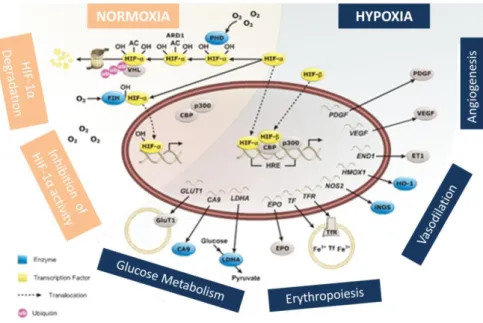
Hypoxia Inducible Factors: the signaling cascade
Hypoxia-inducible factors (HIFs) are a family of transcription factors responsible for regulating homeostasis, by controlling cellular responses, under low oxygen tensions. The HIF family is composed by three fundamental subunits, HIF-1, HIF-2, and HIF-3, and its corresponding heterodimers (alpha and beta). Alpha subunits are oxygen responsive and become stable in low oxygen conditions by binding their corresponding beta subunits in the nucleus or even in the presence of iron chelators [112]. At regular oxygen conditions, the so called normoxia, these alpha subunits are continuously expressed and degraded promptly. On the other hand, the beta
11 subunits, also known as aryl hydrocarbon receptor nuclear translocators (ARNTs), are constitutively expressed in both conditions.
HIFs are known for playing a critical role in a wide range of pathological conditions with a special focus in cancer and ischemic diseases. There is evidence in the literature that many tumors propel strongly hypoxic environments that correlate with the progression of the disease by promoting cell invasion and proliferation, angiogenesis and metastasis [113]. This might be due to the fact that HIFs expression triggers the induction of several crucial genes for glycolysis (glucose transporters and glycolytic enzymes) and formation and maturation of the blood vessels such as VEGF, erythropoietin, angiopoietin and platelet-derived growth factor. The latter also highlights why HIFs can be so promising for preventing or even treating ischemic affected areas [113]. For that, understanding HIFs pathway and the role that each specific subunit plays in oxygen deprived conditions is currently the subject of several studies. By now, it is well accepted that all subunits share diverse similarities especially regarding HIF-1α and HIF-2α, while HIF-3α is thought to have an opposite effect under hypoxic conditions. The basic mechanism in the signaling pathway of HIF-1 and HIF-2 at regular O2 concentrations involves prolyl-hydroxylases that enzymatically induce HIF alpha subunits to bind to von Hippel Lindau (VHL) protein by altering their conformation. The action of the prolyl-hydroxylases occurs in different prolines according to the subunit, hydroxylating them in their respective oxygen-dependent degradation domains (ODD). The lack of O2 in hypoxic conditions leads to an accumulation of the alpha subunits that translocate to the nucleus, where they bind to their respective ARNT subunit. The transcription of several genes is then activated through the bind of the formed heterodimers to the hypoxia-response elements (HRE) [114–116].
12
Figure 95 - HIF pathway for both normoxic and hypoxic conditions. Adapted from: http://being-bioreactive.com/2014/06/27/focus-on-the-hypoxia-pathway/
Both subunits are also distinctive namely by their location, degree of induction controlled by O2 concentration and functionality. While HIF-2α expression appears to be induced by extremely low levels of oxygen and triggered exclusively in endothelial cells and vascularized tissues, HIF-1α is thought to be expressed in all types of cells in mammalian at higher levels of oxygen concentration [113,117].
5.3 The impact of low physiological oxygen tensions
Hypoxia has been widely used in the past few years for research purposes in cell culture. However, the way it affects cell behavior is not yet fully understood or can be even quite controversial. These cultures in low O2 environments have been studied for a long period of time, since 1972 when Allan Richter and his colleagues discovered that culturing cells in hypoxic conditions could help improving the platting efficiency by increasing the amount of cells that adhered to a plastic dish [118].
Oxygen concentrations of 1 % have been shown to be promoters of the osteogenic differentiation in bone marrow mesenchymal stem cells [119]. Pre-culturing human ASCs at 5% O2 concentrations promotes the great ability of these cells to differentiate towards the chondrogenic lineages [120]. Hypoxic conditions of 2% O2 increases also the potential to differentiate into the adipogenic and osteogenic lineages on the same type of cells [121]. Moreover, regarding the stemness and proliferation in the same severe hypoxic conditions, human ASCs were shown to have increased rates at low oxygen levels in comparison with the
13 normoxic. Nevertheless, regarding differentiation, these stress oxygen conditions reduced the capacity of ASCs to differentiate into the adipogenic and osteogenic lineages enhancing the chondrogenic one [122]. Hypoxia pre-conditioning enhanced the survival of hASCs and influenced the behavior of endothelial cells in vitro [123] besides increasing the wound-healing potential of ASCs [124].
Several researchers have also studied the angiogenic potential of hASCs in hypoxic conditions. Severe hypoxia increased the angiogenic paracrine activity of hASCs by increasing the production of VEGF-A and ANG [125]. Long-term hypoxia has been shown to improve the pro-angiogenic potential of hASCs [126]. The addition of growth factors to the culture of stromal cells derived from the adipose tissue is thought to produce the same effect [127]. Nevertheless, there are only a few studies regarding the effects of hypoxia in the whole SVF extract.
14
III.II.
MATERIALS AND METHODS
1.
CULTURE OF SVF CELLS
1.1
Isolation of human adipose stromal cells
In collaboration with Hospital da Prelada, Porto (supplier of lipoaspirate tissues).
Human adipose stromal cells were isolated and collected from subcutaneous fat tissue obtained through liposuction after informed consent of the patients (n = 3), in the scope of an agreement with Hospital da Prelada, Porto, after approval by the ethical committees of both institutions.
Firstly, adipose tissue, processed until 24h after collection, was washed extensively with a great amount of a phosphate-buffered saline solution (PBS) (Sigma Aldrich, USA), and two phases were clearly noticed: an adipose fraction on the top and an aqueous one containing blood, which deposited on the bottom and was removed, by pipetting.
Tissue digestion was then induced by incubating the adipose fraction (1:1) with a solution of PBS containing 0.05% of collagenase type II (Sigma Aldrich, USA) at 37ºC for at least 30 minutes and up to 45 minutes, under agitation, until the tissue was sufficiently loose.
Digested tissue was cleaned of any remnants of connective tissue and blood vessels by percolating it with a nylon mesh strainer with the assistance of forceps, followed by a centrifugation at 4 ºC, 800 g for 10 minutes.
After that period of time, supernatant was discarded and the obtained SVF was incubated with a red blood cells lysis buffer (154 mM of ammonium chloride, 10 mM of potassium bicarbonate and 0.1 mM of ethylenediaminetetraacetic acid – all from Sigma Aldrich, USA – in distilled water) for 10 minutes at room temperature to lyse undesirable erythrocytes.
In order to restore the osmotic equilibrium, α – MEM medium (Life Technologies, United Kingdom) supplemented with 10% FBS (Life Technologies, United Kingdom) and 1% antibiotic/antimycotic (Life Technologies, United Kingdom) was added and the obtained mixture was centrifuged for 5 minutes at room temperature at 300 g.
Lastly, red blood cells-free SVF was resuspended in α – MEM medium (Life Technologies, United Kingdom) supplemented with 10% FBS (Life Technologies, United Kingdom) and 1% antibiotic/antimycotic (Life Technologies, United Kingdom) and filtered by a 100 µm nylon mesh cell strainer (Life Sciences, USA).
15
To perform an accurate cell counting distinguishing nucleated cells from any remaining erythrocytes and debris , 20 µl of cell suspension were incubated with 180 µl of a
solution containing 3% acetic acid (VWR, United Kingdom) in methylene blue 0.05 wt. % in H2O (Sigma Aldrich, USA). Finally, 2x10⁵ cells were distributed for each well of 15.6 mm diameter 24-well culture sterile plates.
1.2 Conditions of culture
Conditions of 21% (normoxia) and 5% of oxygen (hypoxia) were provided to the cultures. Low oxygen conditions were induced in a hypoxia modular incubator chamber (MIC-101, Billups-Rothenberg, USA) which was flushed for 6 minutes with a mixture of 5% O2, 5% CO2 and 90% N2, according to manufacturer’s instructions every day zero, one and five of culture.
The chamber was then placed in an incubator (Galaxy CO-170S, New Brunswick) at 37ºC, 5% CO2 and cells were cultured up to 8 days.
After 5 and 8 days of culture, conditioned medium (for ELISA, matrigel and scratch assays) and cells were collected for posterior characterization.
For matrigel and scratch assays, human dermal microvascular endothelial cells (HDMECs) and hASCs (passage 1-3) were used. A homogeneous culture of hASCs was obtained after the first passage of cells from the SVF since the remaining populations that it contains are lost during the culture process. The process by which cells are detached from the culture surface, changing their passage, happens when they reach approximately 90% of their confluence (percentage of space occupied by cells in the culture surface).
Briefly, after removing the culture medium cells were washed with PBS and then incubated with trypLETM Express (Life Technologies, USA) at 37º C for 10 minutes in the humidified incubator chamber.
After 10 minutes, cells detachment was confirmed on the microscope. The α-MEM medium was added and detached cells were centrifuged at 300g for 5 minutes.
16
1.3 Cryopreservation
Cells cryopreservation is very useful for preserving cells at very low temperatures (-196º C) in liquid nitrogen. Enzymes and biomolecules remain in an inactive state for long periods of time
allowing cells to maintain a viable state and be subsequently thawed without losing its properties.
Firstly, cells were detached from the well by incubation with trypLETM Express X at 37º C for 10 minutes and then resuspended in α-MEM medium.
After a centrifugation to obtain the pellet, cells were resuspended in a solution containing 10% of DMSO (Amresco, USA) in FBS (Life Technologies, United Kingdom) and distributed by cryovials (1 million of cells per each).
The following step, the freezing, was processed slowly to avoid formation of ice crystals: first cells were placed at -20 ° C, then at -80 ° C and finally in liquid nitrogen at -196º C.
2.
IN VITRO CHARACTERIZATION TESTS
2.1
Flow Cytometry
Flow cytometry is a technique used to perform measurements and/or analyze multiple characteristics of cells or particles (cell types in heterogeneous populations, cell surface expression and intracellular molecules, inter alia), as they pass through a laser beam. At that time, light will be scattered and then detected in the form of forward scatter (FS) or side scatter (SS) by appropriate detectors for the purpose. Forward scatter is cell size dependent while side scatter correlates with cellular content. In order to underline, through staining, certain characteristics of the cells, fluorochromes are commonly used. Each fluorochrome, which is connected to an antibody, retains its own emission wavelength, which in turn is going to be excited by the laser with the matching excitation wavelength. Then, by using a wide group of filters and mirrors is possible to steer the light (FS and SS) into defined wavelengths which are going to be captured by different detectors.
As stated by the Mesenchymal and Tissue Stem Cell Committee of the International Society for Cellular Therapy, one of the criteria that define the phenotype of mesenchymal stem cells is the
17 positive expression of surface markers CD105, CD73 and CD90, and lack expression of CD45, CD34, among other surface molecules. However, as a SVF is rich in various nucleated cells (adipocytes, blood cells, and others) it is slightly positive for the hematopoietic surface markers CD34, CD45, and CD31. Thus, after performing a gating strategy with DRAQ5 to discern only
nucleated cells in the SVF, tested markers were CD105 - FITC, CD73 - PE, CD90 - APC, CD45 - FITC, CD34 - PE and CD31 - APC (BD Biosciences, Germany).
Flow cytometry analysis were conducted at day zero, five and eight for each donor as it follows,
2 μl of each fluorochrome-conjugated antibody, mentioned previously, and 100 μl of sample (containing 1x10⁵ cells) were added to each cytometer tube.
Tubes were then vortexed and left for incubation in the dark during 20 minutes at room temperature.
After that period of time, SVF cells and fluorochrome-conjugated antibodies were washed thoroughly with PBS and centrifuged at 350g, room temperature for 5 minutes.
Deposited SVF cells were then resuspended in acquisition buffer, PBS with 1% of formaldehyde (Sigma, USA), and data was then acquired subsequently in a FACSCalibur flow cytometer (BD Biosciences, Belgium) and analyzed using Cyflogic version 1.2.1 (CyFlo Ltd, Finland).
2.2
DNA Quantification on lysed cells
The selected method to quantify DNA in lysed cells (in this case lysed by distilled water and frozen) involves the use of a fluorescent dye - PicoGreen – contained in a kit (Life Technologies, USA) according to manufacturer’s instructions. When bound to dsDNA PicoGreen enhances its fluorescent intensity in accordance with the amount of DNA present in the sample. Thereby, it is often used to determine average dsDNA concentrations of a sample.
The principle of this method is to use a set of samples with known concentrations (standards) to enable an estimate (by comparison) of the fluorescence of the unknown concentrations. Therefore, knowing the concentrations of these standards and by reading their fluorescent intensity it is possible to obtain a calibration curve, from which, values of concentrations are going to be extrapolated for the samples. The procedure was executed as it follows:
18
Standards with concentrations of 0, 0.0018, 0.023, 0.289 and 3,614 μg/ml were prepared by making serial dilutions of the λ DNA in TE 1x which was in turn diluted in ultra-pure water.
To each well of a 96 well-plate, 100 μl of sample or standard plus 100 μl of the solution 1xTE plus Picogreen reagent were added. It is noteworthy that 3 replicates were made for each value of concentration for standards and for each condition of the samples.
The fluorescent intensity was read at 485/528 nm using a microplate reader (SYNERGY HT, BIO-TEK, USA).
2.3 Matrigel Assay
The matrigel assay is a simple tube formation technique to evaluate the capability of vascular endothelial cells to assemble into capillary-like structures by embedding them in a basement membrane murine extract that mimics the extracellular matrix upon certain conditions.
The formation of capillary-like structures was assessed by embedding human dermal microvascular endothelial cells (HDMECs) in a basement membrane extract, Matrigel (Corning, USA). This heterogeneous extract presents a liquid form at 4 °C while at temperatures between 16 and 37°C proteins polymerize solidifying it as a gel.
The liquid Matrigel was casted into 96 sterile well-plates (32 µl per plate) for 30 minutes at 37ºC, 5% CO2 in a humidified incubator chamber.
Immediately after, HDMECs at passage 2 in the respective conditioned mediums were seeded into each well in a density of 1.5 x10⁴/per well and were then placed in the incubator at 37ºC for 24h.
Cells were then stained with calcein-AM (Life Technologies, USA) and visualized under an Axio Observer inverted Microscope with incubation (Zeiss, Germany).
2.4
Scratch Assay
The scratch assay is a widely accepted method in the scientific community to evaluate cell migration rate in vitro. The principle of this technique involves the creation of a "acellular break" in a cell monolayer (through cell removal by scraping the central area of the bottom of the well or by placing a tissue culture insert, which blocks cell growth only in a central space flanked by cells).
Recovered conditioned mediums for both conditions of biological samples used for in vivo testing were thawed and filtered with 0.22 µM strainers.
19
Tissue culture inserts (ibidi, Germany) were attached to the middle of each well of 24 sterile well-plates and 70 µl of cell suspension containing 3,5 x 10⁴ hASCs/well in α-MEM medium were added to each side of the insert.
After 24h, cells were in confluent layer and, for that, the insert was removed and all α-MEM mediums were exchanged for the corresponding conditioned ones.
Cell migration was monitored by capturing images in an Axio Observer Inverted Microscope with Incubation (Zeiss, Germany) every 5 minutes for 24 hours. The obtained images were acquired and processed with Zen 2012 software (Zeiss, Germany).
2.5 ELISA (Enzyme-Linked Immunosorbent Assay)
ELISA (Enzyme-linked Immunosorbent Assay) uses the sandwich principle (antibody-antigen-antibody binding) for colorimetric detection of specific proteins, hormones or other antigens. It can be very useful for measurements of antigen or antibody concentrations. To investigate the angiogenic potential of SVF CS the secretion of VEGF, FGF and PDGF-BB was quantified. Moreover, TIMP1 production was also quantified due to its essential role on promoting proliferation and anti-apoptotic functions in SVF CS. The assay was performed with mini ELISA Developments Kits (Peprotech, USA).
Briefly, the surface of 96 sterile well-plates was coated overnight with a capture antibody followed by a 1h blockage of the unspecific bonds with BSA 1% (Sigma, USA).
All steps in this test were interleaved with several washes.
Conditioned mediums recovered from SVF cultures of all donors (n=3) were added to the wells (100 µl) for 2h.
Antibody-antigen complex was then entrapped by a biotinylated antibody (2h of incubation) which in turn reacted with an avidin-horseradish peroxidase (avidin-hrp) conjugate that was allowed to attach biotin during 30 minutes.
100 µl of ABTS (Sigma, USA), the chromogenic substrate for the enzyme, which changes the intensity of the color in accordance with the amount of antigen present in the standard or sample, were added to the wells.
The color development was then monitored every 5 minutes until 30 minutes in a microplate reader (SYNERGY HT, BioTek) and the absorbance was read at 405 nm with a wavelength correction at 650 nm.