UNIVERSIDADE DE LISBOA
FACULDADE DE CIÊNCIAS
DEPARTAMENTO DE FÍSICA
Development of magnetic responsive nanocomposite scaffolds
for tendon tissue engineering
Ana Rita Soares Tomás
Mestrado Integrado em Engenharia Biomédica e Biofísica
Perfil em Engenharia Clínica e Instrumentação Médica
Dissertação orientada por:
Prof. Dra. Manuela Gomes
Prof. Dr. Hugo Ferreira
Acknowledgments
Let it be known that whosoever ventures in Science, specifically in Research, does not venture alone. That said, all the work developed under the scope of this project is the result of the joint efforts of a group of people who worked very hard to see it through.
I start by thanking Professor Rui Reis for the opportunity of developing my Master Thesis in the 3B’s Research Group from the University of Minho which allowed me to make a small contribution to the research developed in his institute. I thank my supervisor Professor Manuela Gomes for accepting me as her student, making me a part of her research group; for closely following my work and for always being available. I also thank my other supervisor, Professor Hugo Ferreira, for overseeing my thesis and for the much-needed advice, especially during the final period of its development.
To the people with whom I have worked directly, I appreciate all the time that you took to teach me and to help me to whatever extent I needed at that moment. I thank my mentor, Rui Domingues, mostly for the knowledge, and the guidance, and the patience; for it was him directing from behind the curtain and, even though I had a big part to play, it was truly his vision that made this project happen. I also thank Ana Gonçalves for her help with everything involving cells, for always being available and for being a major player in seeing this project through. I thank Sandra Araújo for taking her time to teach me everything when I first started to work in the lab, for all the advice and patience when I was just a newbie. Finally, I thank Mariana Laranjeira for teaching me the ways around electrospinning before I could do anything else.
To the people that have not worked on this project but were somehow important for its outcome, I appreciate, most of all, the time and the energy spent on me. I thank Mahwish Bakht for being the less stress-inducing house mate in the history of house mates, for going out for waffles in the Winter and calling for pizzas in the Spring. I thank all the people who have come through our library in 3Bs, for the Library Crew (it’s what we call ourselves) has made it the most joyful place I could ever hope to sit at and is also a big part of what made last year so fun. I want to acknowledge Helena Almeida, Ana Luísa Silva, Rafael Lemos, Sara Moura, Sofia Oliveira and Vera Barros because they deserve the world and all the good that comes with it, after the countless times waiting for me to have lunch. I thank my friends from university, Cristiana Tiago, Joana Guido, Sara Guerreiro, and the other parts of the tri-couple, Inês Bagulho and Sara Costa, for all their support and their help in surviving five years of Biomedical Engineering. Mostly, I’m thankful for all the time we got to spend together, either while studying or partying, for those are the defining moments that should be remembered. I thank my long-time friends, Beatriz Filipe, Catarina Miao, Claudia Florea, Diogo Diogo, Mariana Galiano, Mariana Gomes, Mariana Guttierrez and Tiago Ferreira, for the support and mood-lifting conversations; for always having time to get together after a long week and for all the adventures we lived together the last couple of years. Finally, I thank Pedro Araújo, the other pea in the pod, for the legendary car rides and for his understanding of whatever physics or math course I may have needed to grasp in just 2 days.
On a different note, I thank my parents for the patience and support; for always, and incessantly, trying to push me to go further. Also, I thank you for all the money you have invested in my education, which, as it turns out, has not been invested in vain.
Let it be known that the decision I made, more than a year ago, to develop my thesis on a subject of which I knew so little about was the right one, for I have learned so much more than I foresaw.
Abstract
Tendons are mechanosensitive tissues that enable skeletal movement by allowing an effective transmission of forces between muscles and bones. Their biomechanical behaviour, particularly their load-bearing capabilities, can be attributed to the anisotropically aligned and hierarchical organization of their extracellular matrix. However, their healing capacity is quite limited, meaning that, in the event of an injury, tendon natural healing will most likely result in a loss of structural and therefore mechanical properties. Thus, the fabrication of scaffolds that synergistically replicate the native tendons structural and mechanical properties and are capable of being remotely actuated to mechanically stimulate cells would be of considerable interest to promote tissue regeneration.
For this purpose, continuous and aligned electrospun nanofibre threads were fabricated and assembled by textile techniques into yarns, 3D hierarchical constructs mimicking the fibrous architecture of native tendons fascicles. These yarns were based on a PCL matrix mechanically reinforced with cellulose nanocrystals (CNCs) decorated with superparamagnetic iron oxide nanoparticles (MNP@CNC). To simultaneously assure these particles chemical and colloidal stability and improve their interfacial compatibility with the polymer matrix, MNP@CNC were coated with thin layers of polydopamine that were further grafted with hydrophobic dodecanethiol (DT-NP). The incorporation of small amounts of DT-NP (0 – 5wt.%) significantly improved the mechanical properties of the nanofibre yarns, simultaneously endowing them with magnetic responsiveness.
Moreover, the biological performance and functionality of the fabricated constructs, namely the capacity to induce tenogenic differentiation, was assessed by culturing human adipose stem cells (hASCs) under the influence or absence of magnetic stimulus. Magneto-mechanical stimulation of cells promoted high expression of tendon-related markers, such as Tenomodulin and Scleraxis, and activation of the YAP/TAZ signalling pathway, that regulates mechanotransduction. Moreover, cells under magneto-mechanical stimulation upregulated genes related with anti-inflammatory cytokines while downregulating pro-inflammatory factors. Overall, the nanofibre yarns were applied to create a magnetically responsive system that, combined with a tendon mimetic architecture, was able to mechanically stimulate cells. As result the proposed system boosted hASCs tenogenic commitment and synergistically had a positive effect on the modulation of their inflammatory response, both of which might play key roles to achieve on tendon regeneration.
Resumo
Atualmente, lesões de tendões e doenças relacionadas representam alguns dos problemas músculo-esqueléticos mais frequentes, afetando tanto a população fisicamente activa como indivíduos mais sedentários. Lesões de tendões prejudicam significativamente a qualidade de vida das pessoas afetadas, dado o papel fundamental destes tecidos para o sistema locomotor. A sua capacidade de regeneração é, no entanto, bastante limitada devido à sua natureza hipovascular e hipocelular. Isto significa que, em caso de lesão, o processo natural de cicatrização muito possivelmente resultará numa perda de propriedades estruturais e, consequentemente, mecânicas. Por outro lado, os tratamentos atualmente disponíveis, dos quais a cirurgia é o procedimento mais comum, são pouco eficazes, resultando frequentemente em tecidos pouco funcionais.
Os tendões são tecidos mecanossensíveis essenciais ao funcionamento do sistema locomotor, visto que transmitem forças entre os músculos e o esqueleto, possibilitando o movimento. O seu comportamento biomecânico, particularmente a capacidade de suporte de carga, deve-se à organização hierárquica e anisotrópica da sua matriz extracelular. As células residentes dos tendões (tenócitos) mantêm a homeostase do tecido, remodelando a matriz extracelular em resposta a forças externas, através de um processo denominado mecanotransdução. Os tendões são maioritariamente compostos por Colagénio I (60 - 85 %) agregado em fibrilas alinhadas paralelamente, que por sua vez se agrupam formando fibras. O próprio tendão é composto por estruturas sucessivamente menores encaixadas umas dentro das outras, tal que existem divisões físicas entre aglomerados de fibras de colagénio (fascículos). Estas divisões não só contribuem para as propriedades mecânicas do tendão, como previnem que uma lesão se estenda a todo o tecido. Posto isto, é de extrema importância a produção de scaffolds que sinergicamente reproduzam as propriedades estruturais e mecânicas de tendões nativos e sejam capazes de ser atuados de forma remota para estimular mecanicamente as células e subsequentemente promover a regeneração dos tecidos.
Dado que os tendões são tecidos fibrosos, scaffolds para regeneração de tendão devem ser igualmente fibrosos e anisotropicamente alinhados. De entre as técnicas de fabrico mais usadas em engenharia de tecidos, electrospinning permite obter fibras com diâmetros desde algumas centenas de nanómetros até alguns micrómetros, de modo a que a escala das estruturas do tecido nativo seja replicada. Assim, linhas contínuas formadas por fibras alinhadas (continuous and aligned fibre threads,) foram fabricadas por electrospinning e posteriormente agregadas em fios (yarns). Estes yarns são estruturas 3D, organizadas hierarquicamente, que mimetizam a arquitetura dos fascículos em tendões nativos, enquanto os threads replicam as respetivas fibras de colagénio. Os threads foram produzidos com uma matriz de PCL reforçada por nanocristais de celulose (cellulose nanocrystals, CNCs) decorados com nanopartículas superparamagnéticas (MNP) de óxido de ferro. Os CNCs são nanopartículas biocompatíveis e com excelentes propriedades mecânicas que foram obtidas por hidrólise ácida da celulose. Por sua vez, as MNPs possuem uma magnetização elevada na presença de um campo magnético externo, sendo também biocompatíveis. Estas foram adsorvidas aos CNCS através de um método de co-precipitação in situ, criando assim um sistema híbrido (MNP@CNC) capaz de reforçar mecanicamente os yarns e permtir a sua atuação remota. Para simultaneamente assegurar a estabilidade química e coloidal destas partículas e também melhorar a sua compatibilidade com a matriz polimérica, MNP@CNC foram cobertas com finas camadas de polidopamina, que foram posteriormente modificadas com dodecanetiol (1-DT). A polidopamina é um biopolímero sintético que resulta da auto-oxidação da dopamina, cuja composição química, contendo grupos amina e catecol, permite a formação de camadas com fortes propriedades adesivas em praticamente qualquer superfície. Por sua vez, a longa cadeia alquila do 1-DT confere às partículas um revestimento hidrofóbico, necessário para a incorporação em PCL.
A morfologia dos threads aqui produzidos por electrospinning, nomeadamente o diâmetro e o alinhamento das fibras, varia consoante a quantidade de nanopartículas incorporadas (0 – 5 wt.%) e a sua velocidade de recolha durante o processo de spinning. Verificou-se que existe um aumento do diâmetro tanto das fibras como do próprio thread com o aumento de MNP@CNC revestidas (DT-NP). Por outro lado, a velocidade de recolha influencia tanto o diâmetro como o alinhamento das fibras: com o aumento da velocidade há uma diminuição do diâmetro das fibras acompanhado por um aumento do grau de alinhamento das mesmas. Assim, os threads produzidos com 0, 2.5 e 5 wt.% DT-NP apresentaram diâmetros mínimos de 44.85 ± 8.26 µm, 65.13 ± 9.99 µm e 62.01 ± 12.27 µm, respetivamente, obtidos com uma velocidade de recolha de 0.73 cm.s-1. As fibras correspondentes apresentaram diâmetros de 0.71 ± 0.28 µm, 0.66 ± 0.25 µm e 1.31 ± 0.44 µm, respetivamente. As fibras e fibrilas de colagénio em tendões nativos têm dimensões médias de 1-20 µm e 20-150 nm, respetivamente. Por esta razão, os threads produzidos e as correspondentes fibras exibiram dimensões mais próximas às dos fascículos (150-1000 µm) e fibras de colagénio em tendões nativos, do que às das fibras e fibrilas de colagénio, respetivamente. Contudo, a literatura sugere que scaffolds produzidos com fibras de dimensões na ordem de alguns micrómetros, não só beneficiam o fenótipo tenogénico como também replicam a topografia de um tendão nativo durante a fase de remodelação do processo regenerativo.
A incorporação de pequenas quantidades (2.5 ou 5 wt.%) de DT-NP nos yarns de PCL melhorou significativamente as suas propriedades mecânicas, sendo o aumento proporcional à quantidade de DT-NP incorporadas. O módulo de Young aumentou de 10.88 ± 0.96 para 21.55 ± 4.68 MPa e o limite de resistência à tração (ultimate tensile strength) de 2.84 ± 0.48 para 4.22 ± 0.50 MPa em yarns com 5 wt.% NP comparativamente a yarns não reforçados. Em particular, a incorporação de 5 wt.% DT-NP conferiu aos yarns propriedades mecânicas na gama fisiológica dos tecidos nativos (20-1200 MPa e 5-100 MPa para o módulo de Young e limite de resistência à tração, respetivamente). Simultaneamente, a presença de DT-NP possibilitaram a atuação remota dos yarns, ao permitirem a sua magnetização em resposta à aplicação de um campo magnético externo. A magnetização foi também proporcional à quantidade de NP incorporadas, sendo, portanto, superior em yarns com 5 wt.% DT-NP.
Posteriormente, o desempenho biológico dos yarns magnéticos, nomeadamente a capacidade de induzir a diferenciação tenogénica, foi avaliada através da cultura de células estaminais do tecido adiposo humano (human adipose stem cells, hASCs) em yarns com 5 wt.% DT-NP, sob a influência ou na ausência de um estímulo magnético. Como foi referido, as células adaptam o seu comportamento em resposta a estímulos físicos transmitidos pela matriz extracelular através de mecanotransdução. Neste caso, o campo magnético aplicado atua sobre as nanopartículas magnéticas, o que causa a sua vibração dentro do material e consequentemente induz pequenas deformações nas respetivas fibras. Verificou-se que a topografia dos yarns, por si só, induz o alongamento do citoesqueleto bem como o Verificou-seu alinhamento segundo a direção longitudinal das fibras. No entanto, a estimulação magneto-mecânica induziu um maior grau de alinhamento anisotrópico de células na direção das fibras, relativamente à cultura estática. O alongamento do citoesqueleto é induzido devido à ativação de reguladores de transcrição relacionadas com a mecanotransdução, sendo que a expressão destas proteínas foi significativamente superior em células mecanicamente estimuladas. Deste modo, houve uma transmissão eficaz de estímulos mecânicos que consequentemente promoveu o alongamento de células, que é particularmente importante para o compromisso tenogénico.
A expressão génica das hASCs foi analisada após 11 dias de cultura com o intuito de avaliar o seu compromisso tenogénico, bem como a capacidade de modulação da resposta inflamatória. Dado que um scaffold induz inevitavelmente inflamação no local de implantação, é importante que o mesmo promova a remodelação do tecido nativo, sem causar inflamação crónica ou fibrose. Posto isto, verificou-se expressão de marcadores tenogénicos e inibição de marcadores osteogénicos e
condrogénicos. Em particular, hASCs cultivadas sob estimulação magneto-mecânica apresentaram uma maior expressão de marcadores relacionados com o compromisso tenogénico de células estaminais,
TNMD (Tenomodulina) e SCX (Scleraxis). Além disso, houve inibição de citocinas pro-inflamatórias e
promoção de citocinas anti-inflamatórias mais acentuadas em hASCs cultivadas sob estímulo mecânico. Em suma, a incorporação de DT-NP em scaffolds cuja estrutura mimetiza a organização da matriz extracelular de tendões, melhorou as suas propriedades mecânicas, colocando-as na gama fisiológica dos tecidos nativos, e conferiu-lhes propriedades superparamagnéticas. A atuação remota dos scaffolds, através da aplicação de um campo magnético, provocou a ativação de vias de sinalização relacionadas com a mecanotransdução em células estaminais. Desta forma, confirmou-se uma transmissão eficaz do estímulo mecânico que se traduziu na diferenciação tenogénica de células estaminais e na modulação da resposta inflamatória. Assim, apresenta-se aqui uma estratégia cujas principais características são, de um ponto de vista clínico, vantajosas não só para a recuperação de um tecido totalmente funcional após uma lesão, mas também para a prevenção de fibrose e inflamações crónicas.
Contents
Acknowledgments ... iii Abstract ... iv Resumo ... v List of Figures ... x List of Tables ... xi List of Equations... xiList of Abbreviations ... xii
Chapter 1. Introduction ... 1
1.1. Incidence of tendon injuries ... 1
1.2. Tendon structure and composition ... 1
1.3. Tendon mechanical behaviour and properties ... 2
1.4. Tendon healing and current treatments ... 2
1.5. Response to mechanical stimuli ... 4
1.6. Tissue engineering strategies for tendon regeneration ... 4
1.6.1. Cell based approaches ... 5
1.6.2. Scaffold-based approaches ... 5
1.6.3. Electrospinning ... 6
1.6.4. 2D vs 3D Scaffolds ... 7
1.6.5. Mechanical stimulation and magnetic actuation ... 11
Chapter 2. Materials and Methods ... 15
2.1. Production and characterization of magnetic nanoparticles ... 15
2.1.1. Cellulose Nanocrystals (CNCs) ... 16
2.1.2. Magnetic nanoparticle hybrid system ... 17
2.1.3. MNP@CNC Surface modification ... 18
2.1.4. Characterization of the produced nanoparticles ... 20
2.2. Fabrication of 3D textile electrospun scaffolds... 22
2.2.1. Poly(ε-caprolactone) (PCL) ... 23
2.2.2. Production of PCL/DT-NP constructs ... 24
2.2.3. Characterization of the produced nanofibre threads ... 24
2.3. Assessment of the biological performance of electrospun scaffolds ... 25
2.3.1. Cell Sources ... 26
2.3.3. Cell Seeding onto electrospun yarns and scaffolds ... 26
2.3.4. Biological Characterization of cell constructs ... 27
Chapter 3. Results and Discussion ... 31
3.1. Characterization of magnetic nanoparticles... 31
3.2. Characterization of electrospun fibre constructs ... 32
3.3. Assessment of the biological performance of electrospun scaffolds ... 39
Chapter 4. Conclusions ... 47
References ... 50
List of Figures
Figure 1.1. Tendon hierarchical structure. ... 1
Figure 1.2. Tendon stress-strain curve. ... 3
Figure 1.3. Diagram illustrating the effect of different loading regimes in different types of injuries. . 4
Figure 1.4. Typical setup of an electrospinning procedure.. ... 6
Figure 1.5. Influence of several processing parameters in the diameter of electrospun fibres.. ... 7
Figure 1.6. Scanning electron microscope images (SEM) of hTDCs cultured on anisotropically aligned electrospun PCL/CHT scaffolds for 10 days. ... 8
Figure 1.7. Mechanical assessment of anisotropically aligned nanofibrous scaffolds of PCL/CHT and PCL/CHT/CNC3 and biological performance of aligned PCL/CHT/CNC3 scaffolds against its random counterpart... 9
Figure 1.8. Characterization of CANT, Yarns and 3D scaffolds... 10
Figure 1.9. Example of a mechanical bioreactor. ... 11
Figure 1.10. Magnetically actuated scaffold. ... 12
Figure 1.11. Magnetically actuated scaffolds for tendon TE applications fabricated from a blend SPCL and iron oxide nanoparticles. ... 13
Figure 2.1. Schematic illustrating the tasks involved in the project. ... 15
Figure 2.2. Schematic illustrating the different steps involved in the production of the magnetic nanoparticle hybrid system. ... 15
Figure 2.3. Production of cellulose nanocrystals through sulphuric acid hydrolysis... 16
Figure 2.4. Schematic illustrating the magnetic nanoparticles surface modification. MNP@CNC were first coated with thin layers of polydopamine (PDA) and further grafted with hydrophobic dodecanethiol (1-DT). ... 18
Figure 2.5. Chemical structure of dopamine and polydopamine (PDA). ... 19
Figure 2.6. Modification of polydopamine (PDA) with a thiol through Michael addition reaction .... 19
Figure 2.7. Schematic illustrating the steps involved in the fabrication of 3D textile electrospun scaffolds. ... 22
Figure 2.8. Chemical structure of Poly(ε-caprolactone) (PCL). ... 23
Figure 2.9. Schematic illustrating the steps involved in the assessment of the biological performance of electrospun scaffolds. ... 26
Figure 3.1. Morphology of the produced nanoparticles. ... 31
Figure 3.2. Characterization of magnetic nanoparticles. ... 33
Figure 3.3. Influence of the nanofiller content on the thread and nanofibre diameters of PCL threads. ... 34
Figure 3.4. Influence of the take-up speed on the thread and nanofibre diameters of PCL threads. ... 35
Figure 3.5. Analysis of nanofibre alignment in threads fabricated at different take up speeds. ... 37
Figure 3.6. Mechanical and magnetic properties of PCL yarns with different nanofiller contents and nanofibre orientation. ... 38
Figure 3.7. Morphometric analysis and metabolic activity of hASCs seeded onto PCL/DT-NP5 yarns. ... 40
Figure 3.8. Analysis of the mechanotransduction mechanism resultant of the mechanomagnetic stimulation of hASCs. ... 41
Figure 3.10. Expression of tendon-related markers. ... 44 Figure 3.11. Immunocytochemistry of COL1A1 and TNMD proteins deposited by hASCs. ... 46
Figure A.1. MATAB script to determine the mechanical properties of the material based on
stress-strain curves. ... 58
Figure A.2. MATAB script to determine the YAP/TAZ nuclear/cytoplasmatic ratio from
immunofluorescent images. ... 59
List of Tables
Table 2.1. Primers used for real-time RT-PCR. ... 28 Table 3.1. Iron oxide nanoparticle crystallite size and respective measurements from the XRD spectra
presented in Figure 3.2. ... 33
List of Equations
List of Abbreviations
1-DT Dodecanethiol F
2D 2 Dimensional FBS Fetal Bovine Serum
2D-FFT 2D-Fast Fourier Transform FTIR Fourier Transform Infrared
3D 3 Dimensional FWHM Full Width at Half
Maximum
α‐MEM α‐Minimal Essential
Medium G A GAPDH Glyceraldehyde-3-phosphate dehydrogenase A/A Antibiotic-antimycotic Solution H
ACAN Aggrecan hASCs Human Adipose Stem Cells
AFM Atomic Force Microscopy hTDCs Human Tendon Derived
Cells
ANOVA One-way Analysis of
Variance I
B IL Interleukin
BMP-12 Bone Morphogenic
Protein-12 M
MCC Microcrystalline Cellulose
C MNP Magnetic Nanoparticle
CANT Continuous and Aligned
Nanofibre Thread MNP@CNC CNC decorated with MNPs
cDNA Complementary DNA mRNA Messenger RNA
CHT Chitosan Ms Magnetization of Saturation
CNC Cellulose Nanocrystals MSC Mesenchymal Stem Cell
COL1A1 Type I Collagen MTS
3-(4,5-Dimethylthiazol-2-yl)- 5(3-carboxymethonyphenol)- 2-(4-sulfophenyl)-2H-tetrazolium
COL3A1 Type III Collagen
COX2 Cyclooxygenase-2
D P
DAPI
4,6-Diamidino-2-phenyindole dilactate PBS Phosphate-buffered Saline
DCN Decorin PCL Poly(ε-caprolactone)
DI Deionized PCR Polymerase-Chain Reaction
DMF Dimethylformamide PDA Polydopamine
DT-NP 1-DT coated PDA-NP PDA-NP Polydopamine coated
MNP@CNC
E PGA Poly(glycolic acid)
ECM EGF
Extracellular Matrix Epidermal Growth Factor
PDGF Platelet- Derived Growth
Factor
PLA Poly(lactic acid)
Q V qPCR Quantitative Polymerase-Chain Reaction VSM Vibrating Sample Magnetometer R X RT-PCR Reverse transcription
Polymerase-Chain Reaction XRD X-Ray Diffraction
RUNX2 Runt-related Transcription
Factor 2 Y
S YAP Yes-associated protein
SCX Scleraxis
SPCL Starch and PCL
SEM Scanning Electron
Microscopy
STEM Scanning Transmission
Electron Microscopy
T
TAZ Transcriptional coactivator
with PDZ binding motif
TE Tissue Engineering TGA Thermogravimetric Analysis THF Tetrahydrofuran TNC Tenascin TNMD Tenomodulin
Chapter 1. Introduction
1.1. Incidence of tendon injuries
Currently, tendon injuries and related disorders represent some of the most frequent musculoskeletal problems, significantly impairing the quality of life of those affected. These injuries not only occur among the physically active population, namely due to sport injuries caused by overuse or direct trauma, but also may affect less active or sedentary individuals 1. In the United States alone,
33 million cases of musculoskeletal problems have been reported annually, in which 50% of these relate to tendon and ligament injuries 2. Thus, considering the vital role of tendons in the overall functioning of the musculoskeletal system, it is important to ensure the correct regeneration of these tissues. However, tendons have a quite limited healing capacity and current treatments for tendon injuries are inefficient, frequently resulting in low tissue functionality and prolonging of the patients suffering 3.
1.2. Tendon structure and composition
Tendons play a vital role in the overall functioning of the musculoskeletal system, given that they connect muscle to bone allowing an effective transmission of forces that enables skeletal movement 4. Tendons are dense connective tissues characterized by an abundant extracellular matrix (ECM) that is repaired, remodelled and maintained, by a small resident cell population comprised mostly of tenocytes and a few stem and progenitor cells 5. The main structural component of the tendon
ECM is Type I collagen, representing 60-85 % of its dry weight. These molecules self-aggregate into collagen fibrils (10-500 nm in diameter) to which the tenocytes bind and order into fibres (1-20 µm in diameter) 3,5,6. Other types of collagen are also present in the ECM in lower quantities. For instance, Type III collagen is involved in tendon repair, while Type V collagen intervenes in Type I collagen self-assembly by stabilizing and ordering its structure. The tendon matrix also accounts for other proteins, such as elastin, which represents 1-2 % of its dry weight and is particularly important for maintaining the matrix structure after mechanical loading, or proteoglycans like decorin or fibromodulin, that play an important role in the collagen fibrils interconnection 3,5.
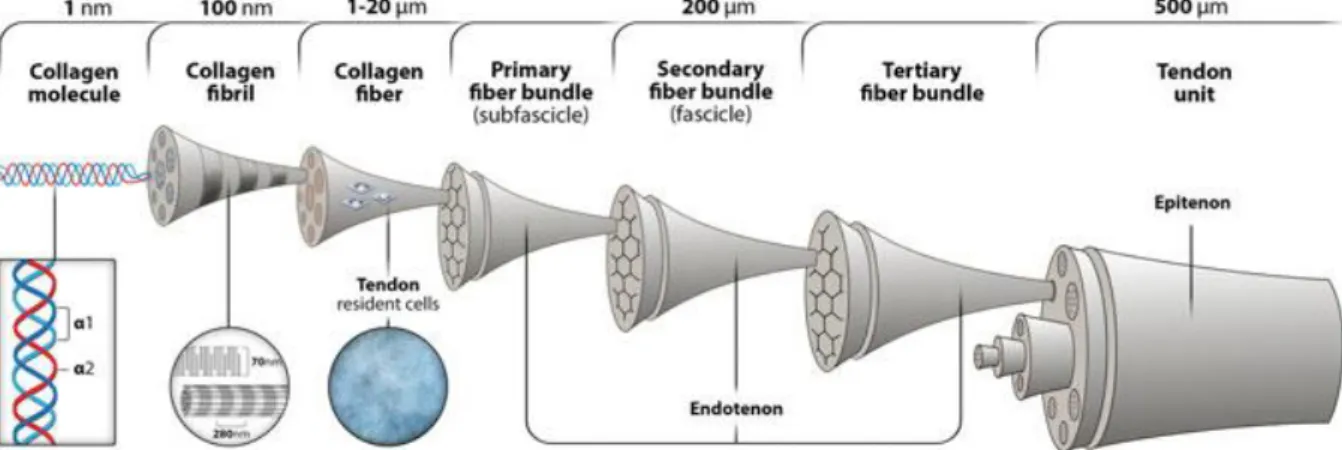
The tendon ECM has a well-organized hierarchical structure in which the tendon itself is comprised of successively smaller structures encased within each other (Figure 1.1). A tendon unit is comprised of several fascicles encased in a thin sheath of connective tissue, the epitenon. Each fascicle is formed by an ensemble of collagen fibres and is bound by the endotenon. Each fibre is composed of parallel collagen fibrils closely packed together, which in turn result from the aggregation of collagen
Figure 1.1. Tendon hierarchical structure. A tendon unit is comprised of several fascicles encased in a thin sheath of connective
tissue, the epitenon. Each fascicle is an ensemble of collagen fibres bound by the endotenon, which in turn are composed of parallel collagen fibrils closely packed together. Collagen molecules self-aggregate into fibrils, to which the tenocytes bind and
molecules 3,7. Collagen fibrils present crimps, a characteristic wavy pattern that is ascribed to the
bending of the fibrils when they change their plane of running. These crimps stretch when a tensile load is applied to the tendon and revert to their original formation once the load is removed, ultimately contributing to the tendon’s flexibility 4,5.
Tendons vary in shape, size and orientation depending on their insertion sites and function of the muscles and bones they are connecting. For instance, extensor muscles like the quadriceps that create powerful forces, have short and wide tendons. On the other hand, muscles like the finger flexors, that are responsible for small and precise movements, have long and thin tendons 6. Despite these differences in morphology, the internal structure of the tendon, namely its characteristic ECM organization, is common throughout the whole body. In particular, the existence of physically separate divisions of fibres not only prevents that localized damage affects the whole tendon but also provides the tendon with its load-bearing capabilities 5. Hence, the tendon ECM is directly responsible for the
mechanical performance of these tissues regarding their ability to successfully carry out musculoskeletal movement.
1.3. Tendon mechanical behaviour and properties
Tendons are capable of sustaining extremely high tensile forces due to their fibres being aligned longitudinally along the tissues mechanical axis and it is their viscoelastic behaviour that allows these tissues to carry out their mechanical role 4,8. In this scope, viscoelasticity, referring to the relationship between stress and strain, is a trait of materials in which this relationship is not constant, but instead depends on the period of load or displacement 1. Namely, when a tensile force is applied, the tendon is stretched and energy is absorbed and stored within the tissue, so when said force is removed, the tissue recovers its original shape and energy is released 1,4. Regarding these tissues tensile properties, it has been reported that natural tendons have tensile strengths, that is the maximum stress a material can sustain without breaking, and Young’s Modulus, which translates the relation between stress and strain, in the range of 5-100 MPa and 20-1200 MPa, respectively 9.
The tendon’s stress-strain curve (Figure 1.2) presents three regions: the toe region, the linear region and the yield and failure region. The toe region encompasses the 0-2 % strain of the collagen fibrils and represents the stretching of their crimps, thus presenting a low stiffness. Once every fibril has been flattened, the tendon enters its linear region. At a strain higher than 2 %, the collagen fibril backbone is pulled and the tendons deformation is caused by the collagen molecules sliding between each other. Thus, the deformation is linear and the slope of the curve represents the Young’s Modulus of the tendon 1,5,8. In normal conditions, most tendons function in the physiological range, that is in the toe region and in the beginning of the linear region, given that at strain lower than 4 %, the deformation is reversible and the tendon is able to recover its shape after loading 1. However, at 4 % strain, individual
fibrils start to tear and the tendon enters the yield region. In this region, micro ruptures accumulate, the tendon itself begins to fail and suffers irreversible deformation. Once the strain is higher than 8-10 %, the tendon suffers macroscopic failure 1,8.
1.4. Tendon healing and current treatments
Tendon natural healing can be divided into three overlapping stages: the inflammatory, the reparative and the remodelling phases. First, in the inflammatory phase, the injury site is covered by a blood clot, whose platelets induce the migration of extrinsic inflammatory cells, such as neutrophils and macrophages that phagocyte the necrotic tissue, by releasing of chemoattractants. Later, in the reparative stage, disorganized collagen is laid in large quantities in the injury site, which leads to scar tissue formation. Also, there is intrinsic fibroblastic proliferation and neovascularization along with
extrinsic fibroblastic migration into the injury site, which results in the production of large quantities of Type III collagen. Finally, during the remodelling phase, Type III collagen is replaced by Type I collagen and the newly formed fibrous tissue is aligned along the direction of the mechanical loading
1,7,8
. This process is guided by two distinct mechanisms that act simultaneously during these stages: the intrinsic healing, which is mainly governed by the tendon resident cell population; and the extrinsic healing, which encompasses the migration of cells from the surrounding tissues to the injury site and is initiated due to the tendons hypocellular nature and the tenocytes poor reparative capabilities 3,7,10.
Overall, tendon healing can evolve into tendon repair or regeneration. During tendon repair there is inflammatory cell infiltration, fibroplasia, disorderly collagen deposition and although, after this process, the vascular, cellular and collagen content of the remodelled tendon are similar to that of a healthy one, its structural and mechanical properties will be most likely inferior 1,4,11. Furthermore, this is an extremely long process that can easily take several months or even years for the tissue to fully remodel 7,11. In contrast, during tendon regeneration there are few inflammatory cells, fibroplasia does not occur and collagen is deposited orderly, so there is no scar tissue formation and the tissues mechanical properties are fully restored 4,5. Thus, in this scope, the preferred approach following an
injury is tissue regeneration rather than tissue repair, so that all the tissue’s structural and mechanical properties may be restored.
Nowadays, tendon injuries are treated surgically or by conservative (non-surgical) therapies. Surgical intervention involves the application of implantable devices that are able to cover the defect at the injury site, such as sutures or tissue grafts 10,12. This type of intervention is followed by a recovery
period during which the tendon is immobilized or different types of mechanical loading are applied, aiming at restoring the tendon load-bearing capacity 13. On the other hand, non-surgical treatment for tendon injuries relies on a combination of different therapies, such as eccentric exercise, extracorporeal shock wave therapy or treatment with low-intensity pulsed ultrasound. Eccentric exercises stimulate tendon remodelling while the latter therapies induce a new trauma to the tendon in order to reinitiate the healing process 14.
Figure 1.2. Tendon stress-strain curve. This curve presents three regions: the toe region, the linear region and the yield and
failure region. The initial toe region (0-2 % strain) represents the stretching of the crimps in the collagen fibrils. The linear region (2-4 % strain) represents the stretching of the fibril backbone and its slope is the tendon’s Young’s Modulus. In the yield region (strain higher than 4 %) microscopic tears accumulate, eventually leading to a macroscopic rupture and subsequent
Surgery has become the treatment of choice for tendon injuries, given that it has proven to be more successful than conservative therapies. Moreover, a large portion of the patients that resort to conservative therapies need to undergo surgery afterwards due to treatment failure. Regardless, none of the current treatments have been able to fully restore tendon function after injury, in spite of its severity
2,10,11.
1.5. Response to mechanical stimuli
Physiological loads are fundamental for the maintenance of tendon tissue homeostasis by influencing cell behaviour in terms of proliferation, differentiation or even healing mechanisms. In fact, tendon injuries frequently occur as a consequence of inadequate mechanical loading, whether excessive or insufficient, resulting in loss of tissue function 13,15. In response to external forces, tenocytes remodel the tendon ECM through mechanotransduction, that is, the process by which mechanical stimulus is translated into biochemical signals. Overall, this process depends on interactions between ECM proteins, tenocytes surface receptors and cytoskeleton and several signalling molecules, resulting in the formation and degradation of ECM proteins 16–18. Also, mechanical loading helps maintain the tenocytes characteristic spindle shape 15. Thus, mechanical stimulation is an important parameter to consider when aiming at maintaining the tenogenic phenotype and promoting stem cell tenogenic differentiation.
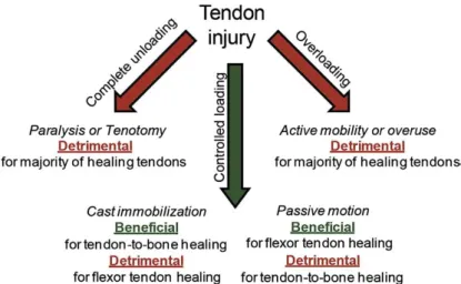
Furthermore, controlled mechanical loading may also be used for clinical purposes to aid the healing process. Although extensive and extended stretching leads to an inflammatory response and promotes osteogenic differentiation of stem cells, the application of forces within the physiologic range can improve the repair process and hence restore most of the normal tissue function 13,15. The most adequate loading regime should vary with the nature and the site of the injury (Figure 1.3) and should also be combined with any additional therapies that may be applied to correct the tendon defect 13,19. For instance, complete unloading of an injured tendon leads to a decrease in structural and mechanical properties, while its overloading may reduce its mechanical properties along with its range of motion. On the other hand, passive motion is particularly effective to reduce the formation of adhesions, which hinder the natural gliding of the tendon 19.
1.6. Tissue engineering strategies for tendon regeneration
In recent years, several tissue engineering (TE) strategies have been developed in order to promote tendon regeneration, thereby aiming at restoring the tendons normal tissue function in a post injury setting, a requirement that current treatments haven’t been able to fulfil. These strategies rely on
Figure 1.3. Diagram illustrating the effect of different loading regimes in different types of injuries. While complete unloading
or overloading impairs the healing process of most injuries, cast immobilization and passive motion are indicated for the
a combination of 3D constructs capable of mimicking the key structural aspects of the tissues ECM and of biological agents able to promote a regenerative process, while also supporting cell proliferation.
1.6.1. Cell based approaches
Given the poor regenerative capacity of the tendon resident cell population and these tissues inherent tendency to generate scar tissue and adhesions as a result of an injury, several therapies based on cell harvesting and seeding have been proposed to overcome the limitations of the tendon natural healing process. Moreover, a population of stem and differentiated cells can give rise to a range of bioactive molecules responsible for regulating different physiological processes. For this reason, delivering cells to the injury site could improve the tissues response to the trauma 4,10.
In this scope, the use of the patients own resident tendon cells is an evident option, since an immune response would be avoided. However, the harvesting process is detrimental for the donor tissue, which may lead to tissue morbidity due to its hypocellular nature. On the other hand, cell populations harvested from another donor are prone to induce an immune response, eventually leading to their rejection 11,20. As an alternative, adult stem cells could be used for this purpose. These cells have the ability to self-renew and are capable of committing and differentiating into different tissue lineages
7
. Also, a population of these cells can be found in almost all tissues of the human body. For instance, adipose tissue is an abundant adult stem cell source, enabling a less invasive harvesting procedure without causing donor tissue morbidity 4,20,21. Importantly, there is no established method to induce
tenogenic differentiation of stem cells, though certain growth factors, such as bone morphogenic protein-12 (BMP-12), epidermal (EGF) and platelet-derived growth factor (PDGF), have promoted the expression of tendon-related markers of stem cells 7,22. Moreover, it has been suggested that tenogenic differentiation may be enhanced by combining biochemical cues, such as growth factor supplementation, with other biophysical cues, such as surface topography or mechanical stimulus 20,22– 24.
Although, stem cells have been extensively investigated for other purposes, the application in tendon TE strategies demands deeper research into the mechanisms underlying tenogenic differentiation. Nonetheless, it is expected that future research may lead to further knowledge on this matter and in particular regarding its role in the regenerative process of tendon tissues.
1.6.2. Scaffold-based approaches
Even though a cell-based approach is an adequate solution for an injured tendon, its individual application may not be sufficient to ensure mechanical properties comparable to that of a native tendon. For this purpose, cell-based approaches can be combined with engineered scaffolds that can provide a suitable environment for cell proliferation while also being able to recapitulate the native tendons mechanical behaviour.
Ideally, scaffolds need to fulfil a few requirements in order to be able to support and guide cell proliferation, eventually originating new and functional tissue. First, and foremost, TE scaffolds must be biocompatible, not to induce an immune response, and biodegradable, degrading at a similar rate as the cells start to produce new ECM. Scaffold architecture is also an important feature to consider: scaffolds should be porous 3D constructs, with a large surface area to allow cell infiltration and nutrient diffusion, and should mimic the overall structure of the tissue ECM. Also, the scaffolds mechanical behaviour should be similar to that of the native tissue, although this requirement depends not only on its architecture but also on the materials with which it is fabricated 12,21,25,26. In particular, the design of 3D scaffolds for tendon TE applications has been a real challenge given the difficulty in recapitulating the tendons mechanical behaviour, especially at the nanoscale, while ensuring that the remaining requirements are met 27.
Considering the application of an engineered scaffold, the selection of suitable biomaterials is of the utmost importance as it will determine its mechanical properties and degradation time. TE scaffolds can be based on a variety of polymers either of synthetic or natural origin or even on a combination of both. Synthetic polymers are easily processed and modified and can satisfy a multitude of structural features required for the scaffolds specific application, presenting higher mechanical properties than natural polymers. Some of the most common synthetic polymers applied in TE strategies are poly(ε-caprolactone) (PCL), poly(ʟ-lactic acid) (PLLA) and poly(glycolic acid) (PGA) 12,28. On the
other hand, natural polymers, like chitosan or silk, are biocompatible, biodegradable and possess higher cell affinity in comparison with synthetic polymers, since its chemical and biological characteristics are similar to that of the tissues ECM 28,29. Thus, combining both natural and synthetic polymers leads to
the fabrication of a scaffold possessing proper mechanical properties while allowing cell attachment and proliferation 4.
As mentioned in the previous section, tendons are fibrous tissues comprised of a very organized ECM of anisotropically aligned collagen fibres. Thus, scaffolds intended for tendon TE are preferably fibre-based and can be fabricated through several fibre processing techniques, such as melt-spinning, wet-spinning or electrospinning 30. Fibres fabricated through these techniques can be further assembled into 3D scaffolds that better mimic the tendons ECM structure 12. Although all these techniques allow the processing of microscaled fibres, through electrospinning one can obtain constructs comprised of fibres scaled from a few hundred nanometres to a few micrometres, therefore allowing the replication of the length scale of the native tendons hierarchical units 30.
1.6.3. Electrospinning
Electrospinning has been among the preferred manufacturing techniques of fibre-based scaffolds for tendon TE applications, given that it allows the production of anisotropically aligned nanofibrous constructions that mimic the highly oriented nanoscale structure of tendon ECM 4. Overall,
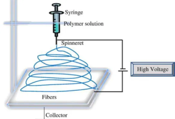
electrospinning is a rather simple procedure in which electrostatic forces are used to produce nanofibres with uniform diameters from a polymer solution. A typical setup (Figure 1.4) encompasses four main components: a syringe holding the spinning solution, which is coupled to a syringe pump, a high voltage power supply and a grounded collector. While the syringe pump forces the solution to flow out of the syringe, a high voltage is applied to its needle. A drop will form at the tip of the needle and, when the stresses caused by the electric field overcome the surface tension, the drop is stretched and a jet is formed. Then, while the jet travels in the direction of the grounded collector in an oscillatory movement due to electrostatic repulsion, the solvent evaporates and a nonwoven fibrous mesh is formed in the grounded collector 26,29.
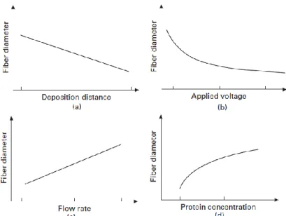
The design of the electrospinning setup significantly impacts the morphology and diameter of electrospun fibres (Figure 1.5), thus several parameters need to be adjusted according to the
requirements of their intended application. Deposition distance (Figure 1.5a), that is, the distance from the tip of the needle to the grounded collector, is known to influence fibre diameter as its increase leads to further stretching of the spinning jet, thereby causing a decrease in the fibre diameter. Also, there is a required minimum distance to allow the solvent to evaporate before the fibres are deposited in the collector 29. The voltage applied to the polymer solution (Figure 1.5b) is an essential requirement of this procedure, since there is a threshold below which jet formation does not occur 31. However, once the applied voltage threshold is reached and the solution is jetted in the direction of the collector, an increase in the applied voltage frequently leads to greater stretching and subsequent decrease of the fibre diameter, although there is a higher probability of causing bead formation 29. On the other hand, a higher flow rate (Figure 1.5c), that is the speed at which the solution is jetted out of the syringe, tends to lead to thicker fibres. Furthermore, a lower flow rate is usually indicated to allow the fibres enough time to dry, thereby preventing the formation of beads 29. Finally, protein or polymer concentration (Figure
1.5d) has to be carefully adjusted since it influences the viscosity of the solution. At lower concentration, beads are formed instead of fibres and at higher concentration continuous formation of fibres is interrupted due to the difficulty in maintaining the flow rate at the tip of the needle. Nonetheless, higher polymer concentration causes an increase in the fibre diameter 29,31.
Another important feature to consider during the electrospinning process is the type of the collector that is used, since it determines the alignment of the electrospun fibres. In a typical electrospinning setup (Figure 1.4), the grounded collector is a flat surface which leads to the formation of randomly aligned fibre meshes 29. However, the ECM from the majority of native tissues has an anisotropic organization that is essential for its overall functioning 26. Therefore, numerous strategies were developed to allow the fabrication of electrospun scaffolds with different types of alignment. Among these, the use of high speed rotating collectors, such as rotating disks, to force an aligned deposition of nanofibres has been the preferred approach to produce anisotropically aligned nanofibrous scaffolds intended to tendon TE 4,26.
1.6.4. 2D vs 3D Scaffolds
In the scope of tendon TE, anisotropically aligned nanofibrous scaffolds showed to be able to provide the necessary topographical cues to guide the adhesion and proliferation of tenocytes, thereby proving its ability to maintain the tendon phenotype 32. In Figure 1.6, human tenocytes seeded onto
Figure 1.5. Influence of several processing parameters in the diameter of electrospun fibres. Fibre diameter as a function of (a)
deposition distance, (b) applied voltage, (c) flow rate and (d) protein or polymer concentration, while keeping the remaining
anisotropically aligned electrospun scaffolds, fabricated from a polymer blend of PCL and chitosan (CHT), clearly show a preferential direction of alignment. Also, these aligned nanofibre mats were able to induce the differentiation of human tendon stem/progenitor cells by promoting an increase in the expression of tendon-specific genes, in regard to random nanofibrous scaffolds. 4,30.
Although these constructs are able to fulfil the biological requirements for tendon TE, their mechanical properties can only ensure the load-bearing capabilities of tendons during the regenerative process to some extent 33. Indeed, scaffolds intended for tendon TE applications should be mechanically
similar to the native tissue, thus minimizing stress-shielding effects, which can cause disorganized tissue growth, and providing a temporary replacement for immediate function that is unlikely to rupture after repair and that enables tissue regeneration at the same time. However, the mechanical properties of electrospun scaffolds are generally much inferior to that of native tendons (1-25 MPa in ultimate tensile strength and 1-350 MPa in Young’s modulus) 4. Therefore, it is necessary to improve the
mechanical properties of electrospun nanofibrous scaffolds, without impairing their biological performance, thereby maximizing their application for tendon TE.
In order to improve the electrospun scaffolds mechanical performance, reinforcement nanofillers, such as cellulose nanocrystals (CNCs) may be incorporated into the fibres . These are biocompatible rod-shaped nanoparticles with exceptional mechanical properties (7.5-7.7 in ultimate tensile strength and 110-220 GPa in axial elastic modulus) and low cytotoxicity, which allows them to be used as load-bearing components for tendon directed scaffolds 34.
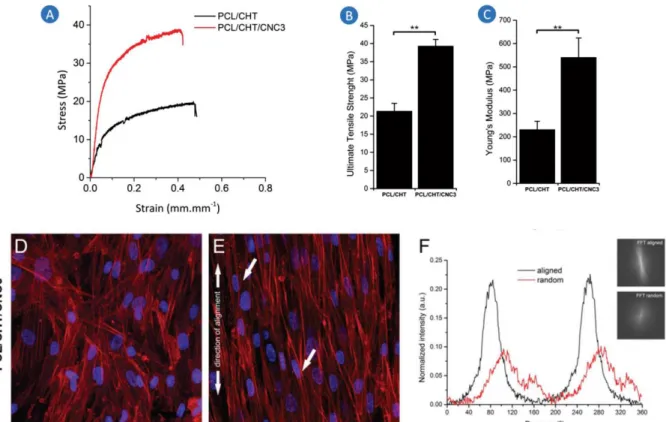
For this purpose, CNCs were incorporated in electrospun nanofibrous scaffolds, both randomly and anisotropically aligned, based on a polymer blend of PCL and chitosan (PCL/CHT) and intended for tendon TE applications (Figure 1.7) 33. The incorporation of small CNC contents up to 3 wt.% (weight percentage) proved to have a significant reinforcement effect on the nanofibrous scaffolds by increasing their mechanical properties to the range of native tendons. In particular, when 3 wt.% CNC content was added to the polymer blend (PCL/CHT/CNC3), the aligned nanofibrous scaffolds suffered a substantial increase in their ultimate tensile strength (Figure 1.7B), from 21.4 ± 2.2 MPa to 39.3 ± 1.9 MPa, and Young’s modulus (Figure 1.7C), from 230.0 ± 2.2 MPa to 540.5 ± 83.7 MPa, which represents an improvement of 132% and 83%, respectively. Moreover, the enhancement of the mechanical performance of the scaffolds was achieved without compromising their biological performance, since the aligned nanofibrous scaffolds reinforced with CNCs were able to promote cell alignment, along the axis of orientation of the fibres, and induce an elongated cell morphology, unlike the random nanofibrous scaffolds (Figure 1.7D, E and F). The stress-strain profiles of the aligned nanofibrous scaffolds (Figure 1.7A), though showing a significant improvement for the PCL/CHT/CNC3 in comparison with the PCL/CHT, lack the characteristic toe region of native tendon mechanical behaviour, which is attributed to the uncramping of the collagen fibres (see Tendon mechanical behaviour section). This region appears at low strains and represents a shock-absorbing feature of
Figure 1.6. Scanning electron microscope images (SEM) of hTDCs cultured on anisotropically aligned electrospun PCL/CHT
scaffolds for 10 days. Magnification (A) X200 and (B) X1000. (C) Confocal microscopy images of hTDCs on aligned
tendons to prevent tissue damage. Nonetheless, these results substantiate the use of CNCs as reinforcement nanofillers for electrospun nanofibrous scaffolds intended for tendon TE 33.
Despite these reinforced aligned nanofibrous scaffolds possessing adequate mechanical properties and biological performance for tendon TE, they lacked the ability to replicate the native tendon tri-phasic biomechanical behaviour and the length scale of their hierarchical units. Furthermore, these scaffolds are usually fabricated as 2D membranes, limiting their dimensions and making them difficult to handle for practical TE approaches. As such, a new electrospinning system has been developed to fabricate scaffolds that may overcome these limitations.
Thus, continuous and aligned nanofibre threads (CANT) were fabricated from a PCL/CHT/CNC blend (with 3wt% CNC content) and were then assembled into 3D hierarchical scaffolds, through different textile techniques (Figure 1.8) 35. CANT are intended to mimic the basic unit of a native tendon, that is, the collagen fibres, while the higher hierarchical constructs mimic different length scale structures from the native tendons ECM. These constructs address several requirements of a tendon directed scaffold, namely regarding its structural, mechanical and biological properties, by presenting an anisotropic hierarchical structure and a nonlinear biomechanical behaviour. Moreover, the newly devised system for the production of CANT provides the necessary conditions to ensure a scalable production of 3D scaffolds while allowing the adaptation of their dimensions to fit the demands of the target tissue.
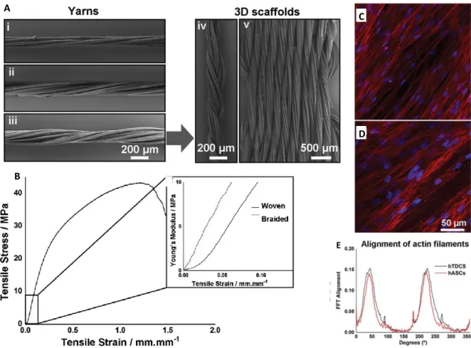
Three textile techniques with different levels of complexity were used to assemble CANTs. First, by twisting 6, 9 or 12 CANT together, nanofibre yarns were obtained in order to replicate the architecture of the native tendons fibre bundles (Figure 1.8A i), ii), and iii)). Mechanical assessment of these yarns showed that an increase in the number of CANT has no significant effect on its Young’s modulus and ultimate tensile strength, as opposed to their tensile load (maximum load sustained before
Figure 1.7. Mechanical assessment of anisotropically aligned nanofibrous scaffolds of PCL/CHT and PCL/CHT/CNC3 and
biological performance of aligned PCL/CHT/CNC3 scaffolds against its random counterpart. (A) Stress-strain profiles; (B) Ultimate tensile strength. (C) Young’s modulus. Confocal microscopy images of the hTDCs seeded on randomly oriented PCL/CHT/CNC3 scaffolds (D) and on anisotropically aligned ones, after 10d of culture (blue: nuclei; red: actin filaments). (F) 2D FFT frequency plots and normalized radial intensity plotted against the angle of rotation for hTDCs cultured on aligned
breaking) and toughness (ability of a material to deform plastically without breaking), which were considerably enhanced. Thus, 3D scaffolds, mimicking the native tendon unit architecture, were fabricated through braiding and weaving of yarns comprised of 12 CANT (Figure 1.8A iv) and v)). Although braided scaffolds presented higher ultimate tensile strength and Young’s modulus in comparison to the woven scaffolds, the latter had a significantly higher toughness and tensile load. Moreover, the woven scaffolds were able to mimic the stress-strain profile of a native tendon, unlike the braided scaffolds, which lacked the characteristic toe region at low strains (Figure 1.8B). Given the superior mechanical performance of woven over braided scaffolds, their biological performance was assessed in terms of tendon-phenotype maintenance and induction of stem cell tenogenic differentiation, by seeding hTDCs (human Tendon Derived Cells) and hASCs (human Adipose Stem Cells), respectively. Indeed, the topography of the woven scaffolds induced an elongated morphology and alignment of the seeded cells, for both cell types (Figure 1.8C and D), after 7 days of culture. The 2D-FFT plots in Figure 1.8E show two distinct peaks at 90° and 180°, which translates the high alignment of cells after 21days, for both cell types. These plots further confirm the effect of these scaffolds topography in the maintenance of the tendon phenotype and induction of tenogenic differentiation of stem cells. Thus, the results herein support the need for target tissue mimicry when developing a scaffold for tendon TE applications 35.
Figure 1.8. Characterization of CANT, Yarns and 3D scaffolds. (A) SEM images of Yarns comprised of i) 6, ii) 9 or iii) 12
CANT, and of iv) braided and v) woven 3D scaffolds. (B) Tensile-strain curve of the woven scaffolds and close up comparing the toe region from woven scaffolds and braided scaffolds. High magnification confocal images of (C) hTDCs and (D) hASCs cultured onto woven scaffolds, at day 7 (nuclei is stained in blue and the cytoskeleton in red). (E) 2D-FFT frequency plots of
actin filaments alignment in hTDCs and hASCs cultured onto woven scaffolds for 21 days. Adapted from ref. 35.
A
B
C
D
1.6.5. Mechanical stimulation and magnetic actuation
As mentioned before, tendons are mechanosensitive load-bearing tissues and, as such, without appropriate mechanical stimulus, collagen alignment and organization are affected during new tissue formation, thereby impairing its load-bearing capabilities 27. Moreover, mechanical stimulation has been linked to enhanced cell proliferation and alignment, as well as tenogenic differentiation and ECM synthesis 36–39.
Nowadays, bioreactors are the preferred approach to mechanically stimulate cells seeded onto scaffolds, therefore guiding tissue remodelling and ultimately enhancing the performance of said scaffold 27,38. A bioreactor system (Figure 1.9) is comprised of a motion component, which grips on both ends of a scaffold and applies physical stimulus; a culture chamber, in which the seeded scaffold is incubated under controlled conditions and continuously perfused with growth media; and a control component that allows the regulation of the parameters for mechanical stimulation and the conditions of incubation 37,40. In short, this equipment applies cyclic tensile strains to a construct by gripping on its ends, while providing a dynamic environment with the suitable conditions to sustain a cell culture.
Indeed, bioreactors have proven to be an important tool to enhance the performance of TE constructs through mechanical stimulation. These systems have not only been able to induce new tissue formation with the characteristic structural organization of native tendon ECM, but also enhance the mechanical properties of loaded tendon TE constructs 21,36,37. However, this approach may only be applied for in vitro studies, due to the need to physically hold onto the ends of the scaffolds to provide the mechanical stimulus 38. Nonetheless, tendon TE directed scaffolds may be mechanically stimulated
in vitro before their implantation in vivo, which enhances their maturation, ECM fibre alignment and
overall mechanical properties 21,40.
The perspective of manufacturing a scaffold capable of replicating the native tendons structural and mechanical properties, while also being able to be remotely actuated to mechanically stimulate cells, thus promoting tissue regeneration, would be of considerable interest. The remote actuation of scaffolds would allow mechanical stimulation of cells without needing to restrict the scaffold when applying external loads. Furthermore, bioreactors would no longer be necessary for this purpose and scaffolds possessing this feature would enable mechanical stimulation in vivo, in a post implantation setting.
Figure 1.9. Example of a mechanical bioreactor. The motion component is highlighted by the red rectangles. The red tubes are
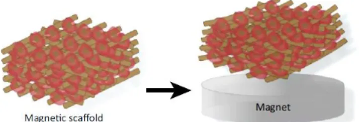
In this scope, magnetic nanoparticles (MNPs) could be combined with the scaffolds biomaterials, thereby creating a mechanically responsive system capable of being magnetically actuated to stimulate cells (Figure 1.10). As previously mentioned, cells adjust their behaviour in response to external mechanical stimuli through mechanotransduction 17. In this sense, physical cues, such as substrate deformation or ECM stiffness, are recognized by cells due to the direct contact between their F-actin cytoskeleton and the ECM, provided by focal adhesions. As such, cells compensate extracellular forces by reorganizing their cytoskeleton, thereby causing tension within the cell. Besides the direct impact on cell geometry, cytoskeletal tension also activates signalling pathways that regulate proliferation and differentiation of stem cells 16,17,41. Thus, the local substrate deformations caused by the magnetic actuation of MNPs 42, should activate mechanotransduction mechanisms that ultimately
drive cellular responses towards the desired behaviour 43,44.
Although the development of magnetic responsive scaffolds for tendon TE is relatively new, this approach has been successfully applied to bone TE strategies. For instance, Singh et al., 2014 45 reported on the osteogenic potential of electrospun nanofibre mats produced from a blend of PCL and iron oxide MNPs exhibiting ferromagnetic behaviour. Results indicate that the incorporation of MNPs enhanced Mesenchymal Stem Cells (MSCs) adhesion and spreading and promoted their osteogenic differentiation in vitro. When implanted in vivo in rat models for 4 weeks, the scaffolds elicited minimal inflammatory reactions, promoted neovascularization and cell migration through the scaffolds and presented a degradation rate comparable to that of the native tissue. Additionally, magnetic responsive scaffolds were rolled in a cylindrical form and applied to rat segmental defects: these scaffolds promoted neobone formation after 8 weeks and exhibited a higher degradation rate than bare PCL scaffolds. Meng
et al., 2013 46 assessed the in vivo osteogenic potential of superparamagnetic nanocomposite electrospun
scaffolds of a blend of PLA (poly lactide acid), iron oxide MNPs and hydroxyapatite nanoparticles (nHA). For this purpose, the scaffolds were implanted in rabbit model defects and an external static magnetic field was applied for 110 days. Results indicate that magnetic actuation of these scaffolds induced earlier and faster bone formation, in comparison to scaffolds implanted in the absence of magnetic field. Also, the degradation rate of the scaffolds was enhanced under magnetic actuation, suggesting higher activation of recruited macrophages. Finally, Hao et al., 2017 47 reported on the use
of the aforementioned superparamagnetic nanocomposite electrospun scaffolds to modulate the function of macrophages related to bone regeneration. Firstly, macrophages were cultured on magnetic responsive scaffolds under a static magnetic field alternatively applied in intervals of 12h. Results indicate that macrophages under magnetic stimulation expressed higher levels of a key marker for the resolving phenotype, than those in static culture, while suppressing a key marker for the pro-inflammatory phenotype. Analogously, macrophages under magnetic stimulation increased the production of anti-inflammatory and wound-healing associated cytokines while supressing the secretion of pro-inflammatory cytokines, in comparison to those in static culture. Moreover, pre-osteoblasts were seeded onto the magnetic scaffolds and cultured on the macrophage-conditioned medium, under magnetic stimulation, to assess the osteogenic potential of macrophages. After 24h there was a higher migration rate, as well as, higher intensity of mineralization, characteristic of the later phases of
Figure 1.10. Magnetically actuated scaffold. In the presence of a magnet, the magnetic field induces structural deformation of
osteoblast maturation, in pre-osteoblasts cultured under magnetic stimulation than in those in static culture. Analogously, when seeded pre-osteoblasts were cultured in a combination of macrophage-conditioned medium and osteogenic induction medium, there was higher intensity of mineralization among pre-osteoblasts under magnetic stimulation than those in static culture. Therefore, magnetic stimulation has a significant impact on the recruitment of pre-osteoblasts and on the induction of osteogenesis mediated by macrophages. Overall, these studies highlight the efficacy of the proposed strategy on the promotion of osteogenesis, both in vitro and in vivo 45,46, and on the modulation of the
inflammatory response by regulating the polarization of macrophages phenotype 47. Moreover, due to the versatility of the electrospinning technique, magnetic responsive scaffolds with different architectures can be designed for TE strategies directed at other tissues, thereby eliciting equivalent responses regarding stem cell differentiation, new tissue formation and modulation of the inflammatory response.
Regardless, magnetically actuated scaffolds, intended for tendon TE, were fabricated through rapid prototyping technology, by incorporating iron oxide nanoparticles into aligned arrangements of fibres based on a polymer blend of starch and PCL (SPCL) (Figure 1.11A) 48. Besides exhibiting a superparamagnetic behaviour, these materials containing iron oxide MNPs had no effect on the viability of seeded cells, thereby proving the MNPs biocompatibility. Magnetic stimulation of the scaffolds had a positive impact on the viability and proliferation of hASCs: the application of an oscillating magnetic field resulted in a significant increase in cellular metabolic activity in both magnetic and non-magnetic constructs, although metabolic activity was higher in magnetic scaffolds under static and dynamic conditions. Thus, cells respond to magnetic stimulus both in the presence and in the absence of MNPs, meaning cellular behaviour can be influenced by indirect magnetic forces. Also, the combination of magnetic stimulation and MNPs seems to have a synergistic effect on cellular mechanisms by benefiting the proliferation of cells 48.
The effect of combining structural cues and magnetic stimuli on guiding hASCs towards a tenogenic lineage was assessed by culturing cells onto SPCL and magnetic SPCL scaffolds under magnetic stimulation. The results showed that hASCs undergo tenogenic commitment, developing a type I collagen matrix, that appears to be more abundant in scaffolds cultured under magnetic stimulation, and especially in magnetic SPCL scaffolds (Figure 1.11B). Also, under magnetic stimulation was observed a more aligned arrangement of collagen fibres that better replicated the ECM structure of native tendons. Furthermore, higher production of Tenascin C, a tendon marker whose expression is upregulated as a result of mechanical loading, was observed in both types of scaffolds
A B
Figure 1.11. Magnetically actuated scaffolds for tendon TE applications fabricated from a blend SPCL and iron oxide
nanoparticles. (A) 3D reconstructed model by micro-CT analysis showing the aligned architecture of these scaffolds (the polymeric matrix is presented in grey and the MNPs in red). (B) Type-I collagen immunolocation in hASCs seeded onto bare and magnetic SPCL (magSPCL) scaffolds, cultured for 7 d under magnetic stimulation or static culture. These images represent 1 or 2 fibres from the aligned scaffolds (blue stains the nuclei while green indicates type-I collagen). Scale bar represents 100