UNIVERSIDADE DA BEIRA INTERIOR
Ciências da Saúde
Development of different drug delivery systems
for skin regeneration
Patrícia Isabel da Cruz Morgado
Master degree thesis in
Biomedical Sciences
(2nd cycle of studies)
Supervisor: Ilídio Joaquim Sobreira Correia, Ph.D.
UNIVERSIDADE DA BEIRA INTERIOR
Ciências da Saúde
Desenvolvimento de diferentes sistemas de
libertação controlada para a regeneração de
feridas cutâneas
Patrícia Isabel da Cruz Morgado
Dissertação para obtenção do Grau de Mestre em
Ciências Biomédicas
(2º ciclo de estudos)
Orientador: Prof. Doutor Ilídio Joaquim Sobreira Correia
iv
Acknowledgments
I would like to thank my supervisor, Professor Ilídio Correia, for all his dedication, trust and support that always showed me, and for making the possible and impossible to gather all the necessary conditions for the development of this study. To him I thank all the knowledge and confidence I gained during this time.
I must thank Professor António Miguel Morão for all his kindness in participating in this project and dedication he showed in its development.
I would also like to thank all my group colleagues for their advice, knowledge sharing and all the good and bad moments we spent together. Thanks for your friendship, kindness and patience.
To my friends for all the support and affection that have always given to me. To everyone with whom I always smile, studied and enjoyed myself.
Finally, I thank my family for the support that always showed in my decisions; to my parents for allowing me to finish another important stage in my life, to my grandmother and brother for the love and confidence that have always given to me and to my boyfriend for all the times that endured my despair and that calmed me in difficult times.
vi
Abstract
The human body has different fluids that are hostile environments for biologically active molecules. To overcome this arbitrary, several drug delivery systems have been developed. These systems not only protect all unstable biological active compounds from enzymatic degradation in the human body but also allow a sustained and targeted release of drugs. They contribute for decreasing drug dose required to achieve the desired therapeutic effect. Hydrogels, with their important characteristics, have been widely used in the development of these systems however, the quantity of drug loaded into them may be limited and the high water content of these polymeric matrices often results in relatively rapid drug release profiles. Nevertheless, due to their good physical and biological properties, hydrogels have been extensively used in the treatment of skin injuries. In order to take advantage of hydrogels for drug delivery and wound healing, different systems (nano and micrometric) have been developed and incorporated into hydrogels matrices. Nano and micro systems exhibit high encapsulation efficiencies of drugs and allow its release for a long period of time. Thus, the main goals of this master thesis work plan were to develop micrometric systems based on chitosan, alginate and a dextran hydrogel, and characterize their applicability in the treatment of skin injuries. Initially, the carriers were characterized according to their size, geometry, swelling behavior and biocompatibility. In vitro release studies allow us to analyze the release profile of a model protein (bovine serum albumin) when encapsulated into the microparticles. The same studies were done for microparticles loaded into a dextran hydrogel. Co-relating the swelling studies with the in vitro protein release studies a mathematical model based on the theory of hindered transport of large solutes in hydrogels was developed for theoretically describing the process of protein release. All the experimental results were interpreted with the aid of the developed model. Moreover it can contribute to decrease the number of experimental studies, reducing costs and saving time for the carrier development. After that, growth factors (vascular endothelial growth factor and endothelial growth factor) were encapsulated into chitosan microparticles that were then loaded into the dextran hydrogel. Subsequently in vivo studies were performed to characterize the applicability of the dextran hydrogel loaded with chitosan microparticles containing growth factors in wound healing. The in vitro and in vivo studies demonstrated that the developed carriers are biocompatible, accelerate the wound healing process and can be used to deliver other bioactive agents.
Keywords
Drug delivery systems, growth factors, in vitro and in vivo studies, mathematical modeling, wound healing.
viii
Resumo
O corpo humano é composto por diferentes fluidos biológicos que são ambientes hostis para diversas moléculas bioactivas. Devido a este ambiente hostil, diversos sistemas de entrega controlada de fármacos têm sido desenvolvidos. Estes, não só protegem os compostos instáveis da degradação enzimática no corpo humano como, também, permitem a libertação desses compostos de uma forma controlada e, muitas vezes, direccionada, diminuindo, assim, a dosagem necessária para se obter o efeito terapêutico desejado. Até ao momento, os hidrogéis têm sido os materiais mais utilizados na produção deste tipo de sistemas. No entanto, a sua baixa eficiência de encapsulação e a elevada capacidade de absorverem água conduz a perfis de libertação do fármaco rápidos. Contudo, estes sistemas têm sido muito utilizados para o tratamento de feridas cutâneas devido às suas propriedades físicas e biológicas. Diferentes estudos têm sido efectuados, de forma a potenciar a utilização dos hidrogéis na libertação de fármacos e na regeneração de feridas. Novos sistemas (nano e micrométricos) têm sido produzidos e introduzidos no interior dos hidogéis. Estes apresentam eficiências de encapsulação de fármacos superiores e permitem que os mesmos sejam libertados durante um maior período de tempo. Assim, o plano de trabalhos deste mestrado teve como objectivos o desenvolvimento de sistemas micrométricos à base de quitosano e alginato e de um hidrogel de dextrano com o intuito de verificar a sua aplicabilidade no tratamento de feridas cutâneas. Deste modo, inicialmente os transportadores foram caracterizados quanto ao seu tamanho, geometria, capacidade de inchaço e biocompatibilidade. Estudos de libertação in vitro permitiram analisar o perfil de libertação de uma proteína modelo (albumina de soro bovino) quando encapsulada no interior das micropartículas. Os mesmos estudos foram efectuados para as micropartículas incorporadas no interior do hidrogel. Tendo por base os estudos de inchaço e os estudos de libertação in
vitro, foi desenvolvido um modelo matemático baseado na teoria de transporte difuso de
macromoléculas para descrever teoricamente o processo de libertação da molécula modelo usada no presente estudo. Todos os resultados experimentais foram interpretados com a ajuda do modelo desenvolvido, o qual pode reduzir o número de estudos experimentais necessários para desenvolver um determinado transportador, reduzindo os custos e o tempo necessário para o desenvolvimento destes novos sistemas para entrega direccionada e controlada de fármacos. Posteriormente, foram efectuados estudos in vivo, onde factores de crescimento (factor de crescimento vascular endotelial e factor de crescimento endotelial) foram encapsulados em micropartículas de quitosano. Através destes estudos verificou-se a aplicabilidade do hidrogel de dextrano e das micropartículas de quitosano com factores de crescimento encapsulados na regeneração de feridas cutâneas. Os estudos in vitro e in vivo demonstraram que os transportadores desenvolvidos são biocompatíveis, aceleram o processo de regeneração de feridas cutâneas e podem futuramente ser usados para libertar outros agentes bioactivos.
ix
Palavras-chave
Cicatrização de feridas, estudos in vitro e in vivo, factores de crescimento, modelação matemática, sistemas de libertação controlada de fármacos.
xi
Index
Chapter 1: Introduction
1. Introduction ... 2
1.1. The Skin: structure and functions ... 2
1.1.1. Epidermis ... 2
1.1.2. Dermis ... 3
1.1.3. Hypodermis ... 4
1.2. Wounds: identification and classifications ... 5
1.3. Wound Healing ... 6
1.3.1. Haemostasis and Inflammation ... 8
1.3.2. New tissue formation ... 9
1.3.3. Remodeling ... 10
1.4. Biomaterials and Tissue Engineering ... 12
1.4.1. The role of biomaterials in skin regeneration ... 13
1.5. The Role of Growth Factors in Wound Healing ... 17
1.6. Hydrogels and Drug Delivery Systems ... 18
1.7. Mathematical Modeling of Drug Delivery ... 23
Chapter 2: Materials and Methods 2. Materials and Methods ... 25
2.1. Materials ... 25
2.1.1. Drug delivery systems produced with alginate and chitosan ... 25
2.1.2. Dextran as a wound dressing ... 26
2.2. Methods... 27
2.2.1. In vitro assays ... 27
2.2.2. In vivo assays ... 29
Chapter 3: Modeling of Drug Release from Polymeric Systems 3. Modeling of Drug Release from Polymeric Systems for Biomedical Applications ... 31
3.1. Abstract ... 31
3.2. Introduction ... 31
3.3. Materials ... 33
3.4. Methods... 33
3.4.1. Alginate and chitosan microparticles preparation... 33
3.4.2. Dextran hydrogel synthesis ... 34
3.4.3. Determination of the encapsulation efficiency ... 34
3.4.4. In vitro protein release study ... 34
3.4.5. Swelling studies ... 35
3.4.6. Scanning electron microscopy analysis ... 35
3.4.7. Proliferation of cells in the presence of the different drug carriers ... 35
xii
3.4.9. Statistical analysis... 36
3.5. Model Development and Theoretical Simulations ... 36
3.6. Results and Discussion ... 39
3.6.1. Morphology and optical properties of the carriers ... 39
3.6.2. Evaluation of the cytotoxic profile of the different carriers ... 41
3.6.3. Modeling of BSA release from alginate and chitosan microparticles ... 43
3.7. Conclusions ... 49
Chapter 4: Drug Delivery Systems for Wound Healing 4. Development of New Carriers to Deliver Bioactive Molecules for Wound Healing ... 52
4.1. Abstract ... 52
4.2. Introduction ... 52
4.3. Materials ... 54
4.4. Methods... 54
4.4.1. Chitosan microparticles production ... 54
4.4.2. Dextran hydrogel synthesis ... 54
4.4.3. Proliferation of cells in the presence of the carriers ... 55
4.4.4. Characterization of the cytotoxic profile of the carriers ... 55
4.4.5. Scanning electron microscopy analysis ... 56
4.4.6. In vivo studies ... 56
4.4.7. Histology study ... 57
4.4.8. Evaluation of the wound size ... 57
4.4.9. Statistical analysis... 57
4.5. Results and Discussion ... 57
4.5.1. Morphology of the carriers ... 58
4.5.2. Evaluation of the cytotoxic profile of the carriers ... 59
4.5.3. In vivo evaluation of the wound healing process ... 62
4.5.4. Histological study ... 65
4.6. Conclusion ... 66
Chapter 5: Concluding Remarks 5. Concluding remarks ... 68
xiv
List of Figures
Chapter 1: IntroductionFigure 1 - The structure of human skin ... 2
Figure 2 – Schematic representation of the different phases of wound healing ... 7
Figure 3 – First stage of wound repair: haemostasis and inflammation ... 8
Figure 4 – Second stage of wound repair: new tissue formation ... 10
Figure 5 – Third stage of wound repair: remodeling ... 11
Figure 6 – Scheme of the therapeutic time window ... 19
Figure 7 – Role of hydrogels in tissue engineering ... 20
Figure 8 – Composite hydrogel containing drug encapsulated ... 21
Figure 9 – Representation of a nano- or micropshere and a nano- or microcapsule ... 22
Figure 10 – Cross section of the human skin with the principal routes of drug penetration ... 23
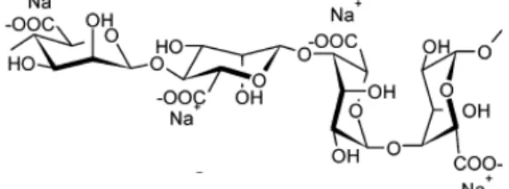
Chapter 2: Materials and Methods Figure 11 – Chemical structure of sodium alginate ... 25
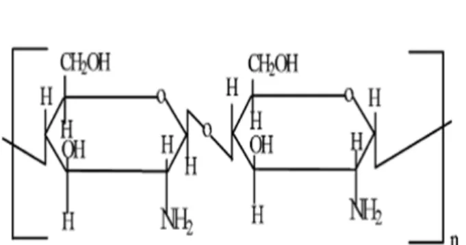
Figure 12 – Chemical structure of chitosan ... 26
Figure 13 – Dextran molecular structure... 26
Figure 14 – Reduction of MTS to formazan ... 28
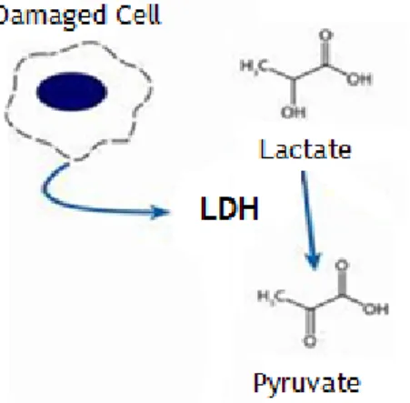
Figure 15 – Schematic representation of the mechanism of LDH activity ... 28
Chapter 3: Modeling of Drug Release from Polymeric Systems Figure 16 – Schematic representation of shrinking and swelling from particles ... 38
Figure 17 – Images of alginate and chitosan microparticles ... 39
Figure 18 – Scanning electron microscopy images of alginate and chitosan microparticles .... 40
Figure 19 – Images of freeze-drying oxidized dextran and dextran hydrogel with microparticles incorporated ... 41
Figure 20 – Microscopic photographs of human fibroblast cells after being seeded in contact with the carriers ... 42
Figure 21 – Cellular activities measured by the MTS assay ... 42
Figure 22 – Swelling ratio for alginate and chitosan microparticles ... 43
Figure 23 – Protein release from alginate and chitosan microparticles ... 44
Figure 24 – Simulation of BSA release from alginate microparticles ... 45
Figure 25 – Simulation of BSA release from chitosan microparticles ... 47
Figure 26 – BSA release from alginate and chitosan microparticles incorporated into dextran hydrogel ... 47
Figure 27 – Proposed model to describe the mass transfer process of the protein from the microparticles placed inside a hydrogel to the outside solution ... 49
Chapter 4: Drug Delivery Systems for Wound Healing Figure 28 – Scanning electron microscopy images of chitosan and of oxidized dextran ... 58
Figure 29 – Microscopic photographs of human fibroblasts cells after being seeded with carriers ... 60
xv Figure 30 – Scanning electron microscopy images of human fibroblast cells in contact with
carriers ... 60
Figure 31 – Cellular activities measured by the MTS assay ... 61
Figure 32 – LDH release activity ... 61
Figure 33 – Typical macroscopic wound-healing panorama over 21 days ... 63
Figure 34 – In vivo evaluation of wound healing process in different groups of animals ... 64
Figure 35 – Effect of oxidized dextran, chitosan microparticles and growth factors on burn wound ... 64
Figure 36 – Hematoxylin and eosin-stained sections of biopsies for the morphological evaluation of skin lesions after 7, 14 and 21 days ... 66
xvii
List of Tables
Chapter 1: IntroductionTable 1 – Comparison of the different layers of the skin ... 4 Table 2 – Tissue-engineered products for skin regeneration ... 16
Chapter 4: Drug Delivery Systems for Wound Healing
xix
List of Abbreviations
0
ρ
initial density of particles 0φ
initial polymer volumetric fractionφ
volume fraction of polymer ATP adenosine tryphosphate AAD adipic acid dihydrazideC
BSA bovine serum albumin
0 initial solute concentration inside the particles
CaCl2
CEA cultured epidermal autograft calcium chloride
Cm
C
concentration near the particle’s surface
out concentration in the bulk of the outside solution
D diffusion coefficient inside hydrogel
D0 diffusion coefficient inside hydrogel in free solution
DexOx+Ch oxidized dextran loaded with chitosan microparticles DexOx oxidized dextran
DexOx+EGF oxidized dextran loaded with chitosan microparticles containing endothelial growth factor
DexOx+VEGF oxidized dextran loaded with chitosan microparticles containing vascular endothelial growth factor
DexOx+VEGF/EGF oxidized dextran loaded with chitosan microparticles containing vascular endothelial growth factor and endothelial growth factor ECM extracellular matrix
DMEM Dulbecco’s modified Eagle’s médium
EGF epidermal growth factor
EDTA ethylenediaminetetraacetic acid
EtOH ethanol
FBS fetal bovine serum FGF fibroblast growth factor FDA Food and Drug Administration GAG glycosaminoglycan
HCl chloride acid
IGF insulin-like growth factor
ISO International Standard Organization K- negative control
K+ positive control Km
LDH lactate dehydrogenase mass transfer coefficient
xx MTS 3-(4,5-dimethylthiazol-2-yl)-2,5-diphenyl-2H-tetrazolium bromide MTT 3-(4,5-dimethylthiazol-2-yl)-2,5-diphenyltetrazolium bromide PDGF platelet-derived growth factor
PBS phosphate buffered saline PLA poly(lactic acid)
PLGA poly(lactide-co-glycolide) R
PMS phenazine methosulfate
calc
R
calculated percentage of protein release
exp experimentally obtained value
rf
R
radii of fibers
part,0
r
initial particle radius
s
S
hydrodynamic radius of the solute
0
SC stratum corneum
size of the full thickness circular skin wound area SEM scanning electron microscopy
S
SDS sodium dodecyl sulphate
N
sw swelling ratio
size of the wound area on the indicated day TBSA total body surface area
TGF transforming growth factor TEM transmission electron microscopy TPP tripolyphosphate
tsamp
V
sampling time
0
VEGF vascular endothelial growth factor
initial volume of the solution outside the particles
VEGF+EGF vascular endothelial growth factor and endothelial growth factor Vpart,0
v
initial total volume of particles
s,p
v
partial specific volumes of polymer
s,w
α ratio of moles of protein transferred due to swelling to the total partial specific volumes of water
number of moles transferred
Chapter 1
2
1.
Introduction
1.1. The Skin: structure and functions
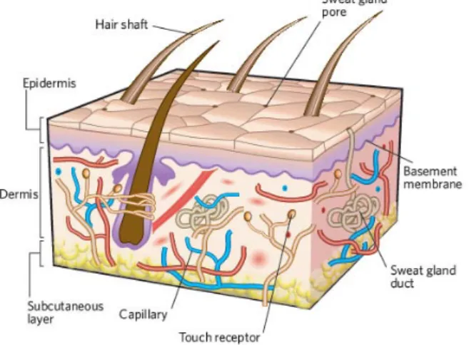
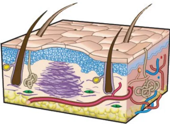
The skin is the largest organ of the body in vertebrates and occupies an area of about 2m2
Anatomically and functionally, skin has two distinctive layers: the epidermis and dermis (Figure 1), (Jones et al., 2002; Metcalfe and Ferguson, 2007a, b; Sheridan and Moreno, 2001). Some authors refer a third layer, the hypodermis or subcutaneous layer, that is mainly composed of fat and a layer of loose connective tissue (Metcalfe and Ferguson, 2007a, b).
, representing approximately one-tenth of the body mass (Balasubramani and RAVI, 2001; Metcalfe and Ferguson, 2007a). This complex organ has highly specialized functions. It protects the organism against toxins and microorganisms, allows regulation of body temperature, provides support to blood vessels and nerves, and prevents dehydration of all non-aquatic animals (Balasubramani and RAVI, 2001; Böttcher-Haberzeth et al., 2010; Clark et al., 2007; Rodrigues et al., 2008). Other critical functions of the skin are related with immune surveillance and sensory detection. Overall, skin is an indicator of general well being and health (Clark et al., 2007; Sachs and Voorhees, 2010). It can be considered an organ associated with physical attractiveness and beauty (Sachs and Voorhees, 2010).
1.1.1. Epidermis
Epidermis, the outermost layer, is relatively thin, 0.1-0.2mm in depth, and totally cellular, but has sufficient thickness to provide vital barrier function (MacNeil, 2008; Metcalfe and Ferguson, 2007a, b; Wong and Chang, 2009). This layer is avascularized and is fed by diffusion from the capillaries of the papillary layer from dermis (Seeley et al., 2003). This layer is primarily composed of stratified squamous epithelium of keratinocytes, which derives from neuroectoderm and comprises over ninety percent of epidermal cells (Wong and Chang, 2009). The keratinocytes produce a protein called keratin, making them responsible for the
3 structural strength and permeability characteristics of the epidermis (Paul and Sharma, 2004; Seeley et al., 2003). The epidermis is also composed of melanocytes that contribute to skin color, Langerhans cells that are part of the immune system, and Merkel cells (specialized epidermal cells associated with nerve endings) responsible for detecting the touch and pressure surface (Seeley et al., 2003; Wong and Chang, 2009).
The cells in the deeper layers of the epidermis are responsible for the constant cell renewal, since they have a higher rate of replication. As there are new cells, older ones are pushed to the surface where they slough off. During this movement, cells change their shape and chemical composition, passing to secrete and accumulate keratin, a process known as keratinization. This process allows the identification of different phases of transition. Based on these stages, the different cells of the epidermis are divided into strata or layers, which are classified into: cornea, translucent, granular, spinous and basal (the top layer to the deepest) (Table 1) (Seeley et al., 2003).
1.1.2. Dermis
The dermis, or middle layer, is a connective tissue situated directly below the epidermis (Metcalfe and Ferguson, 2007a, b; Paul and Sharma, 2004; Wong and Chang, 2009). This layer is responsible for the elasticity and mechanical integrity of the skin, and contains blood vessels that are responsible for the nutrition of the epidermal layer (Jones et al., 2002). It contains receptors for touch, temperature and pain, as well as hair follicles and sweat ducts (MacNeil, 2008). This skin layer is composed of collagen (type I and III), the main type of fiber of your conjunctive tissue, with some elastin and glycosaminoglycans (GAGs). Fibroblasts are the major cell type present in the dermis. They are capable of producing remodeling enzymes such as proteases and collagenases, which play an important role in the wound healing process (Metcalfe and Ferguson, 2007a, b; Wong and Chang, 2009).
The two layers, epidermis and dermis, are connected by a basement membrane that is composed of various integrins, laminins, collagens, and other proteins that make important roles in regulating epithelial-mesenchymal cross-talk (Wong and Chang, 2009).
The dermis is divided into two layers: a deeper layer called reticular and a superficial layer nominated papillary. The reticular layer, the main component of the dermis, is composed of dense and irregular tissue, composed with a set of irregularly arranged fibers that are resistant to stretching in many directions, and is continuous with the hypodermis. Collagen and elastin fibers are, predominantly, oriented in a certain direction and cause lines of tension in the skin (Seeley et al., 2003). In turn, the papillary layer owes its name due to the extensions called papillae that point toward the epidermis. It also contains numerous blood vessels that supply nutrients to the overlying epidermis, removes waste products and help to regulate the body temperature (Seeley et al., 2003; Wong and Chang, 2009).
4
1.1.3. Hypodermis
The hypodermis, which has a deeper localization to the dermis, contains adipose tissue that is well vascularized and contributes to both the mechanical and the thermoregulatory properties of the skin. It separates the dermis from the underlying muscular fascia (Metcalfe and Ferguson, 2007a, b; Wong and Chang, 2009). This skin layer is composed of lax connective tissue with collagen and elastin fibers. The main types of cells formed in the hypodermis are fibroblasts, adipose cells and macrophages. This layer is often regarded as not being part of the skin, being sometimes called the subcutaneous tissue or superficial fascia (Seeley et al., 2003).
About half of the fat stored in the body is found in the hypodermis, although their number and location vary with age, sex and feeding (Seeley et al., 2003).
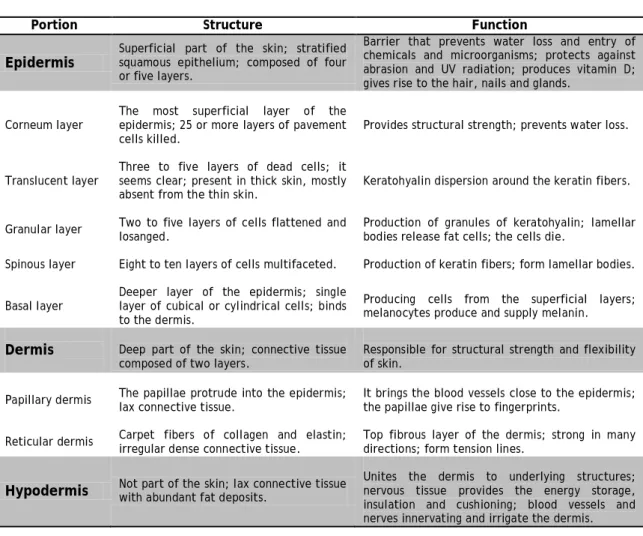
Table 1 – Comparison of the epidermis and dermis with the hypodermis (adapted from (Seeley
et al., 2003)).
Portion Structure Function
Epidermis Superficial part of the skin; stratified squamous epithelium; composed of four or five layers.
Barrier that prevents water loss and entry of chemicals and microorganisms; protects against abrasion and UV radiation; produces vitamin D; gives rise to the hair, nails and glands.
Corneum layer The most superficial layer of the epidermis; 25 or more layers of pavement
cells killed. Provides structural strength; prevents water loss.
Translucent layer Three to five layers of dead cells; it seems clear; present in thick skin, mostly
absent from the thin skin. Keratohyalin dispersion around the keratin fibers. Granular layer Two to five layers of cells flattened and losanged. Production of granules of keratohyalin; lamellar bodies release fat cells; the cells die. Spinous layer Eight to ten layers of cells multifaceted. Production of keratin fibers; form lamellar bodies. Basal layer Deeper layer of the epidermis; single layer of cubical or cylindrical cells; binds
to the dermis.
Producing cells from the superficial layers; melanocytes produce and supply melanin.
Dermis Deep part of the skin; connective tissue
composed of two layers. Responsible for structural strength and flexibility of skin. Papillary dermis The papillae protrude into the epidermis;
lax connective tissue. It brings the blood vessels close to the epidermis; the papillae give rise to fingerprints. Reticular dermis Carpet fibers of collagen and elastin; irregular dense connective tissue. Top fibrous layer of the dermis; strong in many directions; form tension lines.
Hypodermis Not part of the skin; lax connective tissue with abundant fat deposits. Unites the dermis to underlying structures; nervous tissue provides the energy storage, insulation and cushioning; blood vessels and nerves innervating and irrigate the dermis.
5
1.2. Wounds: definition and classifications
As previously mentioned, skin is a complex tissue which provides an essential water, electrolyte, and bacteria-boundary to the outside world (MacNeil, 2008; Shaw and Martin, 2009a). Due to the various functions of the skin, any loss of skin integrity due to injury or illness, in addition to causing physical, mechanical and thermal damage, may also affect the physiological functions, resulting in disorders of the organism and ultimately in significant disability or even death (Clark et al., 2007; Rodrigues et al., 2008). Thus, skin disruption often leads to an increase in fluid loss, infection, scarring, compromised immunity and change in body image (Alemdaroglu et al., 2006). Many reasons can be pointed out for the loss of skin integrity, such as genetic disorders (bullous conditions), acute trauma, chronic wounds (e.g. venous, diabetic and pressure ulcers) or even surgical interventions (Dai et al., 2004; Shevchenko et al., 2010). However, thermal trauma, like burns, is one of the most common cause for major skin loss, where substantial areas of skin can be damaged and the possibility of skin regeneration is unlikely (Shevchenko et al., 2010). The mortality rate caused by burns has been decreasing since the last two decades with the advances in regenerative medicine. However it is still high if more than 70% of the total body surface area (TBSA) is injured or burned (Alemdaroglu et al., 2006).
According to the US Wound Healing Society, a wound can be described as a result of “disruption of normal anatomic structure and function” of the skin, resulting from physical or thermal damage or as a consequence of the presence of an underlying medical or physiological condition (Boateng et al., 2008). Wounds can be classified based on the nature of the repair process, on the number of skin layers and area of skin affected. Based on the type of the repair process, wounds can be classified as acute or chronic wounds (Boateng et al., 2008; Paul and Sharma, 2004).
Acute wounds are characterized by their ability to heal completely with minimal scarring within the expected time frame, normally 8-12 weeks (Boateng et al., 2008). The newly formed tissue has a similar structure and comparable functions to intact skin, however, its regeneration is uncommon (with notable exceptions such as early fetal healing) (Li et al., 2007). Mechanical injuries, due to external factors such as abrasions which are caused by frictional contact between the skin and hard surfaces, are the primary causes of acute wounds (Boateng et al., 2008; Li et al., 2007). Other examples of mechanical injuries include penetrating wounds caused by knives, gun shots and surgical wounds. Burns and chemical injuries (caused by radiation, electricity, corrosive chemicals and thermal sources) are another categories of acute wounds (Boateng et al., 2008). Chronic wounds represent a different kind of challenge for wound healing (Supp and Boyce, 2005). These are characterized by their slow healing, i.e., that have not healed beyond 12 weeks and often reoccur. Repeated tissue insults or underlying physiological conditions such as diabetes and other malignances, persistent infections, poor primary treatment and other patient related
6 factors, contributes to the disability of these wounds to heal faster (Boateng et al., 2008). These wounds involve a large surface area and have a high incidence in the general population, featuring an enormous medical and economic impact (Supp and Boyce, 2005). Pressure ulcers and leg ulcers (venous, ischaemic or of traumatic origin) are known as the most common chronic wounds (Boateng et al., 2008; Supp and Boyce, 2005).
Based on the number of layers and the area of affected skin, wounds can be divided into: epidermal, superficial and deep partial-thickness, and full-thickness ones (Paul and Sharma, 2004; Shevchenko et al., 2010). Epidermal injuries are normally caused by sunburns, light scalds or grazing, characterized by erythema and minor pain. In this type of lesions, only the epidermis is affected. As so, such injuries do not require specific surgical treatment and regenerate rapidly without any scarring (Shevchenko et al., 2010). Superficial partial-thickness wounds affect the epidermis and superficial parts of the dermis, with epidermal blistering and severe pain accompanying this type of injury, especially in the case of thermal trauma. The wound healing in this type of wounds is featured by epithelialization from the margins of the wound where basal keratinocytes change into a proliferating migratory cell type and cover the damaged area (Shevchenko et al., 2010). In the other hand, deep partial-thickness injuries involve greater dermal damaged that results in fewer skin appendages remaining and, therefore, they take longer to heal. Scarring and fibroplasia are more pronounced and intensive in this depth of injury when compared with superficial partial-thickness wounds (Shevchenko et al., 2010). Finally, in full-partial-thickness injuries all epithelial-regenerative elements are completely destroyed. Wound healing is characterized by contraction, with epithelialization from only the edge of the wound, leading to cosmetic and functional defects. When the wound has more than 1 cm in diameter a skin grafting is usually required. These types of wounds cannot epithelialize on their own and may lead to extensive scarring, resulting in limitations in joint mobility and severe cosmetic deformities (Shevchenko et al., 2010).
1.3. Wound Healing
The wound healing process is extremely dynamic, interactive and difficult. It involves complex interactions of extracellular matrix (ECM) molecules, soluble mediators, various resident cells, and infiltrating leukocyte subtypes which, together, act to reestablish the integrity of damaged tissue and replace the lost one (Boateng et al., 2008; Branski et al., 2006; Eming et al., 2007). If this response is successful and the injury does not cause organism dead, these processes must be shut down in a precise sequence in the ensuing days for the recovery takes place (Gurtner et al., 2008).
It can be considered that a “healed wound” is one in which the connective tissues have been repaired, the wound is completely re-epithelialized and the skin has returned to its
7 normal anatomical structure and function without the need for continued drainage or dressing (Beldon, 2010; Enoch and Leaper, 2008).
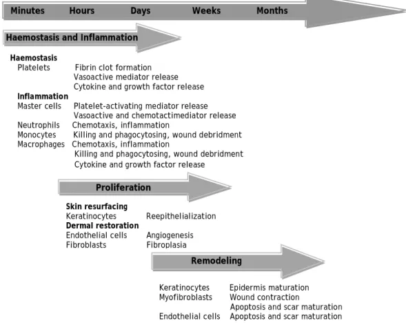
In order to achieve the primary goal of wound healing (tissue integrity and haemostasis), there are five overlapping stages that need to be considered: haemostasis, inflammation, migration, proliferation and maturation (Boateng et al., 2008). These can be summarized in three main phases like haemostasis and inflammation, new tissue formation (proliferation) and tissue remodeling as shown in Figure 2 (Eming et al., 2007; Gurtner et al., 2008).
In contrast, embryonic wound healing is essentially a regenerative process, characterized by the absence of scarring and fibrosis (Böttcher-Haberzeth et al., 2010; Metcalfe and Ferguson, 2007b). Embryonic wounds exhibit a number of major differences from adult ones which scar, lack of fibrin clots and platelet degranulation, markedly reduce inflammatory response and distinctly elevate levels of molecules involved in skin morphogenesis and growth (Metcalfe and Ferguson, 2007b).
Keratinocytes Epidermis maturation Myofibroblasts Wound contraction
Apoptosis and scar maturation Endothelial cells Apoptosis and scar maturation
Skin resurfacing
Keratinocytes Reepithelialization
Dermal restoration
Endothelial cells Angiogenesis Fibroblasts Fibroplasia
Haemostasis
Platelets Fibrin clot formation
Vasoactive mediator release Cytokine and growth factor release Inflammation
Master cells Platelet-activating mediator release
Vasoactive and chemotactimediator release Neutrophils Chemotaxis, inflammation
Monocytes Killing and phagocytosing, wound debridment Macrophages Chemotaxis, inflammation
Killing and phagocytosing, wound debridment Cytokine and growth factor release
Figure 2 – Schematic representation of the main phases of wound healing, the major cell types and their
effects (adapted from (Li et al., 2007)).
Minutes Hours Days Weeks Months
Proliferation
Remodeling Haemostasis and Inflammation
8
1.3.1 Haemostasis and Inflammation
Unless there is a severe arterial hemorrhage, haemostasis is achieved initially by the formation of a platelet plug, followed by a fibrin matrix, which becomes the scaffold for infiltrating cells (Auger et al., 2009; Gurtner et al., 2008; Ousey and McIntosh, 2009; Strodtbeck, 2001). The platelets degranulate and release their alpha granules, which secrete several growth factors, such as: platelet-derived growth factor (PDGF), insulin-like growth factor-1 (IGF-1), epidermal growth factor (EGF), transforming growth factor-β (TGF-β) and platelet factor-IV. They also contain dense bodies that store vasoactive amines, like serotonin, which increase the microvascular permeability (Enoch and Leaper, 2008). Owing to growth factors, the wound healing cascade is initiated by attracting and activating fibroblasts, endothelial cells and macrophages (Enoch and Leaper, 2008). Subsequently, four major amplification systems are activated (complement cascade, clotting mechanism, kinin cascade, plasmin generation), which contribute to haemostasis and the other staged of healing process. Finally, the clot (comprising fibrin, fibronectin, vitronectin, von Willbrand factor, thrombospondin) provides the provisional matrix for cell migration (Enoch and Leaper, 2008).
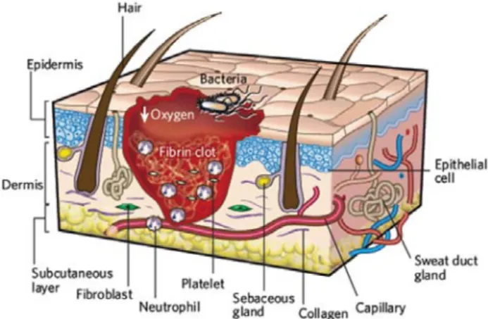
The inflammatory phase begins almost simultaneously with haemostasis, sometimes from within a few minutes of injury to 24h, and lasts for about 3 days. It plays a central role in wound healing, not only protecting the wound from invading microbes, but also by participating in the tissue repair processes (Boateng et al., 2008; Shaw and Martin, 2009b; Tsirogianni et al., 2006). In this phase, components of the coagulation cascade, inflammatory pathways and immune system are needed to prevent ongoing blood and fluid losses, to remove dead and dying tissues and to avoid infection (Figure 3) (Gurtner et al., 2008).
Figure 3 – First stage of wound repair: haemostasis and inflammation. The process of inflammation lasts
until 48h after injury. The wound is characterized by a hypoxic (ischaemic) environment in which a fibrin clot has formed. Bacteria, neutrophils and platelets are abundant in the wound. Hair follicles and sweat duct glands, the normal skin appendages, are still present in the skin outside the wound (adapted from (Gurtner et al., 2008)).
9 Inflammation can be divided into two stages (early and late inflammatory phases) depending either on the time and duration of the response and the type of inflammatory cell involved (Enoch and Leaper, 2008; Li et al., 2007). In the early inflammatory phase (days 1-2), neutrophil granulocytes (polymorphonuclear leukocytes) are recruited to the wound in response to the activation of complement, the degranulation of platelets and the products of bacterial degradation, such as: fragments of ECM protein, TGF-β, complement components (e.g. C3a, C5a) and formyl-methionyl peptide products from bacteria (Enoch and Leaper, 2008; Gurtner et al., 2008). The main function of polymorphonuclear leukocytes is to minimize bacterial contamination of the wound, thus preventing infection (Enoch and Leaper, 2008). After 2-3 days in the late inflammatory phase, monocytes appear in the wound area and differentiate into macrophages (Enoch and Leaper, 2008; Gurtner et al., 2008). Monocytes are attracted to the wound by a variety of chemoattractants like complement, clotting components, fragments of immunoglobulin G, breakdown products of collagen and elastin and cytokines (Enoch and Leaper, 2008). Macrophages are thought to be crucial for coordinating later events in response to injury and they function as phagocytic cells. They are the primary producers of growth factors responsible for the proliferation and production of the ECM by stimulating fibroblasts, endothelial and smooth muscle cells (Enoch and Leaper, 2008; Gurtner et al., 2008). These cells of the immune system also release proteolytic enzymes (e.g. collagenase) that help to debride the wound (Enoch and Leaper, 2008). Neutrophils are known to produce vascular endothelial growth factor (VEGF), while macrophages can also release PDGF, EGF, TGF-α, TGF-β in addition to IGF (Auger et al., 2009).
At the end of this phase, collagen fibers are vertically oriented and do not connect the incision’s edges; epithelial cell proliferation continues, yielding a thickened epidermal covering layer (Enoch and Leaper, 2008).
1.3.2. New tissue formation
The second stage of wound healing, which takes place 2-10 days after injury, is characterized by fibroblast migration, deposition of the ECM and formation of granulation tissue (Figure 4) (Branski et al., 2006; Enoch and Leaper, 2008; Gurtner et al., 2008).
This phase starts with the migration of keratinocytes over the injured dermis (Branski et al., 2006; Gurtner et al., 2008). Then, new blood vessels are formed (angiogenesis), and the sprouts of capillaries associated with fibroblast and macrophages restore the fibrin matrix with granulation tissue, thus forming a new substrate for keratinocyte migration at later stages of the repair process (Gurtner et al., 2008). These cells proliferate, mature and restore the barrier function of the epithelium (Gurtner et al., 2008). In the last part of this stage, fibroblasts are attracted to the wound by a number of factors, including PDGF and TGF-β, and some differentiate into myofibroblasts, a type of contractile cells that, over time, bring the edges of the wound together (Enoch and Leaper, 2008; Gurtner et al., 2008).
10 Fibroblasts and myofibroblasts interact, proliferate and produce the ECM proteins fibronectin, hyaluronan and, later, collagen and proteoglycans (Enoch and Leaper, 2008; Gurtner et al., 2008). Fibroblasts also secrete fibroblast growth factor (FGF) that can, along with the VEGF secreted by platelets and neutrophils, act as an angiogenic factor to stimulate endothelial cell proliferation and migration, thus promoting vascularization at the healing site (Auger et al., 2009). Epithelialization of the wound represents the final stage of the proliferative phase (Enoch and Leaper, 2008).
1.3.3. Remodeling
Remodeling is the last step of normal acute wound healing, also called maturation, during which all of the processes activated after injury cease. It begins 2-3 weeks after injury and lasts for a year or more (Auger et al., 2009; Gurtner et al., 2008). During this stage, the ECM components are modified by the balanced mechanisms of proteolysis and new matrix formed, and wound becomes gradually less vascularized (Auger et al., 2009). Most of endothelial cells, macrophages and myofibroblasts undergo apoptosis or exit from the wound, leaving a mass that contains few cells and consists mostly of collagen and other ECM components (Figure 5) (Gurtner et al., 2008). Although the initial granulation tissue is weak, during this stage, it will eventually gain strength over time due to the gradual replacement of immature type III collagen by mature type I collagen (Auger et al., 2009; Gurtner et al., 2008). This process is carried out by matrix metalloproteinases that are secreted by fibroblasts, macrophages and endothelial cells (Gurtner et al., 2008).
Further remodeling of the wound causes a decrease in the activity of metalloproteinases, an increase in activity of tissue inhibitors of enzymes, reduction in the density of macrophages and fibroblasts, stop in the outgrowth of capillaries, reduction in the blood flow and metabolic activity and decrease in size of the underlying contractile
Figure 4 – Second stage of wound repair: new tissue formation. It is characterized by the formation of
an eschar (scab). Most cells from the previous stage of repair have migrated from the wound, and new blood vessels grew. Epithelial cells under the eschar can be observed (adapted from (Gurtner et al., 2008)).
11 connective tissue (Enoch and Leaper, 2008). Finally, the granulation tissue evolves into an avascular scar composed of inactive fibroblasts, fragments of elastic tissue, dense collagen and other components of the ECM. As the scar matures, fibronectin and hyaluronan are broken down and collagens bundles rise in diameter, increasing the tensile strength of the wound (Enoch and Leaper, 2008). However, the tissue never recover the properties of uninjured skin (Enoch and Leaper, 2008; Gurtner et al., 2008).
Sometimes, due to local and systemic factors, the normal process of healing is disrupted and this process can be divided into two types: non-healing or chronic wounds (as previously described in the text) and excessive wound healing (Enoch and Leaper, 2008). In cases of excessive wound healing, hypertrophic scars and keloids are formed resulting from overproduction of all components of the healing process, such as fibroblasts, collagen, elastin and proteoglycans (Enoch and Leaper, 2008). Hypertrophic scars are elevated and may fall down with time, while keloids continue to grow behind the margin of the original wound, like a benign tumor. Other type of scars can be defined as atrophic ones, that cause a valley or depression in the skin (Metcalfe and Ferguson, 2007b). All types of scarring can occur on all areas of the body, but some of them such as chest, knees and elbows are more susceptible to scarring (Metcalfe and Ferguson, 2007b). The main feature of the phenomenon scarring is called wound contraction. This is caused, at least in part, by the presence of myofibroblasts which develop characteristics of smooth muscles cells under the influence of TGF-βs (Böttcher-Haberzeth et al., 2010).
Figure 5 – Third stage of wound repair: remodeling. This stage lasts for a year or longer. It can be
observed that disorganized collagen has been laid down by fibroblasts. The wound has contracted near its surface. The re-epithelialized wound is slightly higher that the surrounding surface; the healed region doesn’t have normal skin appendages (adapted from (Gurtner et al., 2008)).
12
1.4. Biomaterials and Tissue Engineering
Tissue engineering is emerging as an interdisciplinary field in biomedical engineering that integrates many concepts of science and engineering in order to design and develop biological substitutes that restore, maintain, or improve damaged tissues and organs functions (Clark et al., 2007; Desai, 2000; Harrison and Atala, 2007; Metcalfe and Ferguson, 2007b; Norman and Desai, 2006; Zhang and Webster, 2009). It has appeared as a solution to the a number of clinical problems that were not properly treated with the use of permanent replacement devices (Huebsch and Mooney, 2009). Major advances in transplantation of cells and organs, as well as in materials science and engineering have contributed for the continuous development of tissue engineering and regenerative medicine (Desai, 2000; Harrison and Atala, 2007; Norman and Desai, 2006).
Usually, there are three aspects considered in the production of any device for tissue engineering – the cells, the scaffold or biomaterial, and cell-material interaction: isolation of the cells from the patient by a small biopsy and their multiplication by cell culture techniques; amplification of the cell number and impregnation of these cells into a biodegradable scaffold or biomaterial; introduction of the construct cell-biomaterial into a functional tissue mass (Desai, 2000; Eisenbarth, 2007). It is know that the extracellular environment influences cell behavior in relation to morphology, cytoskeletal structure and function. Thus, when studying cell function and development of tissue substitute, it is important to reproduce as much as possible, the size, configuration and composition of the environment of cells, creating physiologically relevant models of cells and tissues (Desai, 2000; Ito et al., 2005).
Biomaterials, traditionally defined as materials used in medical devices, have been used since antiquity, but recently their degree of sophistication has increased significantly (Huebsch and Mooney, 2009). It gives the cells a temporary structure in which they further proliferate and differentiate in order to build the injured tissue, maintains the shape of the defect until the reconstruction process is finished and prevents the infiltration of the tissue in contact with the defect (Eisenbarth, 2007). The biomaterials used in tissue engineering applications are used in the form of carriers, hydrogels or scaffolds, and they can be derived from biological or synthetic origins (Eisenbarth, 2007; Lutolf and Hubbell, 2005). Usually, materials from natural sources (polypeptides, hydroxyapatites, hyaluronan, GAGs, fibronectin, collagen, chitosan, alginate and dextran) are advantageous because of their inherent properties of biological recognition, including presentation of receptor-binding ligands and susceptibility to cell-triggered proteolytic degradation and remodeling. Despite of these advantages, many issues have led the development of synthetic biomaterials as cellular substrates, including complexities associated with purification, immunogenicity and pathogen transmission. Examples of these materials include polyglycolide, polylactide, polytetrafluoroethylene and others (Lutolf and Hubbell, 2005; Metcalfe and Ferguson, 2007b).
13 A major disadvantage of synthetic materials is the lack of cell-recognition signals. However, new manufacturing processes are now being developed, which will allow the incorporation of cell-adhesion peptides into biomaterials, that are known to be involved in cellular interactions (Metcalfe and Ferguson, 2007b).
The biomaterials must have favorable properties in order to promote cell adhesion, proliferation and differentiation for the formation of the desired tissue (Eisenbarth, 2007; Huebsch and Mooney, 2009; Roach et al., 2007):
a. They should present similar mechanical properties to the injured tissue;
b. Must enable or support cell metabolism that build up a new tissue with suitable surface chemistry for cell attachment, proliferation and differentiation;
c. Have a three dimensional structure with interconnective pores for cell growth and nutrients flow. The substructure must be vascular supportive for almost all tissues and has to maximize the space for cellular adhesion, growth, ECM secretion, revascularization and adequate nutrient and oxygen supply;
d. It must be biodegradable along with the reconstruction of the newly build tissue and the products resultant of degradation must not affect the tissue regeneration and remodeling process;
e. It must be biocompatible. Biocompatibility is dependent on the site of implantation, the function and size of the implant and the duration of implantation with a key issue being the time-scale required for material-host tissue interactions to become established.
1.4.1. The role of biomaterials in skin regeneration
Large full-thickness skin defects, with more than 4 cm in diameter, resulting from burns, soft tissue trauma and disease leading to skin necrosis, will not heal well without a graft and represent a significant clinical problem that is far from being solved (Böttcher-Haberzeth et al., 2010; MacNeil, 2007). In the past, wounds were treated by allowing them to dry and hence, acquire a hard protective coating, the scab (Dos Santos et al., 2006). About 30 years ago, the wounds treatment was revolutionized with the discovery that a wound would heal faster when a moist dressing was applied rather than the traditional dry dressings such as gauze type materials produced from cotton or lint (Dos Santos et al., 2006). Other reasons to develop a material capable of promote skin regeneration are the following two: first, it is difficult to get autologous skin transplants when the defect exceeds 50-60% of the TBSA; second, most conventional skin grafting techniques in order to provide autologous defect coverage are based on transplanting split-thickness skin which is comprised throughout the epidermis but only for a portion of the dermis, and that frequently leads to scarring (Böttcher-Haberzeth et al., 2010; MacNeil, 2008). These problems could be reduced with the development of dermo-epidermal skin substitutes (Böttcher-Haberzeth et al., 2010).
14 There are many types of skin substitutes and they can be classified in a number of ways depending on their function in the wound (debridement, antibacterial, occlusive, absorbent, adherence), type of material used to produce the dressing (e.g. hydrocolloid, alginate, collagen), their origin (synthetic or natural), the physical form of the dressing (ointment, film, foam, gel) and according to the presence or absence of living cells (the use of autologous, allogeneic or xenogenic cells or tissues) (Auger et al., 2009; Boateng et al., 2008). They can also be classified into primary, secondary and island dressings (Boateng et al., 2008). Primary dressings are the ones that are in physical contact with the wound, while secondary dressings cover the primary ones. Island dressings possess a central absorbent region which is surrounded by an adhesive portion (Boateng et al., 2008).
The concern for the development of skin substitutes to improve wound healing is valid for extensive wounds that cannot heal spontaneously, since they are deep and do affect a high percentage of TBSA, and for smaller wounds which cannot heal due to inflammation or a deficiency in the other steps of wound healing process. Dressings can also be used in wounds that would heal by themselves, however very slowly, and the objective is a faster and/or better quality of healing (Auger et al., 2009).
Three factors are of paramount importance in the development of tissue-engineered materials: the safety of the patient, clinical efficacy and convenience of use (MacNeil, 2007; MacNeil, 2008). Tissue-engineered skin substitutes need to provide a barrier layer of renewable keratinocytes (the cells that form the upper barrier layer of skin), which is firmly bound to the underlying dermis. They must also attach well to the wound bed, maintain a moist environment at the wound/dressing interface, absorb the excess exudates without leakage to the surface of the dressing, be supported by new vasculature, provide thermal insulation, mechanical and bacterial protection, allow gaseous and fluid exchange, absorb wound odor, be non-adherent to the wound and easily removed without trauma, not be rejected by the immune system and be capable of self repair throughout a patient’s life (Eldin et al., 2010; MacNeil, 2007; MacNeil, 2008). It should also have the ability to replace all the structures and functions of skin (Supp and Boyce, 2005).
The first milestones in skin research and skin tissue engineering focused on the enzymatic separation of the epidermis and dermis and the in vitro culture of keratinocytes (Böttcher-Haberzeth et al., 2010). Rheinwald and Green, in 1975, manipulated the growth of human primary epidermal cells on a layer of lethally irradiated 3T3 murine fibroblasts. After the first clinical application, cultured epidermal autografts (CEAs) were tested in almost all leading burn centers worldwide (Böttcher-Haberzeth et al., 2010; Green and Rheinwald, 1975; MacNeil, 2007). After that, Bell et al (1981) generated a dermo-epidermal substitute which was tested in an animal model and, subsequently, transformed into the product Apligraft® (Table 2) (Bell et al., 1981). This substitute contains both allogeneic fibroblasts and keratinocytes derived from neonatal foreskin (Böttcher-Haberzeth et al., 2010; Supp and Boyce, 2005). Another useful dermo-epidermal substitute is Integra®, a bilayered artificial
15 skin, composed of porous bovine collagen and chondroitin-6-sulfate glycosaminoglycan with a silicone membrane that functions as a temporary carrier (Böttcher-Haberzeth et al., 2010; MacNeil, 2007; Supp and Boyce, 2005). Futhermore, donor (cadaveric) skin is commonly used for managing serious burns and treating chronic wounds. Donor skin is currently an underused resource that could be especially valuable in the developed world (MacNeil, 2007). However, skin allografts isolated from cadavers, provide only temporary coverage because of host rejection (Clark et al., 2007). The most effective treatment will be based on the decellularization of each tissue and organ and it is dependent of many factors, including the tissue’s cellularity, density, lipid content and thickness. It should be understood that every cell removal agent and method will alter ECM composition and cause some degree of ultrastructure disruption (Crapo et al., 2011). Triton X-100 and sodium dodecyl sulfate (SDS) are two typical detergents used to remove cells from tissues. SDS is typically more effective to remove cell residues from tissue compared to other detergents, but it is also more disruptive to ECM (Badylak et al., 2010; Crapo et al., 2011).
However, the most advanced skin substitute still do not incorporate many of the innate features of native skin such as glands, dermal microvascularization, pilosity and others specialized cells, responsible for the perception of heat, cold, pressure, vibration and pain. Other factor is the skin pigmentation that is not only a matter of aesthetics and quality of life, but also it provides protection against the UV radiation (Auger et al., 2009). For example, some researchers concluded that skin pigmentation can be controlled by keratinocytes and by the addition of melanocytes into skin substitutes (Auger et al., 2009). It is also possible to improve nerve regeneration in wound healing by controlling the amount of laminin present in the skin substitute (Auger et al., 2009). Other important factor in skin substitutes is that they can contribute for wound vascularization, preventing necrosis and loss of the epithelial covering. Some studies indicate that endothelial cells can spontaneously form into capillary-like structures and microvascular networks when cocultured with fibroblasts.
Another approach to improve vascularization is the use of angiogenic recombinant factors like VEGF and FGF. With the same idea, other factors such as PDGF or products like platelet-rich plasma (PDGF, EGF, VEGF, IGF-1 and TGF-β) can promote wound healing when added to the skin substitutes matrix improving the speed and the quality of healing (Auger et al., 2009). Finally, the emerging field of regenerative medicine aims to develop skin substitutes that can promote skin regeneration without scarring. It is believed that the inclusion of some factors and cells responsible for scar less healing in an embryo (e.g. TGF-β3) into collagen composite scaffolds, will optimize the healing process by providing an aesthetic and functional skin (Auger et al., 2009; Balasubramani and RAVI, 2001).
16
Table 2 – Tissue-engineered products, with or without living cells (Auger et al., 2009;
Böttcher-Haberzeth et al., 2010; MacNeil, 2008; Pham et al., 2007; Supp and Boyce, 2005).
Commercial Product Description Application
Epidermal substitutes
Epicel® Cultured epidermal autograft (autologous keratinocytes grown in the presence of murine
fibroblasts)
Full- and partial-thickness burns and chronic ulcers treatment
Epidex® Cultured epidermal autograft (autologous outer root sheet hair follicle cells) Full- and partial-thickness burns and chronic ulcers treatment
Laserskin® (Vivoderm®) Subconfluent autologous keratinocytes seeded on esterified laser-perforated hyaluronic acid matrix Full- and partial-thickness burns and chronic ulcers treatment BioSeed-S® Autologous oral mucosal cells on a fibrin matrix Partial-thickness burns and chronic ulcers treatment
Myskin® Cultured epidermal autograft (autologous keratinocytes grown in the presence of irradiated
murine fibroblasts)
Partial-thickness burns and chronic ulcers treatment
CellSpray® Preconfluent autologous keratinocytes delivered into a suspension for spray Partial-thickness burns and chronic ulcers treatment
Dermal substitutes
Dermagraft® Bioabsorbable polyglactin mesh scaffold seeded with human allogeneic nenonatal fibroblasts Full-thickness diabetic foot ulcers treatment
Alloderm® Acellular donated allograft human dermis Full- and partial-thickness wounds treatment
EZ-Derm® Aldehyde-crosslinked porcine dermal collagen Full- and partial-thickness wounds treatment
Repliform® Acellular human dermal allograft Urological plastic surgery applications
Cymetra® Micronized particulate acellular cadaveric dermal matrix Wound filler in plastic surgery
Biobrane® Porcine collagen chemically bound to silicone/nylon membrane Temporary covering of partial-thickness burns and wounds
Hyalograft 3D® Esterified hyaluronic acid matrix seeded with autologous fibroblasts Full- and partial-thickness wounds treatment
Matriderm® Bovine dermal collagen type I, III, V and elastin Full- or partial-thickness wounds treatment
ICX-SKN® Allogeneic fibroblasts set in a natural human collagen matrix Phase II trial pending
Bilayer substitutes
Integra® Thin polysiloxane (silicone) layer; cross-linked bovine tendon collagen type I and shark
glycosaminoglycan (chondroitin-6-sulfate)
Full- or partial-thickness wounds treatment
OrCel® Human allogeneic neonatal keratinocytes on gel-coated non-porous side of sponge; bovine collagen
sponge containing human allogeneic neonatal fibroblasts
Treat skin graft donor sites and mitten-hand surgery for epidermolysis bullosa
Apligraf®
Human allogeneic neonatal keratinocytes; bovine collagen type I containing human allogeneic neonatal fibroblasts
Venous and diabetic foot ulcers treatment
TissueTech® autograft system
Combination of Hyalograft 3D® and Laserskin® Full- and partial-thickness burns and chronic ulcers treatment
17
1.5. The Role of Growth Factors in Wound Healing
As previously stated, regeneration is characterized by a constant change in the environment to which cells are exposed to (Metcalfe and Ferguson, 2007b). Thus, the regeneration of any type of tissue requires an interaction between different phenotypes of cells, local and systemic functioning mediators such as growth factors and hormones, and the ECM within which these events occur (Chen et al., 2006). Growth factors are proteins that induce a change in the cellular function by inducing proliferation or differentiation. They play a role in the modulation of tissue growth and development (Karakeçili et al., 2008) and bind to specific high-affinity receptors on the cell-surface to stimulate cell growth (Alemdaroglu et al., 2006). Exogenous application of growth factors has been extensively used for the treatment of chronic wounds, where growth factors are present in lower concentrations (Branski et al., 2006). Despite such clear rationale suggesting the probability of enhancing wound healing trajectories with topical growth factor application, clinical success has been limited when a single growth factor is applied. This is due to the fact that the application of a single growth factor only has a transient effect in the enhancement of wound healing (Branski et al., 2006).
Among the various growth factors involved in skin regeneration, EGF and VEGF play an important role in this process. VEGF is a multifunctional molecule with several important biological activities that depend on both the stage of development and physiological function of the organ in which it is expressed (Breen, 2007). VEGF has many beneficial functions involved in regulating physiological angiogenesis associated with exercise and metabolism, ovarian follicular development and function, and wound healing (Breen, 2007). It acts as an endothelial cell mitogen, chemotactic agent, induce vascular permeability, maintenance and protection of both vascular endothelial and non-endothelial cells in mature mammals (Bao et al., 2009; Breen, 2007). Compared with other angiogenic growth factors (e.g. bFGF and TGF-β) VEGF is unique for its effects on multiple components of the wound-healing cascade; it promotes the closure of chronic wounds exhibiting hypoxia and compromised vascularity (Bao et al., 2009; Barrientos et al., 2008; Metcalfe and Ferguson, 2007a, b).
As previously described, VEGF is produced by many cell types that participate in wound healing mechanism, such as endothelial cells, fibroblasts, smooth muscle cells, platelets, neutrophils, and macrophages (Bao et al., 2009). The addition of VEGF alone or in combination with other growth factors may be important for the treatment of nonhealing wounds and other ischemic processes. It may also have a future role as adjunctive therapy to accelerate healing and prevent combinations in surgical revascularization, anastomoses and plastic surgeries (Bao et al., 2009).
EGF is a small polypeptide of 53 amino acid residues that is secreted by platelets, macrophages and fibroblasts. It acts in a paracrine fashion on keratinocytes (Alemdaroglu et al., 2006; Barrientos et al., 2008; Karakeçili et al., 2008). It has been implicated in wound healing and homeostasis in a number of tissues including colon, skin, mammary gland and
18 liver (Metcalfe and Ferguson, 2007b). It is also known to be present at high levels in saliva and may accelerate healing of skin lesions in animals when they lick their wounds (Metcalfe and Ferguson, 2007b). In vivo studies have shown that EGF is effective for the acceleration of epithelization in human and animal wounds. It stimulates the proliferation of keratinocytes in culture, and its topical administration accelerates dermal regeneration of partial thickness burns or split-thickness incisions in vivo (Alemdaroglu et al., 2006). It has been reported that repeated treatment with EGF increases the epithelial cell proliferation in a dose dependent manner, and accelerates the wound healing process, whereas a single EGF treatment has no noticeable effect on the wound-healing rate (Kwon et al., 2006). Therefore, EGF may still be useful to individuals with chronic wounds if delivered by a system, polymers, or electrospun nanofibers since such techniques maintain a continuous growth factor concentration, sustaining its presence in the wound and preventing its rapid degradation (Barrientos et al., 2008).
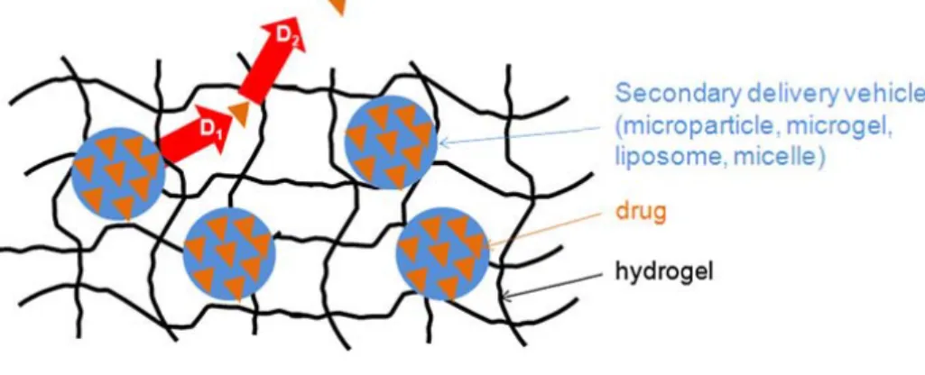
1.6. Hydrogels and Drug Delivery Systems
Several biological active molecules such as peptides, proteins (e.g. growth factors), oligonucleotides and genes, are easily degraded in the body fluids (Bhavsar et al., 2009); (Guse et al., 2006; Prabaharan and Mano, 2008). Usually, these compounds must be administrated several times in order to have the desired therapeutic effect. Drug delivery systems have been developed in order to allow a sustained and targeted release, decreasing the number of therapeutic doses administrated and also contributing to increase the efficacy of the therapy (Guse et al., 2006; Hiemstra et al., 2007; Kenawy et al., 2009; Kim et al., 2009; Reddy and Swarnalatha, 2010; Siepmann and Siepmann, 2008). The goal of an ideal drug delivery system is to deliver a drug to a precise site, in specific time and with a right release pattern. The traditional medical forms (tablets, injection solutions, etc.) provide drug delivery with peaks, often above the required dose (Figure 6). The constant drug level in blood or sustained drug release to avoid multiple doses and bypassing the hepatic “first-pass” metabolism are the main challenges for every delivery system (Stamatialis et al., 2008). The therapeutic time window can be applied for the tissue engineering based on drug delivery. To obtain specialized function in the body, normal tissues also require temporally specified stimulation during their development (Enoch and Leaper, 2008; Kim et al., 2009).
19 As an extension of the pulsatile release system, a temporally programmed drug release system is an excellent solution to supply multiple drugs (Kim et al., 2009). Drug delivery systems can be categorized into two major groups: time-controlled systems and stimuli-induced release systems. In this last case the drug is released in response to internal/external stimuli such as changes in pH, temperature or glucose levels in blood, for example (Caldorera-Moore and Peppas, 2009; Hamidi et al., 2008). The drug can also be released at the site of action, through the functionalization of the system structure with specific ligands that are recognized by membrane receptors like integrins (Petrie and García, 2009).
The interest in hydrogels, three-dimensional polymeric networks capable of absorbed high amounts of water (Hamidi et al., 2008), has been growing after the establishment of the first synthetic hydrogels by Wichterle and Lim in 1954 (Lin and Metters, 2006; Wichterle and Lim, 1960). Since then, the growth of hydrogel technologies have increased in many fields (Lin and Metters, 2006) of tissue engineering and regenerative medicine such as diagnostics, cellular immobilization, separation of biomolecules or cells, and barrier materials to regulate biological adhesions (Hoare and Kohane, 2008).
The hydrogels affinity to absorb high amounts of water is attributed to the presence of hydrophilic groups such as –OH, -CONH-, -CONH2-, and –SO3H in polymers forming hydrogel
structures. Because of these groups, the polymer is thus hydrated to different degrees (sometimes more than 90%wt) depending on the nature of the aqueous environment and polymer composition (Hamidi et al., 2008). However, the hydrogel can be formed from polymers with hydrophobic characteristics (e.g., poly(lactic acid) (PLA) or poly(lactide-co-glycolide) (PLGA)) which have limited water absorbing properties (< 5-10%) (Hamidi et al., 2008). Hydrogels can also be classified based on a variety of characteristics, such as the nature of side groups (neutral or ionic), mechanical and structural features (affine and phantom), method of preparation (homo- or co-polymer), physical structure (amorphous, semicrystalline, hydrogen bounded, supermolecular, and hydrocolloidal), and responsiveness
Figure 6 – Scheme of the therapeutic time window. The various cases; maximum, minimum, traditional