1. Cardiology Department, Rehabilitation Hospital, “Iuliu Hatieganu” University of Medicine and Pharmacy, Cluj-Napoca, Romania 2. Rheumatology Clinic, “Iuliu Hatieganu” University of Medicine and Pharmacy, Cluj-Napoca, Romania.
3. Department of Internal Medicine, Second Medical Clinic, “Iuliu Hatieganu” University of Medicine and Pharmacy, Cluj-Napoca, Romania
4. “Niculae Stancioiu” Heart Institute, Cluj-Napoca, Romania
Conclusion: Arrhythmias and conduction disorders are common in patients with scleroderma. Patients with such disorders are older, have a higher prevalence of pulmonary hypertension, more severe mitral and tri-cuspid regurgitation, left atrial and right ventricular di-lation on echocardiography.
Keywords: Scleroderma; Conduction disorders; Car-diac arrhythmias.
IntroductIon
Arrhythmias and conduction disorders are commonly found among patients with scleroderma1-7. However,
their reported prevalence varies depending on the dia -gnostic tool used – surface Electrocardiogram (ECG), 24-hour Holter ECG monitoring, electrophysiological study1, 8, with an abnormal resting ECG being found
between 25 - 75% of scleroderma patients1, 2.
Myocardial causes, especially ventricular arrhyth-mias, represents the third most common cause of death in patients with scleroderma, after pulmonary fibrosis and pulmonary arterial hypertension, accounting for 6% of the total causes of death9. An early diagnosis of
arrhythmias is therefore important, patients with scle-roderma and arrhythmias having a 2-fold higher risk of death compared with scleroderma patients without ar-rhythmias10.
Several studies identified risk markers for mortality in scleroderma patients, including the presence of an abnormal ECG1, 2, right bundle branch block6and clini
-cally significant arrhythmias10. However, the
cardio-vascular profiles of scleroderma patients with various kinds of arrhythmias and conduction disorders have not been described so far. Therefore, the aim of this study was to outline the cardiovascular profiles of scle-roderma patients with different types of arrhythmias and conduction disorders.
Cardiovascular profiles of scleroderma patients
with arrhythmias and conduction disorders
Muresan L1, Petcu A2, Pamfil C2, Muresan C3, Rinzis M2, Mada RO4, Gusetu GN1, Pop D1, Zdrenghea D2, Rednic S2
ACTA REUMATOL PORT. 2016;41:26-39
AbstrAct
Introduction: Arrhythmias and conduction disorders are common among patients with scleroderma. Their early identification is important, since scleroderma pa-tients with arrhythmias have a higher mortality risk compared with scleroderma patients without arrhyth-mias. The aim of this study was to characterize the car-diovascular profiles of scleroderma patients with dif-ferent types of arrhythmias and conduction disorders. Methods: One hundred and ten consecutive patients with a diagnosis of systemic sclerosis according to the ACR criteria were included in the study. Patients un-derwent a 12-lead ECG and a 24-hour Holter ECG mon-itoring for arrhythmias and conduction disorders iden-tification. Blood sample testing, echocardiography, spirometry, chest X-ray and, when considered appro-priate, high resolution chest CT were also performed. A subgroup of 21 patients underwent NT-pro BNP le vel measurements. Patients’ clinical and paraclinical cha -racteristics were compared according to the pre sence or absence of arrhythmias and conduction di sorders. Results: The prevalence of arrhythmia and conduction disturbances was 60.9%. Patients with such disorders were older (54.4 ± 13.3 vs. 49.7 ± 10.1 years, p=0.05), had a higher prevalence of pulmonary hypertension (p=0.008), valve disease (p < 0.001), especially mitral and tricuspid regurgitation, chamber enlargement on echocardiography (left atrial and right ventricular, p = 0.012 and 0.005, respectively), as well as, higher NT-pro BNP levels: 265.5 ± 399.7 vs. 163 ± 264.3 pg/ml, p=0.04.
Methods
PAtIent PoPulAtIon And dAtA collectIon From October 2011 to December 2014, 113 conse -cutive patients, males and females, with a diagnostic of systemic sclerosis according to the American College of Rheumatology (ACR)/criteria, were admitted to the Rheumatology Clinic of Cluj-Napoca, Romania. Only patients with the diffuse cutaneous subtype and limi -ted cutaneous subtype were included in the study. Three patients with scleroderma sine scleroderma were excluded from the study and therefore the final popu-lation consisted of 110 patients. The research protocol was approved by the Local Ethics Committee.
After giving informed consent, all patients underwent a complete physical examination and blood sample tes -ting. Afterwards, a cardiovascular evaluation was per-formed for each patient, including a standard 12-lead resting ECG, 24-hour Holter ECG monitoring and transthoracic Doppler echocardiography. Lung evalua-tion included spirometry and a standard chest X-ray and, in cases where pulmonary fibrosis was suspected based on the patient’s history, physical exam and chest X-ray, a high resolution computer tomography (CT).
In order to define their cardiovascular profiles, pa-tients’ clinical and para-clinical characteristics were compared according to the presence or absence of ar-rhythmias and conduction disorders as follows: pa-tients with arrhythmias (either supraventricular or ventricular) and/or conduction disturbances were compared to patients without arrhythmias and/or con-duction disorders; patients with ventricular arrhyth-mias to patients without ventricular arrhytharrhyth-mias; pa-tients with ventricular arrhythmias and high-risk markers (ventricular couplets, nonsustained ventricu -lar tachycardia, right bundle branch block) to patients without these markers; patients with supraventricular arrhythmias to patients without supraventricular ar-rhythmias; patients with conduction disorders to pa-tients without conduction disorders.
Clinical data were collected from the patients’ clini -cal records. The 12-lead ECG, 24-hour Holter ECG monitoring result and the echocardiographic images were interpreted by a full time cardiologist.
evAluAtIon of PAtIents
12-
LEADECG
The resting ECGs were recorded using an Esaote P8000 electrocardiograph, with an ECG amplifier sen-sitivity of 10 mm/mV, at a speed of 25 mm/s. The
as-sessed parameters were: rhythm, heart rate, QRS axis, the presence of atrial or ventricular hypertrophy, signs of myocardial ischemia, the PR interval, the duration of the QRS complex, the QT and the QTc interval.
Bradycardia was defined as a heart rate of < 60 bpm, while tachycardia as a heart rate of > 100 bpm.
The presence of left anterior fascicular block was defined as a QRS axis deviation between -30 and – 90 degrees and of left posterior facicular block as a QRS axis deviation between +120 and +180 degrees. Com-plete right bundle branch block (RBBB) was defined as a QRS duration of >120 ms, with a RSR’ pattern in V1--V3 and a wide S wave in lead I, aVL and V5, V6, ± ST depression and T wave inversion in V1-V3. Incom-plete RBBB was defined as a QRS duration of 100 – 120 ms, with a rSr’ aspect in V1. Left bundle branch block (LBBB) was considered present when the QRS duration exceeded 120 ms and a dominant S wave in V1 was present giving an rS or QS aspect, broad R wave in I, aVL, V5,V6 + ST segment depression in these leads. Incomplete LBBB was defined as a QRS aspect suggestive of LBBB, with a QRS duration of between 100 and 120 ms. Non-specific intraventricular con-duction defects were defined as QRS morphology changes incompatible with any of the above mentioned conduction disorders.
The corrected QT (QTc) interval was considered prolonged if > 440 ms for males and > 460 ms for fe-males. Ischemia was defined as the presence of negative T waves, or ST depression of ≥1 mm or the presen -ce of Q waves (as a marker of myocardial infarction) in at least 2 contiguous leads.
T
HE24-
HOURH
OLTERECG
MONITORINGRecordings were made using a 7-lead BTL CardioPoint H600 device, with a 2000Hz sampling frequency and 16bit digital resolution.
The assessed parameters were: maximum, average and minimum heart rate, average heart rate while awake, average heart rate while asleep, the presence of supraventricular and ventricular arrhythmias, the presen ce of paroxysmal conduction disorders, QT and QTc interval.
Bradycardia was defined as an average heart rate of < 60 bpm while awake and tachycardia as a heart rate of > 100 bpm while awake or asleep.
All supraventricular and ventricular ectopic beats were recorded. Supraventricular arrhythmia was de-fined as the presence of > 100 PAC/24 hours and ven-tricular arrhythmia as the presence of > 1 PVC/hour.
T
HE TRANSTHORACIC ECHOCARDIOGRAPHYAll echocardiographic examinations were performed with an Esaote MyLabTM X-View 50 machine, with a 7.5 - 10 MHz transducer.
The assessed parameters were: chamber size, inter-ventricular septum (IVS) and posterior wall (PW) of the left ventricle (LV) thickness, systolic and diastolic function of the LV, systolic function of the right ventricle (RV), leftsided filling pressures, global and regio -nal motion abnormalities, systolic, mean and diastolic pulmonary aterial pressure (sPAP, mPAP, dPAP), the presence of pericardial effusion, the presence of valve disease (stenosis and regurgitations).
LV hypertrophy was defined as an increased thick-ness of IVS and PW (>11 mm), LV dilation as an end-diastolic dimension > 60 mm, end systolic dimension > 40 mm, RV dilation as a diameter of the RV > 26 mm in the parasternal long axis view, LV systolic dysfunc-tion as an ejecdysfunc-tion fracdysfunc-tion of < 55%, RV systolic dys-function as TAPSE < 17 mm.
Pulmonary hypertension was defined as mild when sPAP was 35-49 mmHg, moderate when between 50--69 mmHg and severe if ≥ 70 mmHg. sPAP, mPAP and dPAP were calculated using the simplified Bernoulli ecuation (4 x vmax2), based on the tricuspid
regurgi-tation jet velocity (for sPAP) and on the pulmonary in-sufficiency jet velocity (for mPAP and dPAP).
L
ABORATORY ANALYSESBlood sample testing included a complete blood count, ESR, blood urea nitrogen, creatinine, electrolites (Na+, K+, Ca2+, Cl-), glucose, total cholesterol, HDL-choles-terol, LDL-cholesHDL-choles-terol, triglycerides, uric acid, coagu-lation parameters (Quick time, INR, activated partial thromboplastin time), GOT, GPT, alkaline phos-phatase, gamma-glutamyl transferase, bilirubin, total protein and albumin. Complement levels, C3, C4, cir-culating immune complexes, IgA, IgG, IgM, rheuma-toid factor, antinuclear antibodies and anti-topoiso-merase I levels were also measured.
In a subgroup of 21 randomly selected patients, the NT-pro BNP levels (pg/ml) were measured.
stAtIstIcAl AnAlysIs
Statistical analysis was performed using the Statistical Package for the Social Sciences (SPSS Inc. Chicago, Illi-nois) version 20. Descriptive statistics were used to summarize patients’ characteristics. Normality was as-sessed for all continuous variables using the Shapiro_Wilk test. When the assumption held, results
were expressed as mean ± standard deviation (SD) or otherwise by median ± interquartile range. Categorical variables were presented as counts and proportions (%). The Chi square test was used to compare different scleroderma characteristics, associated diseases, cate-gorical echocardiographic elements and medical treat-ment of patients, with and without arrhythmias, and conduction disorders. According to the sample size of the compared patient populations, the t-test for inde-pendent samples or Mann-Whitney U test were used to compare the age, duration of clinical manifestation, the skin score, the levels of NT-pro BNP, the LV ejection fraction and the left-sided filling pressures of sclero-derma patients with and without arrhythmias and con-duction disorders.
Spearman’s correlation coefficient was used to assess the relationship between electrocardiographic findings and echocardiographic abnormalities, such as pul-monary hypertension and right ventricular dilation.
Receiver operating characteristic (ROC) curves were used to analyze the accuracy of several ECG parameters in predicting the existence of important echocardio-graphic abnormalities (pulmonary hypertension, right ventricular dilation).
A p value of < 0.05 was considered statistically sig-nificant.
results
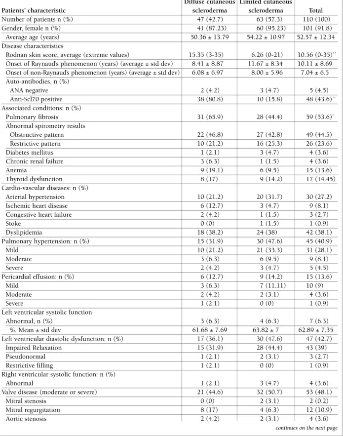
GenerAl chArActerIstIcs of the PAtIents The general characteristics of the patients are presen -ted in Table I.
The main features that distinguished patients with diffuse cutaneous scleroderma from patients with limi -ted cutaneous scleroderma were a higher Rodnan skin score (15.35 ± 8.5 vs 6.26 ± 6.28, p<001), a higher number of positive anti Scl70 antibodies tests (80.8% vs. 15.8%, p<0.001) and a higher prevalence of pul-monary fibrosis (65.9% vs. 44.4%, p=0.02). There were no statistically significant differences in what con-cerns the onset of Raynaud’s and non-Raynaud’s phe-nomenon, other significant associated conditions or cardiovascular diseases, echocardiographic and labo-ratory parameters between the 2 subgroups of patients (Table I).
PrevAlence of ArrhythMIAs And conductIon dIsorders
tAble I. GenerAl chArActerIstIcs of the PAtIents, AccordInG to the scleroderMA subtyPe Diffuse cutaneous Limited cutaneous
Patients’ characteristic scleroderma scleroderma Total
Number of patients n (%) 47 (42.7) 63 (57.3) 110 (100)
Gender, female n (%) 41 (87.23) 60 (95.23) 101 (91.8)
Average age (years) 50.36 ± 13.79 54.22 ± 10.97 52.57 ± 12.34 Disease characteristics
Rodnan skin score, average (extreme values) 15.35 (3-35) 6.26 (0-21) 10.56 (0-35)** Onset of Raynaud’s phenomenon (years) (average ± std dev) 8.41 ± 8.87 11.67 ± 8.34 10.11 ± 8.69 Onset of non-Raynaud’s phenomenon (years) (average ± std dev) 6.08 ± 6.97 8.00 ± 5.96 7.04 ± 6.5 Auto-antibodies, n (%)
ANA negative 2 (4.2) 3 (4.7) 5 (4.5)
Anti-Scl70 positive 38 (80.8) 10 (15.8) 48 (43.6)**
Associated conditions: n (%)
Pulmonary fibrosis 31 (65.9) 28 (44.4) 59 (53.6)*
Abnormal spirometry results
Obstructive pattern 22 (46.8) 27 (42.8) 49 (44.5)
Restrictive pattern 10 (21.2) 16 (25.3) 26 (23.6)
Diabetes mellitus 1 (2.1) 3 (4.7) 4 (3.6)
Chronic renal failure 3 (6.3) 1 (1.5) 4 (3.6)
Anemia 9 (19.1) 6 (9.5) 15 (13.6)
Thyroid dysfunction 8 (17) 9 (14.2) 17 (14.45)
Cardio-vascular diseases: n (%)
Arterial hypertension 10 (21.2) 20 (31.7) 30 (27.2)
Ischemic heart disease 6 (12.7) 3 (4.7) 9 (8.1)
Congestive heart failure 2 (4.2) 1 (1.5) 3 (2.7)
Stoke 0 (0) 1 (1.5) 1 (0.9) Dyslipidemia 18 (38.2) 24 (38) 42 (38.1) Pulmonary hypertension: n (%) 15 (31.9) 30 (47.6) 45 (40.9) Mild 10 (21.2) 21 (33.3) 31 (28.1) Moderate 3 (6.3) 6 (9.5) 9 (8.1) Severe 2 (4.2) 3 (4.7) 5 (4.5) Pericardial effusion: n (%) 6 (12.7) 9 (14.2) 15 (13.6) Mild 3 (6.3) 7 (11.11) 10 (9) Moderate 2 (4.2) 2 (3.1) 4 (3.6) Severe 1 (2.1) 0 (0) 1 (0.9)
Left ventricular systolic function
Abnormal, n (%) 3 (6.3) 4 (6.3) 7 (6.3)
%, Mean ± std dev 61.68 ± 7.69 63.82 ± 7 62.89 ± 7.35
Left ventricular diastolic dysfunction: n (%) 17 (36.1) 30 (47.6) 47 (42.7)
Impaired Relaxation 15 (31.9) 28 (44.4) 43 (39)
Pseudonormal 1 (2.1) 2 (3.1) 3 (2.7)
Restrictive filling 1 (2.1) 0 (0) 1 (0.9)
Right ventricular systolic function: n (%)
Abnormal 1 (2.1) 3 (4.7) 4 (3.6)
Valve disease (moderate or severe) 21 (44.6) 32 (50.7) 53 (48.1)
Mitral stenosis 0 (0) 2 (3.1) 2 (0.2)
Mitral regurgitation 8 (17) 4 (6.3) 12 (10.9)
Aortic stenosis 2 (4.2) 2 (3.1) 4 (3.6)
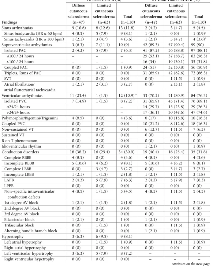
monitoring, a high prevalence of arrhythmias and con-duction disorders was found (Table II). All but 4 pa-tients had some sort of supraventricular or ventricular ectopy and/or conduction disorder. Conduction disor-ders were encountered in 30% of patients (n=33). A number of 35 patients (31.8%) had at least 100 pre-mature atrial contractions/24 hours and 50 patients (45.4%) had at least 1 premature ventricular contrac-tion/hour at the Holter ECG monitoring. There was an important overlap of patients with arrhythmias and conduction disorders, 19% of patients (n=21) having both supraventricular and ventricular arrhythmias and 10% (n=11) of patients having both types of arrhyth-mias plus some type of conduction disorder.
Taking the cut-off values mentioned above, a total of 67 patients had supraventricular arrhythmia, ventricu -lar arrhythmia or a conduction disorder, the prevalence of arrhythmias and conduction disturbances being 60.9%.
When compared according to the scleroderma sub-type, on the 12-lead ECG, patients with the diffuse cu-taneous form had a significantly higher prevalence of ventricular arrhythmias compared to patients with the limited cutaneous form. However, on the 24-hour
Holter ECG monitoring, there were no significant dif-ferences between the 2 groups of scleroderma subtypes in what concerns the prevalence of the different ar-rhythmias and conduction disorders.
According to the presence or absence of pulmonary hypertension, a significantly higher number of patients from the pulmonary hypertension group had arrhyth-mias and conduction disorders on the 12-lead ECG (22.2% vs. 7.7%, p=0.035, and 44.4% vs. 20%, p=0.009, respectively (Table 3). During the 24-hour Holter ECG monitoring, patients with pulmonary hy-pertension had a significantly higher number of pre-mature ventricular contractions (74 ± 327 vs. 4 ± 71, p= 0.01) compared to patients without pulmonary hy-pertension (Table III).
According to the type of arrhythmia and conduction disorders found, we outlined the cardiovascular pro-files of the different scleroderma patients.
the scleroderMA PAtIent wIth
ArrhythMIAs And conductIon dIsorders Several features distinguished scleroderma patients with arrhythmias and conduction disorders from the other scleroderma patients: they were older (p=0.05) tAble I. contInuAtIon
Diffuse cutaneous Limited cutaneous
Patients’ characteristic scleroderma scleroderma Total
Aortic regurgitation 1 (2.1) 3 (4.7) 4 (3.6)
Pulmonary regurgitation 1 (2.1) 2 (3.1) 3 (2.7)
Tricuspid regurgitation 9 (19.1) 19 (30.1) 28 (25.4)
Hypertrophy/Dilation (echocardiography): n (%)
Left ventricular hypertrophy 7 (14.8) 11 (17.4) 18 (16.3)
Right ventricular dilation 6 (12.7) 3 (4.7) 9 (8.1)
Left atrial dilation 5 (10.6) 6 (9.5) 11 (10)
Right atrial dilation NA NA NA
NT-pro BNP (pg/ml) 265.5 ± 274 185 ± 63.5 207 ± 129
Medication: n (%)
Beta blockers 3 (6.3) 5 (7.9) 8 (7.2)
ACE inhibitors/ARBs 15 (31.9) 12 (19) 27 (24.5)
Calcium channel blockers 15 (31.9) 17 (26.9) 32 (29)
Digoxin 1 (2.1) 0 (0) 1 (0.9)
Propafenone 3 (6.3) 1 (1.5) 4 (3.6)
Amiodarone 2 (4.2) 0 (0) 2 (1.8)
PAH-specific medication 2 (4.2) 1 (1.5) 3 (2.7)
*p< 0.05; **p<0.01; Arrhythmia was defined as the presence of > 100 PAC/24 hours and/or the presence of > 24 PVC/ 24 hours. ANA = Antinuclear antibodies; Anti SCL70 = anti topoisomerase I; ACE Inhibitors = Angiotensin converting enzyme inhibitors; ABRs = Angiotensin receptor blockers; PAH = Pulmonary arterial hypertension; NA = not available.
tAble II. tyPes of ArrhythMIAs, conductIon dIsorders And other AbnorMAlItIes dIAGnosed wIth 12 leAd ecG And 24 hour holter ecG MonItorInG
12 Lead ECG n (%) 24 Hour Holter ECG n (%) Diffuse Limited Diffuse Limited
cutaneous cutaneous cutaneous cutaneous
scleroderma scleroderma Total scleroderma scleroderma Total
Findings (n=47) (n=63) (n=110) (n=47) (n=63) (n=110) Sinus arrhythmias 5 (10.6) 8 (12.6) 13 (11.8) 2 (4.2) 3 (4.7) 5 (4.5) Sinus bradycardia (HR ≤ 60 bpm) 4 (8.5) 5 (7.9) 9 (8.1) 1 (2.1) 0 (0) 1 (0.9)a Sinus tachycardia (HR ≥ 100 bpm) 1 (2.1) 3 (4.7) 4 (3.6) 1 (2.1) 3 (4.7) 4 (3.6)b Supraventricular arrhythmias 3 (6.3) 7 (11.1) 10 (9) 42 (89.3) 57 (90.4) 99 (90) Isolated PAC 2 (4.2) 5 (7.9) 7 (6.3) 41 (87.2) 56 (88.8) 97 (88.1) ≤100 / 24 hours – – – 25 (53.1) 37 (58.7) 62 (56.3) >100 / 24 hours – – – 16 (34) 19 (30.1) 35 (31.8) Coupled PAC 0 (0) 1 (1.5) 1 (0.9) 24 (51) 32 (50.8) 56 (50.9)
Triplets, Runs of PAC 0 (0) 0 (0) 0 (0) 31 (65.9) 42 (62.6) 73 (66.3)
SVT 0 (0) 0 (0) 0 (0) 0 (0) 1 (1.5) 1 (0.9)
Atrial fibrillation/ 1 (2.1) 2 (3.1) 3 (2.7) 0 (0) 2 (3.1) 2 (1.8) atrial flutter/atrial tachycardia
Ventricular arrhythmias 11 (23.4) 1 (1.5) 12 (10.9)** 33 (70.2) 51 (80.9) 84 (76.3) Isolated PVC 7 (14.9) 1 (1.5) 8 (7.2)** 31 (65.9) 45 (71.4) 76 (69.1) ≤24/24 hours – 14 (29.7) 15 (23.8) 29 (26.3) >24/24 hours – 17 (36.1) 30 (47.6) 47 (74.6) Polymorphic/Bigemini/Trigemini 4 (8.5) 0 (0) 4 (3.6) 8 (17) 10 (15.8) 18 (16.3) Coupled PVC 0 (0) 0 (0) 0 (0) 10 (21.2) 8 (12.6) 18 (16.3) Non–sustained VT 0 (0) 0 (0) 0 (0) 6 (12.7) 1 (1.5) 7 (6.3) Sustained VT 0 (0) 0 (0) 0 (0) 0 (0) 0 (0) 0 (0) R on T phenomenon 0 (0) 0 (0) 0 (0) 0 (0) 0 (0) 0 (0) Idioventricular rhythm 0 (0) 0 (0) 0 (0) 1 (2.1) 0 (0) 1 (0.9) Conduction disorders 18 (38.2) 16 (25.4) 34 (30.9) 19 (40.4) 16 (25.4) 35 (31.8) Complete RBBB 4 (8.5) 0 (0) 4 (3.6) 4 (8.5) 0 (0) 4 (3.6) Incomplete RBBB 5 (10.6) 4 (6.2) 9 (8.1) 5 (10.6) 4 (6.2) 9 (8.1) Complete LBBB 0 (0) 3 (4.7) 3 (2.7) 0 (0) 3 (4.7) 3 (2.7) Incomplete LBBB 1 (2.1) 1 (1.5) 2 (1.8) 1 (2.1) 1 (1.5) 2 (1.8) LAFB 2 (4.2) 5 (7.9) 7 (6.3) 2 (4.2) 5 (7.9) 7 (6.3) LPFB 0 (0) 0 (0) 0 (0) 0 (0) 0 (0) 0 (0) Non–specific intraventricular 4 (8.5) 1 (1.5) 5 (4.5) 4 (8.5) 1 (1.5) 5 (4.5) conduction defects 1st degree AV block 1 (2.1) 1 (1.5) 2 (1.8) 1 (2.1) 1 (1.5) 2 (1.8) 2nd degree AV block 0 (0) 0 (0) 0 (0) 0 (0) 0 (0) 0 (0) 3rd degree AV block 0 (0) 0 (0) 0 (0) 0 (0) 0 (0) 0 (0) Bifascicular block 1 (2.1) 0 (0) 1 (0) 1 (2.1) 0 (0) 1 (0.9) Trifasciular block 0 (0) 1 (1.5) 1 (0) 0 (0) 1 (1.5) 1 (0.9)
Alterning bundle branch block 0 (0) 0 (0) 0 (0) 1 (2.1) 0 (0) 1 (0.9)
Hypertrophy 3 (6.3) 6 (9.5) 9 (8.1)
Left atrial hypetrophy 0 (0) 1 (1.5) 1 (0.9) 0 (0) 1 (1.5) 1 (0.9) Right atrial hypertrophy 0 (0) 0 (0) 0 (0) 0 (0) 0 (0) 0 (0)
Left ventricular hypertrophy 3 (6.3) 5 (7.9) 8 (7.2) – – –
Right ventricular hypetrophy 0 (0) 0 (0) 0 (0) – – –
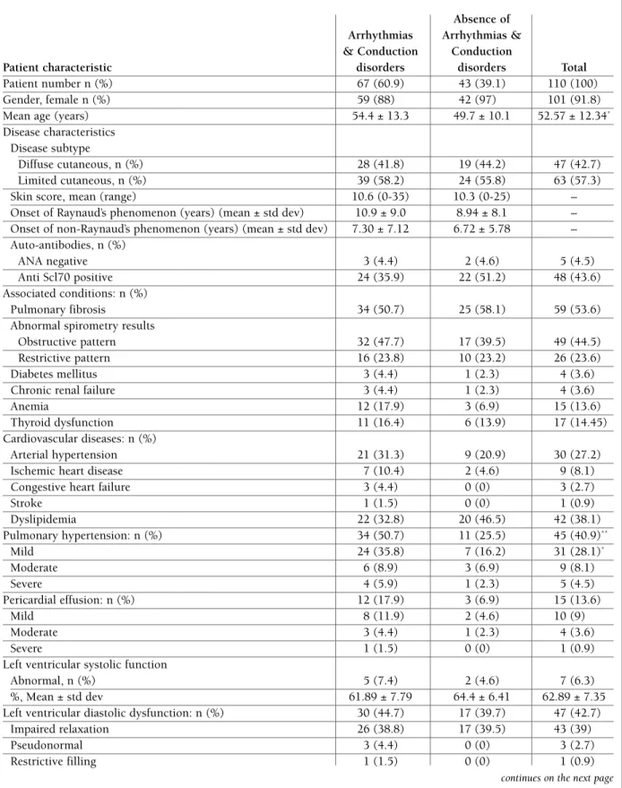
and had a higher prevalence of pulmonary hyperten-sion (p=0.008), valve disease (p < 0.001), especially mitral and tricuspid regurgitation, and chamber en-largement on echocardiography (left atrial and right ventricular, p = 0.012 and 0.005, respectively). These patients also had significantly higher levels of NT-pro -BNP, of 265.5 ± 399.7 pg/ml vs. 163 ± 140.1 pg/ml (p=0.047). There were no statistically significant dif-ferences in what concerns the type of scleroderma, the presence of relevant associated diseases, cardiovascu-lar diseases, left ventricucardiovascu-lar systolic and diastolic func-tion, right ventricular systolic function and cardio-vas-cular medication used. The characteristics of sclero-derma patients with arrhythmias and conduction dis-orders are presented in Table IV.
the scleroderMA PAtIent wIth suPrAventrIculAr ArrhythMIAs
There were more men in this group of patients com-pared to the “absence of supraventricular arrhythmias” group (17.2% vs. 4%, p=0.019). Patients with these type of arrhythmia were older (p=0.019), had a hi gher skin score (p=0.04) and a higher prevalence of anaemia (p=0.011).
Their cardiovascular profile was characterized by a higher prevalence of arterial hypertension (p=0.012), pulmonary hypertension (p=0.017), left ventricular dias tolic dysfunction (p=0.03), increased left–sided fill-ing pressure (p=0.02), significant valve diseases (p=0.01) especially tricuspid regurgitation (p=0.054) and left atrial dilation (p=0.016). The use of beta--blo ckers was also significantly higher in this group
(p < 0.01).
The characteristics of scleroderma patients with supraventricular arrhythmias are summarized in Table V. the scleroderMA PAtIent wIth ventrIculAr ArrhythMIAs
The characteristics of scleroderma patients with ven-tricular arrhythmias are summarized in Table VI. Com-pared to patients without ventricular arrhythmias, scle-roderma patients with ventricular arrhythmias were older (p=0.05). Their cardiovascular profile was cha -racterized by increased left-sided filling pressures (p=0.05), a higher prevalence of significant valve disea -se (p<0.001) especially tricuspid regurgitation (p=0.02) and right ventricular enlargement (p<0.01), and significantly higher NT-pro BNP levels (p=0.02).
There was no statistically significant difference in what concerns the left ventricular ejection fraction va -lues and associated cardiovascular diseases between these patients and scleroderma patients with no ven-tricular arrhythmias.
the scleroderMA PAtIent wIth hIGh ArrhythMIA rIsk MArkers
There were 21 patients (18 females) who had either ventricular couplets or non-sustained ventricular tachycardia on Holter ECG monitoring, or right bun-dle branch block. Their average age was 54.4 ± 13.6 years.
Their cardiovascular profile was characterized by a higher prevalence of left ventricular systolic dysfunc-tion (4 patients vs. 3 patients, p<0.01). The prevalence tAble II. contInuAtIon
12 Lead ECG n (%) 24 Hour Holter ECG n (%) Diffuse Limited Diffuse Limited
cutaneous cutaneous cutaneous cutaneous
scleroderma scleroderma Total scleroderma scleroderma Total
Findings (n=47) (n=63) (n=110) (n=47) (n=63) (n=110) Ischemia 3 (6.3) 7 (11.1) 10 (9) 4 (8.5) 7 (11.1) 11 (9.9) ↑ QTc interval 1 (2.1) 1 (1.5) 2 (1.8) 7 (14.9) 8 (12.7) 15 (13.6) Rotation 3 (6.3) 7 (11.1) 10 (9) – – – Clockwise 1 (2.1) 4 (6.3) 5 (4.5) – – – Counterclockwise 2 (4.2) 3 (4.7) 5 (4.5) – – –
*p< 0.05; **p<0.01; a – average heart rate while awake; b – average heart rate while awake or asleep. HR = Heart rate; PAC = Premature atrial contraction; SVT = Supraventricular tachycardia; PVC = Premature ventricular contraction; VT = Ventricular tachycardia; RBBB = Right bundle branch block; LBBB = Left bundle branch block; LAFB = Left anterior fascicular block; LPFB = Left posterior fascicular block; AV = Atrio-ventricular.
of significant valve disease was also higher among this subgroup of patients (18 patients vs. 34 patients, p<0.001), especially mitral regurgitation (p=0.03) and tricuspid regurgitation (p<0.01). Right ventricular di-lation was also more frequently encountered (6 patients vs. 3 patients, p<0.01).
the scleroderMA PAtIent wIth conductIon dIsorders
Thirtythree patients (30%) had some type of condu -ction disorder. Most were females (n=30), with an ave rage age of 53.1 ± 12.4 years. These patients had a higher prevalence of pulmonary hypertension (p<0.01) and of significant valve disease (p<0.001), namely
mi-tral (p=0.02) and tricuspid regurgitation (p=0.02), and NT-pro BNP values: 452 ± 725 pg/ml vs. 174 ± 98.5 pg/ml, p<0.01.
relAtIonshIP between
electrocArdIoGrAPhIc fIndInGs And severIty MArkers of scleroderMA
We found statistically significant but weak associations between several Holter ECG abnormalities and esta -blished echocardiographic risk markers of scleroder-ma. Most importantly, the total number of premature ventricular complexes/24 hours correlated with the sPAP and the right ventricular diameter (p=0.01, r=0.24 and p=0.008, r=0,266, respectively) and the tAble III. PrevAlence And tyPes of ArrhythMIAs, conductIon dIsorders And other AbnorMAlItIes dIAGnosed wIth 12-leAd ecG And 24-hour holter ecG MonItorInG, AccordInG to the Presence or Absence of PulMonAry hyPertensIon
12–lead ECG
Patients with pulmonary Patients without pulmonary
Finding Hypertension (n=45) Hypertension (n=65) p
Sinus rhythm, n (%) 42 (93.3) 65 (100%) ns
Average heart rate 77.2 ± 12.5 74.1 ± 11.3 ns
Left ventricular hypertrophy, n (%) 6 (13.3) 3 (4.6) ns
Ischemia, n (%) 6 (13.3) 3 (4.6) ns
Arrhythmias, n (%) 10 (22.2) 5 (7.7) 0.035
Conduction disorders, n (%) 20 (44.4) 13 (20) 0.009
QT interval 363 ± 36 365 ± 31 ns
QTc interval 408 ± 27 403 ± 23 ns
24-hour Holter ECG
Patients with pulmonary Patients without pulmonary
Finding Hypertension (n=45) Hypertension (n=65) p
Maximum heart rate 132 ± 29 134 ± 17 ns
Average heart rate 78 ± 10 76 ± 10 ns
Minimum heart rate 56 ± 9 56 ± 8 ns
Supraventricular arrhythmias Total PAC 76 ± 206 17 ± 77 ns Isolated PAC 63 ± 164 15 ± 66 ns Couplets 1 ± 5 0 ± 4 ns Runs 0 ± 1 0 ± 0 0.047 Ventricular arrhythmias Total PVC 74 ± 327 4 ± 71 0.01 Isolated PVC 74 ± 319 3 ± 52 0.01 Couplets 0 ± 1 0 ± 0 ns Number of PVC morphologies 1 ± 1 1 ± 2 ns QT interval 396 ± 9 395 ± 23 ns QTc interval 440 ± 19 434 ± 19 ns
tAble Iv. chArActerIstIcs of scleroderMA PAtIents wIth And wIthout ArrhythMIAs (suPrAventrIculAr And ventrIculAr) And conductIon dIsorders
Absence of Arrhythmias Arrhythmias & & Conduction Conduction
Patient characteristic disorders disorders Total
Patient number n (%) 67 (60.9) 43 (39.1) 110 (100)
Gender, female n (%) 59 (88) 42 (97) 101 (91.8)
Mean age (years) 54.4 ± 13.3 49.7 ± 10.1 52.57 ± 12.34*
Disease characteristics Disease subtype
Diffuse cutaneous, n (%) 28 (41.8) 19 (44.2) 47 (42.7)
Limited cutaneous, n (%) 39 (58.2) 24 (55.8) 63 (57.3)
Skin score, mean (range) 10.6 (0-35) 10.3 (0-25) –
Onset of Raynaud’s phenomenon (years) (mean ± std dev) 10.9 ± 9.0 8.94 ± 8.1 – Onset of non-Raynaud’s phenomenon (years) (mean ± std dev) 7.30 ± 7.12 6.72 ± 5.78 – Auto-antibodies, n (%)
ANA negative 3 (4.4) 2 (4.6) 5 (4.5)
Anti Scl70 positive 24 (35.9) 22 (51.2) 48 (43.6)
Associated conditions: n (%)
Pulmonary fibrosis 34 (50.7) 25 (58.1) 59 (53.6)
Abnormal spirometry results
Obstructive pattern 32 (47.7) 17 (39.5) 49 (44.5)
Restrictive pattern 16 (23.8) 10 (23.2) 26 (23.6)
Diabetes mellitus 3 (4.4) 1 (2.3) 4 (3.6)
Chronic renal failure 3 (4.4) 1 (2.3) 4 (3.6)
Anemia 12 (17.9) 3 (6.9) 15 (13.6)
Thyroid dysfunction 11 (16.4) 6 (13.9) 17 (14.45)
Cardiovascular diseases: n (%)
Arterial hypertension 21 (31.3) 9 (20.9) 30 (27.2)
Ischemic heart disease 7 (10.4) 2 (4.6) 9 (8.1)
Congestive heart failure 3 (4.4) 0 (0) 3 (2.7)
Stroke 1 (1.5) 0 (0) 1 (0.9) Dyslipidemia 22 (32.8) 20 (46.5) 42 (38.1) Pulmonary hypertension: n (%) 34 (50.7) 11 (25.5) 45 (40.9)** Mild 24 (35.8) 7 (16.2) 31 (28.1)* Moderate 6 (8.9) 3 (6.9) 9 (8.1) Severe 4 (5.9) 1 (2.3) 5 (4.5) Pericardial effusion: n (%) 12 (17.9) 3 (6.9) 15 (13.6) Mild 8 (11.9) 2 (4.6) 10 (9) Moderate 3 (4.4) 1 (2.3) 4 (3.6) Severe 1 (1.5) 0 (0) 1 (0.9)
Left ventricular systolic function
Abnormal, n (%) 5 (7.4) 2 (4.6) 7 (6.3)
%, Mean ± std dev 61.89 ± 7.79 64.4 ± 6.41 62.89 ± 7.35
Left ventricular diastolic dysfunction: n (%) 30 (44.7) 17 (39.7) 47 (42.7)
Impaired relaxation 26 (38.8) 17 (39.5) 43 (39)
Pseudonormal 3 (4.4) 0 (0) 3 (2.7)
Restrictive filling 1 (1.5) 0 (0) 1 (0.9)
tAble Iv. contInuAtIon
Absence of Arrhythmias Arrhythmias & & Conduction Conduction
Patient characteristic disorders disorders Total
Right ventricular systolic function: n (%)
Abnormal 2 (2.9) 2 (4.6) 4 (3.6)
Valve disease (moderate or severe) 45 (67.4) 8 (18.6) 53 (48.1)**
Mitral stenosis 2 (3) 0 (0) 2 (1.8) Mitral regurgitation 11 (16.4) 1 (2.3) 12 (10.9)* Aortic stenosis 4 (5.9) 0 (0) 4 (3.6) Aortic regurgitation 3 (4.4) 1 (2.3) 4 (3.6) Pulmonary regurgitation 2 (3) 1 (2.3) 3 (2.7) Tricuspid regurgitation 24 (35.8) 4 (4.6) 28 (25.4)**
Hypertrophy / Dilation (echocardiography): n (%)
Left ventricular hypertrophy 13 (19.4) 5 (11.6) 18 (16.3)
Right ventricular dilation 9 (13.4) 0 (0) 9 (8.1)*
Left atrial dilation 11 (16.4) 0 (0) 11 (10)**
Right atrial dilation NA NA NA
NT-pro BNP (pg/ml) 265.5 ± 399.7 163 ± 140.1 207 ± 129*
Medication: n (%)
Beta blockers 7 (10.4) 1 (2.3) 8 (7.2)
ACE inhibitors/ARBs 17 (25.3) 10 (23.2) 27 (24.5)
Calcium channel blockers 18 (26.8) 14 (32.5) 32 (29)
Digoxin 1 (1.5) 0 (0) 1 (0.9)
Propafenone 4 (5.9) 0 (0) 4 (3.6)
Amiodarone 2 (3) 0 (0) 2 (1.8)
PAH-specific medication 3 (4.4) 0 (0) 3 (2.7)
number of ventricular couplets on Holter ECG corre-lated with the right ventricular diameter (p=0.003, r=0.298). A number of ≥ 119 premature ventricular complexes/24 hours was associated with the presence of a dilated right ventricle on echocardiography, with a sensitivity of 77% and a specificity of 76% (area under the curve = 0.807). Also, the presence of ≥ 3.5 mor-phologies of premature ventricular contractions/24 hours had a sensitivity of 44% and a specificity of 90% in predicting the presen ce of a dilated right ventricle on echocardiography (area under the curve = 0.711).
dIscussIon
The present study describes the cardiovascular profiles of scleroderma patients with arrhythmia and condu
-ction disorders. Based on the nature of the arrhythmias (supraventricular, ventricular, high risk ventricular) and conduction disturbances identified with the 12--lead ECG and 24-hour Holter ECG monitoring, pa-tients were compared to scleroderma papa-tients without these findings. Several clinical and echocardiographic features distinguished these patients from the other scleroderma patients, which merit to be commented upon.
There was a high prevalence of arrhythmias and con-duction disturbances in the present population, of 60.9%, a prevalence comparable to the one reported in other studies1-5,7. Using the same diagnostic tools,
Roberts et al1found a very similar prevalence of
con-duction defects and arrhythmias, of 32% using the ECG and of 62% using the Holter ECG. However, several points need to be made. First, there is no
unanimous-*p< 0.05; *unanimous-*p<0.01; Arrhythmia was defined as the presence of > 100 PAC/24 hours and/or the presence of >24 PVC/ 24 hours. ANA = Antinuclear antibodies; Anti SCL70 = Anti topoisomerase I; ACE Inhibitors = Angiotensin converting enzyme inhibitors; ABRs = Angiotensin receptor blockers; PAH = Pulmonary arterial hypertension; NA = not available.
ly accepted low threshold for defining the presence of supraventricular and ventricular arrhythmias, since a low number of premature atrial and ventricular con-tractions can be found in the healthy population as well11, 12. In one study performed on healthy military
in-dividuals13, using the 12-lead ECG, the prevalence of
PVC was 0.8% (between 0.2% in individuals younger than 20 years and 2.2% in individuals older than 50 years). However, in contrast to individuals with struc-turally normal hearts, in whom the presence of a low number of PVC is considered benign14,15, in the pre
sence of structural heart disease, the prognostic signi -ficance of PVC is different16. The majority of patients
with scleroderma have subclinical cardiac disease17,
represented mainly by myocardial fibrosis18as a result
of repeated ischemic episodes involving the small coro-nary arteries19. Therefore, we chose a low cut-off value
for defining the presence of ventricular arrhythmia, of one PVC/hour.
Regarding supraventricular arrhythmias, 90% of pa-tients had at least one isolated PAC on Holter ECG monitoring. Given the frequent occurrence of isolated PAC in normal individuals, the cutoff value for defi -ning the presence of supraventricular arrhythmia was
100 PAC/24 hours, as in other studies20. Using this
threshold, we identified 35 patients (31.8%) with supraventricular arrhythmias.
It should be mentioned, however, that only 6 pa-tients (5.5%) required class I or III antiarrhythmic medi cation and that no patient fulfilled, at the time of enrolment in the study, the current recommendations for ICD or pacemaker implantation.
There were no statistically significant differences be-tween the 2 groups of scleroderma patients regarding the prevalence and type of arrhythmias found using the 24 hour Holter ECG monitoring, nor between the prevalence and type of the different conduction disor-ders found using the 12 lead ECG and the 24 hour Holter ECG monitoring. Similar findings have been re-ported before. For instance, in their study, Ferri et al7
found that the prevalence and severity of ventricular arrhythmias did not correlate with the clinical variants of scleroderma.
Among patients with arrhythmias and conduction disorders, there was a higher prevalence of pulmonary hypertension and right ventricular dilation, findings that have previously been reported. Based on data ana -lysis form the EULAR and EUSTAR databases, among
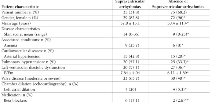
*p< 0.05; **p<0.01; Supraventricular arrhythmia was defined as the presence of > 100 PAC/24 hours at Holter ECG monitoring. E = E wave of the transmitral flow on Doppler echocardiography; Em = Myocardial E wave on tissue Doppler echocardiography; std dev = standard deviation
tAble v. chArActerIstIcs of scleroderMA PAtIents wIth And wIthout suPrAventrIculAr ArrhythMIAs Supraventricular Absence of
Patient characteristic arrhythmias Supraventricular arrhythmias
Patient number n (%) 35 (31.8) 75 (68.2)
Gender, female n (%) 29 (82.8) 72 (96)*
Mean age (years) 57.0 ± 13.1 50.4 ± 11.4*
Disease characteristics
Skin score, mean (range) 14 (0-35) 9 (0-25)*
Associated conditions: n (%)
Anemia 9 (25.7) 6 (8)*
Cardiovascular diseases: n (%)
Arterial hypertension 15 (42.8) 15 (20)*
Pulmonary hypertension: n (%) 20 (57.1) 25 (33.3)*
Left ventricular diastolic dysfunction 20 (57.1) 27 (36)*
E/Em 7.84 ± 4.04 6.11 ± 1.89*
Valve disease (moderate or severe) 23 (65.7) 30 (40)*
Chamber dilation (echocardiography): n (%)
Left atrial dilation 7 (20) 4 (5.3)*
Medication: n (%)
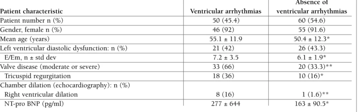
tAble vI. chArActerIstIcs of scleroderMA PAtIents wIth And wIthout ventrIculAr ArrhythMIAs Absence of Patient characteristic Ventricular arrhythmias ventricular arrhythmias
Patient number n (%) 50 (45.4) 60 (54.6)
Gender, female n (%) 46 (92) 55 (91.6)
Mean age (years) 55.1 ± 11.9 50.4 ± 12.3*
Left ventricular diastolic dysfunction: n (%) 21 (42) 26 (43.3)
E/Em, n ± std dev 7.2 ± 3.5 6.1 ± 1.9*
Valve disease (moderate or severe) 33 (66) 20 (33.3)**
Tricuspid regurgitation 18 (36) 10 (16)*
Chamber dilation (echocardiography): n (%)
Right ventricular dilation 8 (16) 1 (1.6)**
NT-pro BNP (pg/ml) 277 ± 644 163 ± 90.5*
scleroderma patients who died of arrhythmias, 47% had a history of PAH9. Elevated pulmonary arterial
pressure can trigger ventricular arrhythmias21. As a
con-sequence of an increased pulmonary vascular resis-tance, there is an increase in right ventricular afterload, leading to right ventricular hypertrophy and dilatation, which contributes to the initiation of ventricular tachy -arrhythmias.
Patients with arrhythmias and conduction disorders from the present studies were also older, a fact which is not surprising, since the prevalence of ventricular ar-rhythmias increases with age11,22,23.
In our study, a significantly higher prevalence of moderate and severe valve disease (especially mitral and tricuspid regurgitation), left atrial and right ventri -cular dilation on echocardiography was found among patients with arrhythmias and conduction distur-bances. The prevalence of echocardiographic abnor-malities is high in scleroderma patients. Smith et al24
re-ported a prevalence of 69%, the most common being high right ventricular systolic pressure, right ventricu-lar dilation, left atrial enventricu-largement and the presence of pericardial effusion. Ferri et al7 also found that
ven-tricular arrhythmias were more likely to be present in patients with echocardiographic abnormalities. How-ever, the abnormalities found on echocardiography (asymmetric septal hypertrophy, impaired ventricular function, congestive cardiomyopathy, mitral prolapse and pericardial effusion) were mostly different than the ones from the present study.
Another important finding was that scleroderma
pa-tients with ventricular arrhythmias also had signifi-cantly higher NT-pro BNP levels. Currently little is known about the relationship between NT-pro BNP levels and ventricular arrhythmias in patients with scle-roderma. However, in other populations of patients, there is evidence linking increased levels of NT-pro BNP and the occurrence of ventricular arrhythmias, both in patients with a severely reduced LV ejection fraction25, and a normal ejection fraction26. Given the
scarcity of data in patients with scleroderma, this pos-sible association needs further investigation.
Among the 50 patients with ventricular arrhythmias, 21 (42%) had markers of high cardiovascular risk, namely complex ventricular arrhythmias10,16,27,28and
right bundle branch block6. These patients had a
hi gher prevalence of left ventricular systolic dysfun -ction. As recognized before, the association of a re-duced systolic function of the LV and the presence of complex ventricular arrhythmias caries a poor progno -sis27,29. Right ventricular dilation, also more frequently
encountered among these patients, leads to right heart failure, which represents one of the most important causes of death in scleroderma patients9.
In the present study, scleroderma patients with supraventricular arrhythmias were significantly older compared to scleroderma patients without tricular arrhythmias. A higher prevalence of supraventricular arrhythmias in the elderly, especially atrial fi -brillation and atrial flutter has been reported before30-32.
The higher prevalence of arterial hypertension, left ventricular diastolic dysfunction, increased leftsided fil
-The presence of ventricular arrhythmia was defined as > 24 PVC/ 24 hours at Holter ECG monitoring. E = E wave of the transmitral flow on Doppler echocardiography; Em = Myocardial E wave on tissue Doppler echocardiography; std dev = standard deviation.
ling pressure and left atrial dilation among this sub-group of scleroderma patients is not surprising, since one of the most frequent causes of supraventricular ar-rhythmias is arterial hypertension, the underlying mechanism being diastolic dysfunction, with subse-quent increased left–sided filling pressures and left atrial enlargement33. Pulmonary arterial hypertension
is another important cause of supraventricular arrhy -thmias21, which can explain its higher prevalence found
in this subgroup of patients. The higher levels of NT--pro BNP found in these patients did not reach statis-tical significance (p > 0.5).
Among the 33 patients (30%) with conduction disor ders, 4 patients (13.3%) had RBBB. Pulmonary hypertension and significant valve disease were also more frequently encountered compared to the other scleroderma patients. Both pulmonary hypertension and RBBB are predictors of mortality among scleroder-ma patients6, 9. NT-pro BNP values were significantly
higher in this group of patients, but given the low num-ber of patients with conduction disorders in which NT--pro BNP levels were measured (n=5), these results should be interpreted with caution.
The present study also demonstrated a positive cor-relation between electrocardiographic abnormalities (the total number of isolated and coupled premature ventricular complexes/24 hours) and echocardio-graphic abnormalities, more exactly the diameter of the right ventricle. Right ventricular dysfunction is an es-tablished risk marker for adverse cardiovascular events in patients with scleroderma9. Therefore, simple Holter
ECG findings suggesting its presence would be useful in clinical practice. We found that a number of ≥ 119 premature ventricular complexes/24 hours has an acce ptable sensitivity and specificity for detecting the pre -sence of a dilated right ventricle and that the pre-sence of multiple morphologies of premature ventricular con-tractions (≥3.5) has a low sensitivity but good speci-ficity for detecting the presence of right ventricular di-lation. However, due to the small-sample size of the population included in the study, these findings should be prospectively tested on larger populations of patients. lIMIts of the study
The most significant limitation of the present study is the absence of a clear cut-off value, which separates pa-tients with supraventricular and ventricular arrhyth-mia from healthy individuals. The cut-off value of 100 PAC/24 hours and 1 PVC/hour at the Holter ECG moni toring chosen in this study influenced the way
pa-tients were defined as having an arrhythmia or not. Based on these definitions, patients were categorized into arrhythmia positive and arrhythmias free, which markedly influences the detected prevalence of ar-rhythmias among scleroderma patients and consecu-tively the described cardiovascular profiles.
The small subgroup of patients in which NT-pro BNP levels were measured is another important limi-tation which might have influenced the results, by sometimes not identifying or overestimating the possi-ble correlation between the NT-pro BNP levels and the presence of ventricular arrhythmias and conduction disorders.
conclusIon
Arrhythmias and conduction disorders are common in patients with scleroderma. Their prevalence and types do not differ significantly according to the scleroderma subtype (diffuse cutaneous and limited cutaneous). Pa-tients with such disorders are older, have a higher prevalence of pulmonary hypertension, more severe mitral and tricuspid regurgitation, left atrial and right ventricular dilation on echocardiography and increased levels of NT-pro BNP. Since the prevalence of arrhyth-mias and conduction disorders is high in scleroderma patients, all patients with symptoms and signs sug-gesting their presence (palpitations, syncope, chest pain, dyspnea, fatigue, peripheral edema) should have a thorough cardiovascular examination.
corresPondence to
Lucian Muresan
Rehabilitation Hospital, Cardiology Department, no. 46 – 50 Viilor Street,
400347 Cluj-Napoca, Cluj, Romania. E-mail: lmure_san@yahoo.com
references
1. Roberts NK, et al. The prevalence of conduction defects and cardiac arrhythmias in progressive systemic sclerosis. Ann In-tern Med 1981. 94(1): p. 38-40.
2. Kostis, J.B., et al., Prognostic importance of cardiac arrhythmias in systemic sclerosis. Am J Med, 1988. 84(6): p. 1007-1015. 3. Morelli, S., et al., Relationships between electrocardiographic
and echocardiographic findings in systemic sclerosis (sclero-derma). Int J Cardiol, 1996. 57(2): p. 151-160.
4. Nordin, A., et al., Electrocardiography in 110 patients with sys-temic sclerosis: a cross-sectional comparison with population-based controls. Scand J Rheumatol, 2014. 43(3): p. 221-225. 5. Vacca, A., et al., Cardiac arrhythmias and conduction defects in
systemic sclerosis. Rheumatology (Oxford), 2014. 53(7): p. 1172-1177.
6. Draeger, H.T., et al., Right bundle branch block: a predictor of mortality in early systemic sclerosis. PLoS One, 2013. 8(10): p. e78808.
7. Ferri, C., et al., Noninvasive evaluation of cardiac dysrhythmias, and their relationship with multisystemic symptoms, in pro-gressive systemic sclerosis patients. Arthritis Rheum, 1985. 28(11): p. 1259-1266.
8. Rokas, S., et al., Electrophysiologic abnormalities of cardiac function in progressive systemic sclerosis. J Electrocardiol, 1996. 29(1): p. 17-25.
9. Tyndall, A.J., et al., Causes and risk factors for death in syste-mic sclerosis: a study from the EULAR Scleroderma Trials and Research (EUSTAR) database. Ann Rheum Dis, 2010. 69(10): p. 1809-1815.
10. Assassi, S., et al., Clinical and genetic factors predictive of mor-tality in early systemic sclerosis. Arthritis Rheum, 2009. 61(10): p. 1403-1411.
11. DePaula, R.S., et al., Cardiac arrhythmias and atrioventricular block in a cohort of asymptomatic individuals without heart di-sease. Cardiology, 2007. 108(2): p. 111-116.
12. Wajngarten, M., et al., Frequency and significance of cardiac rhythm disturbances in healthy elderly individuals. J Electro-cardiol, 1990. 23(2): p. 171-176.
13. Hiss, R.G. and L.E. Lamb, Electrocardiographic findings in 122,043 individuals. Circulation, 1962. 25: p. 947-961. 14. Hinkle, L.E., Jr., S.T. Carver, and M. Stevens, The frequency of
asymptomatic disturbances of cardiac rhythm and conduction in middle-aged men. Am J Cardiol, 1969. 24(5): p. 629-650. 15. Kennedy, H.L., et al., Long-term follow-up of asymptomatic
healthy subjects with frequent and complex ventricular ectopy. N Engl J Med, 1985. 312(4): p. 193-197.
16. Huikuri, H.V., et al., Prediction of sudden cardiac death: ap-praisal of the studies and methods assessing the risk of sudden arrhythmic death. Circulation, 2003. 108(1): p. 110-115. 17. Candell-Riera, J., et al., Comprehensive noninvasive assessment
of cardiac involvement in limited systemic sclerosis. Arthritis Rheum, 1996. 39(7): p. 1138-1145.
18. Deswal, A. and W.P. Follansbee, Cardiac involvement in sclero-derma. Rheum Dis Clin North Am, 1996. 22(4): p. 841-860. 19. Varga, J. and D. Abraham, Systemic sclerosis: a prototypic
mul-tisystem fibrotic disorder. J Clin Invest, 2007. 117(3): p. 557--567.
20. Chong, B.H., et al., Frequent premature atrial complexes pre-dict new occurrence of atrial fibrillation and adverse cardiovas-cular events. Europace, 2012. 14(7): p. 942-947.
21. Rajdev, A., H. Garan, and A. Biviano, Arrhythmias in pulmona-ry arterial hypertension. Prog Cardiovasc Dis, 2012. 55(2): p. 180-186.
22. Manolio, T.A., et al., Cardiac arrhythmias on 24-h ambulatory electrocardiography in older women and men: the Cardiovas-cular Health Study. J Am Coll Cardiol, 1994. 23(4): p. 916-925. 23. Marcus, F.I., J.N. Ruskin, and B. Surawicz, Cardiovascular di-sease in the elderly. Arrhythmias. J Am Coll Cardiol, 1987. 10(2 Suppl A): p. 66A-72A.
24. Smith, J.W., et al., Echocardiographic features of progressive systemic sclerosis (PSS). Correlation with hemodynamic and postmortem studies. Am J Med, 1979. 66(1): p. 28-33. 25. Levine, Y.C., et al., B-type natriuretic peptide is a major
pre-dictor of ventricular tachyarrhythmias. Heart Rhythm, 2014. 11(7): p. 1109-1116.
26. Sutovsky, I., et al., Relationship between brain natriuretic pep-tide, myocardial wall stress, and ventricular arrhythmia severi-ty. Jpn Heart J, 2004. 45(5): p. 771-777.
27. Bigger, J.T., Jr., et al., The relationships among ventricular arr-hythmias, left ventricular dysfunction, and mortality in the 2 years after myocardial infarction. Circulation, 1984. 69(2): p. 250-258.
28. Ruberman, W., et al., Ventricular premature complexes and sud-den death after myocardial infarction. Circulation, 1981. 64(2): p. 297-305.
29. Ruberman, W., et al., Repeated 1 hour electrocardiographic mo-nitoring of survivors of myocardial infarction at 6 month inter-vals: arrhythmia detection and relation to prognosis. Am J Car-diol, 1981. 47(6): p. 1197-1204.
30. Brembilla-Perrot, B., Age-related changes in arrhythmias and electrophysiologic properties. Card Electrophysiol Rev, 2003. 7(1): p. 88-91.
31. Camm, A.J., et al., 2012 focused update of the ESC Guidelines for the management of atrial fibrillation: an update of the 2010 ESC Guidelines for the management of atrial fibrillation—de-veloped with the special contribution of the European Heart Rhythm Association. Europace, 2012. 14(10): p. 1385-1413. 32. Go, A.S., et al., Prevalence of diagnosed atrial fibrillation in
adults: national implications for rhythm management and stro-ke prevention: the AnTicoagulation and Risk Factors in Atrial Fibrillation (ATRIA) Study. JAMA, 2001. 285(18): p. 2370--2375.
33. Lutas, E.M., et al., Increased cardiac performance in mild essen -tial hypertension. Left ventricular mechanics. Hypertension, 1985. 7(6 Pt 1): p. 979-988.