1
Original Article
Diastolic Dysfunction in Diabetic Normotensive
Patients, Regardless of the Presence of
Microangiopathy
João Carlos Ferreira Braga, Fábio Villaça Guimarães Filho, Carlos Roberto Padovani,
Beatriz B. Matsubara
Faculdade Estadual de Medicina de Marília e Faculdade de Medicina de Botucatu
Marília, SP - Botucatu, SP
Objective
To assess the Doppler-echocardiographic changes in normo-tensive patients with type II diabetes mellitus, in the presence or absence of signs of microangiopathy.
Methods
Patients with type II diabetes mellitus were submitted to funduscopy contrasted with fluorescein and dosage of micro-albuminuria for diagnose of microangiopathy and divided into two groups: DMII (patients without microangiopathy, n=19) and DM+A (patients with microangiopathy, n=13). All of them were submitted to a Doppler-echocardiography and the results were compared with normotensive patients of same sex and age (group C, n=20), by using the ANOVA, followed by the test of Tukey. In all comparisons the significance level p<0.05 was adopted.
Results
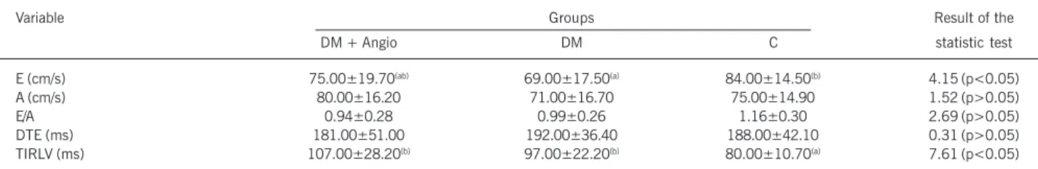
There were no differences among the groups regarding the systolic function indicators or left ventricular mass. Differences compatible with diastolic dysfunction in the two groups of dia-betic were observed, regardless of the presence of microangio-pathy, which showed significantly higher values of the times of isovolumetric relaxation of the left ventricle (TIRLV, ms): (DMII= 97±22.2; DM+A= 107±28.2 and C= 80±10.7; p<0.05), and lower values of the maximum speeds of the wave of fast ventri-cular filling (E, cm/s): (DMII= 69±17.5; DM+A= 75±19.7 and C= 84±14.5, p<0.05 between DMII and C). There was no diffe-rence among the groups concerning the E/A rate.
Conclusion
Normotensive patients with type II diabetes mellitus and without clinical signs of cardiovascular compromising showed signs of diastolic dysfunction, non-associated to the presence of microangiopathy.
Key words
echocardiogram, left ventricle, cardiomiopathy, diabetes mellitus
Mailing address: João Carlos Ferreira Braga - Rua Ametistas, 55 - 17516-080 - Marília, SP - Brazil
E-mail: [email protected] Sent for publishing on 04/29/2004 Accepted on 11/19/2004
Clinical, epidemiological and hystopathologic evidences indicate the existence of a specific cardiopathy related to the diabetes mellitus1. Indeed, non-invasive assessments of the cardiac function
in diabetic individuals frequently show the existence of abnormalities in both systolic and diastolic functions of the left ventricle, which can be evinced by means of many diagnostic methods. However, despite the regular use of the term diabetic cardiomiopathy, there is still a considerable debate on the exact nature and cause of the cardiac dysfunction found in diabetic individuals, non-carriers of coronary atherosclerosis and hypertension, which are pathologic conditions frequently associated to diabetes mellitus and that can lead to a cardiac dysfunction, according to the literature.
The existence of diabetic cardiomyopathy was first suggested by Rubler et al.1, who described myocardial hypertrophy and
fi-brosis, in addition to prominent endothelial and sub-endothelial proliferation, indicating that the changes in the small vessels could be involved in the pathogenesis of the myocardial dysfunction. From that initial description, and to this moment, there have still been disputes concerning the importance of the disease in the small vessels, of interstitial fibrosis, and the metabolic distur-bance in the pathogenesis of diabetic cardiomyopathy2-15.
Rossen15 has debated that, although the correlation of
car-diomyopathy with microangiopathy is doubtful, it may exist, having in mind the similarities among the abnormalities in the coronary microvascular function, which are observed in the diabetes mellitus and the idiopathic dilated cardiomyopathy.
Gutierrez and Higuchi 14, in our milieu, accept three hypotheses
to explain what they call of contingent causal effect of the diabetes as for the cardiac disease: 1) microangiopathy; 2) direct action on the cardiac fiber of the metabolic disturbances caused by the hyperglycemia, among other factors; 3) changes in the extracellular matrix, caused by a greater level of glycation in its components, with consequent changes in the muscular structure.
So far, the studies that have compared the ventricular function of diabetic patients with or without microangiopathy10,15-20 have
shown conflicting results. Recently, Liu et al.21 has found a relation
2
coronary artery disease, and left ventricle mass index. Once the microalbuminuria is a endothelial dysfunction marker in the glo-merular arteriole, Bell22 though valid to postulate that the
myo-cardial endothelial dysfunction causes an increase of healing and rigidity, also considering that such low-cost urinary test could anticipate a Doppler echocardiographic examination, which, al-though more sensitive to detect the diastolic dysfunction, it has a higher cost. After the diastolic dysfunction was documented, the therapy aiming at preventing the progression of the disease to cardiac insufficiency should have started.
By having in view that the echocardiographic examination is described as a sensitive method for the early detection of diabetic cardiomyopathy8, the present study aims at identifying whether
the presence of microangiopathy is associated to cardiac structural and functional changes in non-hypertensive and asymptomatic dia-betic patients, from the cardiovascular point of view.
Methods
All procedures were submitted to and approved by an Ethics Committee in Research Involving Human Beings.
A control-case cross examination was carried out involving 32 patients with diabetes mellitus, aged between 40 and 65 years old, and 20 normal voluntaries, with comparable age and sex. All participants in the study were submitted to a complete clinical assessment with a cardiologist.
The exclusion criteria were as follows: systolic blood pressure
≥ 140 mmHg or diastolic blood pressure ≥ 90 mmHg; signs or symptoms of cardio-respiratory disease; use of cardiovascular action drugs; acoustic fenestra unsuitable for the analysis of the echo-cardiogram.
The diabetic patients were submitted to laboratory tests for the assessment of renal function and of lipidic profile, and also a funduscopy contrasted with fluorescein and an examination of urine collected in 12 hours for the detection of microalbuminuria. The diagnosis of retinopathy and/or microalbuminuria was the criterion to define the presence of microangiopathy.
The DM+A group consisted of patients with signs of microan-giopathy (n=13); and the DM group for patients without mi-croangiopathy (n=19); and the control group of clinically normal voluntaries (n=20).
The funduscopy consisted of direct and indirect ophthalmoscopy under mydriasis; retinography with aneritra light, fluorescenic an-giography, through the injection 2.5 ml of intravenous sodic fluorscein at 10%. For the documentation of fluoresceinic angio-graphy and retinoangio-graphy, the TRC-FE model Topcon equipment and a 400-ASA Tri-X Pam Kodak film were used. The diagnosis of retinopathy was based on the presence of at least one microaneu-ryism in one of the eyes, or in the other changes such as he-morrhages, hard exudates, soft (cotton-like) exudates, and fibro-vascular proliferation23.
The urinary excretion of albumina was determined by means of a turbidimetry, by using the Behring Turbidimeter and the
0.3µg/ml sensitivity Turbiquant reactive agents. The examination
was performed in urine collected for 12 hours in a sterile flask and without any preservatives. The samples containing bacteriuria > 105 /mm3 were disregarded. The result was regarded as positive
for values greater than 15µ/min.
All procedures were carried out by a single qualified echocar-diographer, who did not have any knowledge on the group the individuals belonged to. The examinations were done by means of an ATL Apogee CX 200, equipped with a 2.0-3.5MHz multifre-quency ultrasonic transducer and image record system. During the procedure, the patient stayed in left lateral decubitus, with the left upper member slightly flexed under the head. An electro-cardiographic derivation was continuously monitored.
The images were obtained by following the recommendations of American Society of Echocardiography24, from usual
echocar-diographic sections. The sequence of measurements was: final systolic dimension of the outlet way of the LV (OWLV), anteropos-terior diastolic diameter of the left atrium (LA), final diastolic diameter (dLV), final systolic diameter (sLV), diastolic thicknesses of the IVS and of the posterior wall of LV (PW). Such dimensions were used for the calculation of:
LV mass index (LVMI, g/m2) =
{[(dIVS + dPWLVd + DdLV)3 - DDLV3]x1.04}-13.6
BS
Fraction of ejection (FE) =
[7/(2.4+DdLV)] x DdLV3 - [7/(2.4+DsLV)] x DsLV3
[7/(2.4+DdLV)] x DdLV3
Percentage of variation of the ventricular diameter (%∆D)= [(dLV - sLV) / deV] x 100
The evaluations of the flows followed the recommendations of the Canadian Consensus25 for the Doppler-echocardiographic
measurements, obtaining the following variables: c1) maximum speed of fast ventricular filling (peak of E wave, cm/s); c2) maximum speed of tardive filling, after atrial contraction (peak of A wave, cm/s); c3) deceleration time of E wave (DTE, ms), corresponding to the time interval between the peak of E wave and its extrapo-lation to the base line. Also with the pulsated Doppler, simultaneous flow curves at the outlet and inlet ways of the LV were obtained. The time interval between the end of the systolic flow and the beginning of the transmitral flow corresponded to the isovolumetric relaxation time of the LV (VIRT). The analysis of the systolic flow in the OWLV allowed for the calculation of the following variables: systolic index (SI, mL/BS) = (OWLV2 x 0.785 x IOWLV)/BS, in which OWLV is the measurement of the outlet way of the LV, IOWLV is the integral of the systolic flow in the OWLV and BS is the body surface; cardiac output (CO, L/min)= SV x HR.
The comparisons among the groups were done through ANOVA, followed by the test of Tukey. The level of significance p<0.05 was considered in all cases.
Results
Diabetic patients with microangiopathy showed average values of microalbuminuria of 36 mcg/min. In the same way, the most frequent changes observed in the contrasted fundoscopy were the presence of microaneurysms, a clinical picture classified as non-proliferative diabetic retinopathy23. From the 13 patients of DM+A
group, 9 showed isolated eyegrounds change, 2 only microalbu-minuria and 2 showed both changes.
microan-3
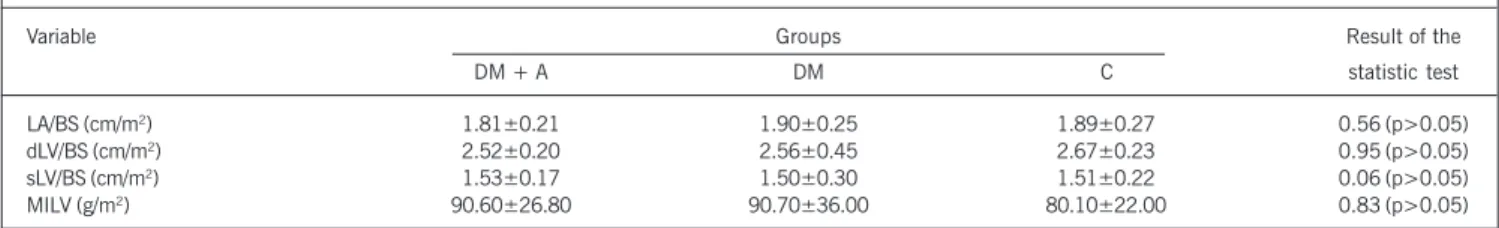
giopathy (DM + A), diabetes mellitus without microangiopathy (DM)and control (C) groups. The values of the means, their respective standard deviations, and the results from the statistic test are shown. Concerning the age, body weight and body surface variables, the 3 groups were statistically similar among each other. The comparison of hemodynamic data evinced that the groups do not differ in relation to the systolic blood pressure and diastolic blood pressure. Concerning the heart rate, the DM group showed a value, statistically significant, greater than the C group value (74±10.30 bpm vs. 65±9.40 bpm, p< 0.05). In table II, the cardiac morphometric variables, normalized for the body surface were observed. They were obtained through the M-mode echocardiogram, from the diabetes mellitus with microangio-pathy (DM + A), diabetes mellitus without microangiomicroangio-pathy (DM) and control (C) groups. Significant differences among those three groups were not observed.
The variables of systolic function, obtained through Doppler echocardiography, from the three groups studied are in table III. There were no statistic differences among them and, in table IV, the variables of diastolic function. The maximum sped of fast ventricular filling (E) was similar in the DM + A (75±19.70 cm/s)
and DM (69±17.50 cm/s) groups. The value obtained in that last group was significantly lower than that observed in the control group (84±14.50 cm/s). The isovolumetric relaxation time of the left ventricle (IRTLV) showed statistically similar values in the DM + A (107±28.20 ms) and DM (97±22.20 ms) patients. Both groups were different from the control group, which showed a significantly lower value (80±10.70 ms).
Discussion
The absence of differences among the patient groups, in relation to the morphometric and systolic function variables, was not sur-prising. The inclusion and exclusion criteria were very specific and restricted the patient sampling, which was already discussed. That fact, despite having contributed to decrease the power of evincing potential among the groups, was in accordance to the proposal of assessing the association between the presence of signs of diabetic microangiopathy and cardiac morphofunctional changes in groups that are similar in all the other aspects, except the lesion of small vessels. It was also necessary to restrict the
Table I - Age, body weight, body surface and hemodynamic variables of the individuals from diabetes mellitus with microangiopathy (DM + A), diabetes mellitus without microangiopathy (DM) and control (C) groups. The values of the means, their respective standard deviations and the results of the statistic test are shown
Variable Groups Result of the
DM + A DM C statistic test
Ag (years) 55.00±6.90 50.90±8.47 51.20±9.41 1.06 (p>0.05)
BW (Kg) 71.60±11.24 72.30±12.54 68.50±14.97 0.45 (p>0.05)
BS (Kg/m2) 1.81±0.18 1.79±0.18 1.76±0.24 0.27 (p>0.05)
SBP (mmHg) 120.00±13.60 ±11.50 112.00±10.57 2.11 (p>0.05)
DBP (mmHg) 79.00±8.20 ±7.90 76.00±5.90 1.24 (p>0.05)
HR (bpm) 71.00±8.40(ab) 74.00±10.30(b) 65.00±9.40(a) 4.82 (p<0.05)
Ag - age; BW - body weight; BS - body surface; SBP and DBP - systolic and diastolic blood pressure, respectively; HR - heart rate. Small letters represent statistically significant difference among the groups (ANOVA and Tukey).
Table II - Cardiac morphometric variable, normalized for the body surface (BS), obtained through the M - mode echocardiogram, in the individuals from the diabetes mellitus with microangiopathy (DM + A), diabetes mellitus without microangiopathy (DM) and control (C) groups. The values of the means,
their respective standard deviations and the results of the statistic test are shown
Variable Groups Result of the
DM + A DM C statistic test
LA/BS (cm/m2) 1.81±0.21 1.90±0.25 1.89±0.27 0.56 (p>0.05)
dLV/BS (cm/m2) 2.52±0.20 2.56±0.45 2.67±0.23 0.95 (p>0.05)
sLV/BS (cm/m2) 1.53±0.17 1.50±0.30 1.51±0.22 0.06 (p>0.05)
MILV (g/m2) 90.60±26.80 90.70±36.00 80.10±22.00 0.83 (p>0.05)
LA - left atrium; dLV and sLV - diastolic and systolic diameters of the left ventricle, respectively; MILV - mass index of the left ventricle; ANOVA.
Table III - Variables of systolic function, obtained through Doppler-echocardiography in the individuals from the diabetes mellitus with microangiopathy (DM + A), diabetes mellitus without microangiopathy (DM) and control (C) groups. The values of the means, their respective standard deviations and the results of the statistic test are shown
Variable Groups Result of the
DM + A DM C statistic test
Delta d 0.39±0.06 0.41±0.05 0.43±0.07 1.91 (p>0.05)
FE 0.69±0.07 0.72±0.05 0.74±0.07 2.23 (p>0.05)
CO (L/min) 4.25±0.98 4.20±1.26 3.94±1.29 0.33 (p>0.05)
SI (mL/m2) 32.80±6.00 32.30±10.80 34.00±6.90 0.23 (p>0.05)
FSS (g/cm2) 51.60±12.20 49.40±12.90 47.00±14.10 0.49 (p>0.05)
4
analysis to the diabetic patients non-carrier of other cardiovascular diseases. Besides, that has been the problem faced by researchers interested in the subject. For example, many studies with greater casuistry have concluded on left ventricular dysfunction associated with diabetes, by including normotensive and hypertensive patients in the same group16,26,27. Cosson et al.8 attributed the published
contradictory results to the lack of homogeneity of the population studied and the uniformity of the echocardiographic indexes used. So, our conclusions are only applicable to groups of individuals with similar characteristics to those assessed in the present study. The study by Framingham22 showed that diabetic women had a
left ventricle mass 10% greater than those non-diabetic. The Tay-side study28 showed that the ventricular hypertrophy was present
in 32% of the normotensive diabetic patients, who did not use drugs that inhibited the enzyme of conversion of the angiotensin and did not have coronary disease. Besides, hypertensive and dia-betic women showed a greater level of left ventricular hypertrophy and increase of the left atrium, when compared to non-diabetic hypertensive ones29. While the myocardial fibrosis seems to be
related to the hyperglycemia, the left ventricular hypertrophy has been more related to the insuline-resistence syndrome30,31. Although
the left ventricular hypertrophy is more prevailing in diabetic patients and such fact is related to ventricular dysfunction 22,32, there was
no variation in the left ventricular mass in our casuistry.
Despite the rest-preserved systolic function, a considerable proportion of those patients showed diastolic changes at the Doppler-echocardiographic examination. Those findings are in ac-cordance to the clinical observations by other authors who showed the diastolic diabetes mellitus8,32,33.
The study by Framingham convincingly showed that diabetic patients show addition risk for the development of cardiomyopathy and cardiac insufficiency34. However, the nature of such association
reported in that classic epidemiological study wa.s not completely clear. The identification, in which many of the patients with clinical signs of cardiac failure objectively showed preserved systolic func-tion, caused a huge interest in researchers and clinicians on the condition of the diastolic ventricular function in those patients8,32,33.
This diastolic compromising that precedes the systolic changes in the evolution of the functional changes in the diabetic heart has been observed even in the absence of coronary artery disease and left ventricular hypertrophy. Many reports have shown preva-lence’s from 30 to 60% of diastolic dysfunction in well-controlled and normotensive diabetic individuals22,32,35-37.
An interesting finding in the present study was the increase of
the isovolumetric relaxation time in the diabetic patients, when compa-red to the controls. Despite such increase, in the average, is not of sufficient magnitude to extrapolate the values regarded as normal, it can be acknowledged that some level of compromising of the myocar-dial relaxation process must be current in those patients.
The myocardial relaxation happens as a consequence of the removal of the ion Ca2+ from the cytoplasm to the inside of the
sarcoplasmic reticule, after the myocardial contraction, in a complex and active process, with great consumption of ATP and involving many proteins. Therefore, the extension of the myocardial relaxation time can be the result from a large quantity of potential subcellular changes that may impair the relaxation.
The extension of the myocardial relaxation time influences the fast ventricular filling and could be the mechanism subjacent to the observation of decreased values at the peak of the E wave. The ventricular filling depends fundamentally on the atrioventricular pres-sure gradient, during that stage in the cardiac cycle. That prespres-sure gradient is directly influenced by the intra-atrial pressure and, there-fore, by its volume, and inversely influenced by the intraventricular pressure. So, the delayed or incomplete myocardial relaxation promo-tes an increase in the ventricular diastolic pressure, a reduction in the transmitral gradient and a decrease of the speed of the initial diastolic flow. There is also the possibility that myocardial interstitial changes, with the increase of collagen concentration, have taken part in the induction process of the diastolic function38.
On the contrary we expected, we did not observe significant changes between the two groups of diabetic patients. Although in the literature, the presence of albuminuria in diabetic individuals identifies that high risk of presence of cardiovascular disease, our data suggest, that its presence is not related to diastolic dysfunction of the left ventricle. Such change is regarded as the initial mani-festation of diabetic cardiomyopathy21,33. In that aspect, it is
re-levant to consider the size of the sample. That means, differences among the groups could be statistically demonstrated, if the number of patients were greater. However, given the similarity among the average values of the diastolic function index in the DM and DM+A groups, only the inclusion of hundreds of patients in the study would allow for the demonstration. Despite such limitations, we regarded as valid the disclosure of the results in the present study, by having in view that similar studies can be carried out and, together, allow for in the future solving that relevant matter con-cerning the physiopathology of the cardiomyopathy of the diabetic individual.
Finally, we regard as great importance the observation that
Table IV - Variables of diastolic function, obtained through Doppler-echocardiography in the individuals from the diabetes mellitus with microangiopathy (DM + A), diabetes mellitus without microangiopathy (DM) and control (C) groups. The values of the means, their respective standard deviations and the results of the statistic test are shown
Variable Groups Result of the
DM + Angio DM C statistic test
E (cm/s) 75.00±19.70(ab) 69.00±17.50(a) 84.00±14.50(b) 4.15 (p<0.05)
A (cm/s) 80.00±16.20 71.00±16.70 75.00±14.90 1.52 (p>0.05)
E/A 0.94±0.28 0.99±0.26 1.16±0.30 2.69 (p>0.05)
DTE (ms) 181.00±51.00 192.00±36.40 188.00±42.10 0.31 (p>0.05)
TIRLV (ms) 107.00±28.20(b) 97.00±22.20(b) 80.00±10.70(a) 7.61 (p<0.05)
5
1. Rubler S, Dlugash J, Yuceoglu YZ, Kumral T, Branwood AW, Grishman A. Newtype of cardiomyopathy associated with diabetic glomerulosclerosis. Am J Cardiol 1972;30:595-602.
2. Uusitupa MI, Mustonen JN, Airaksinen KE. Diabetic heart muscle disease. Ann Med 1990;22:377-86.
3. AronsonD, Rayfield E. Diabetes. In: Topol E, editor.Textbook of Cardiovascular Me-dicine. 2nd edition. Philadelphia, PA; Lippincott Williams & Wilkins, 2002:171-94. 4. Nesto RW, Libby P. Diabetes mellitus and the cardiovascular system. In: Braunwald E, Zipes DP, Libby P, editors. Heart Disease. A Textbook of Cardiovas-cular Medicine. 6th edition. Philadelphia, PA; WB Saunders, 2001:2133-50.
5. Uusitupa MI, Mustonen JN, Laakso M et al. Impairment of diastolic function in middle-aged type 1 (insulin-dependent) and type 2 (non-insulin-dependent) diabe-tic patients free of cardiovascular disease. Diabetologia 1988;31:783-91. 6. Uusitupa MI, Siitonen O, Pyorala K, Lansimies E. Left ventricular function in newly
diagnosed non-insuldependent (type 2) diabetics evaluated by systolic time in-tervals and echocardiography. Acta Med Scand 1985;217:379-88.
7. Vered A, Battler A, Segal P et al. Exercise-induced left ventricular dysfunction in young men with asymptomatic diabetes mellitus (diabetic cardiomyopathy). Am J Cardiol 1984; 54:633-7.
8. Cosson S, Kevorkian J. Left ventricular diastolic dysfunction: an early sign of dia-betic cardiomypathy ? Diabetes Metab. 2003;29:455-66.
9. Codinach HP, Freixa PR. Diabetic cardiomyopathy: concept, heart function, and pathogenesis. An Med Interna 2002;19:313-20.
10. Brown HB, Waugh NR, Jennings PE. Microangiopathy as a prognostic indicator in dia-betic patients suffering from acute myocardial infarction. Scott Med J 1992; 37:44-6. 11. Bauters C, Lamblin N, Mc Fadden EP, Van Belle E, Millaire A, De Groote P. Influ-ence of diabetes mellitus on heart failure risk and outcome. Cardiovascular Diabetology 2003,2:1.
12. Wolfenbuttel B, Boulanger C, Crijns FR et al. Breakers of advanced glycation end products restore large artery properties in experimental diabetes. Proc Natl Acad Sci USA 1998; 95:4630-4.
13. Chaves FR, Jorge PAR. Miocardiopatia diabética. Arq Bras Endocrinol Metab 1998; 42:134-9.
14. Gutierrez PS, Higuchi ML. Alterações cardíacas e vasculares no diabete: aspectos anatomopatológicos. Rev Soc Cardiol Estado São Paulo 1998;8:1020-4. 15. Rossen JD. Abnormal microvascular function in diabetes: relationship to diabetic
cardiomyopathy. Coron Artery Dis 1996;7:133-8.
16. Devereux RB, Roman MJ, Paranicas M et al. Impact of diabetes on cardiac stru-ture and function; the strong heart study. Circulation 2000;101:2271-6. 17. Zarich SW, Arbuckle BE, Cohen LR. Diastolic abnormalities in young asymptomatic
diabetic patients assessed by pulsed Doppler echocardiography. J Am Coll Cardiol 1988;12:114-20.
18. Nitenberg A, Ledoux S, Valensi P, Sachs R, Attali JR, Antony I. Impairment of coro-nary microvascular dilatation in response to cold pressor-induced symphatetic sti-mulation in type 2 diabetic patients with abnormal stress thallium imaging. Diabetes 2001;50:1180-5.
19. Di Bonito P, Cuomo S, Moio N, Sibilio G, Sabatini D, Capa B. Diastolic dysfunction in patients with non-insulin-dependent diabetes mellitus of short duration. Diabet Med 1996; 13: 321-4.
20. Annonu AK, Fattah AA, Mokhtar MS, Ghareeb S, Elhendy A. Left ventricular
sys-References
tolic and diastolic functional abnormalities asymptomatic patients with non-insu-lin-dependent diabetes mellitus. J Am Soc Echocardiogr 2001;14:885-91. 21. Liu JE, Robbins DC, Palmieri V et al. Association of albuminuria with systolic and
diastolic left ventricular dysfunction in type 2 diabetes. The Strong Heart Study. J Am Coll Cardiol 2003;41:2022-8.
22. Bell DSH. Diabetic Cardiomyopathy. Diabetes Care 2003;26:2949-50. 23. American Diabetes Association. Clinical Practice Recommendations 1995. Screening
for diabetic retinopathy. Diabetes Care 1995;18:21-3.
24. Sahn DJ, DeMaria A, Kisslo J, Weyman A. Recommendations regarding quantita-tion in M-mode echocardiography: results of a survey of echocardiographic meas-urements. Circulation 1978;58:1072-83.
25. Rakowski H, Appleton C, Chan Kl et al. Canadian consensus recommendations for the measurement and reporting of diastolic dysfunction by echocardiography. J Am Soc Echocardiogr 1996; 9: 736-60.
26. Mustonen JN, Uusitupa MI, Tahavanainen K et al. Impaired left ventricular systo-lic function during exercise in middle-aged insulin-dependent and noninsulin-de-pendent diabetic subjects without clinically evident cardiovascular disease. Am J Cardiol 1988;62:1273-9.
27. Tischler MD. Clinical abnormalities of cardiac function and echocardiographic tis-sue characterization in diabetes mellitus. Coron Artery Dis 1996;7:139-42. 28. Struthers AD, Morris AD. Screening for and treating left-ventricular abnormalities
in diabetes mellitus: a new way of reducing cardiac deaths. Lancet 2002;359: 1430-32.
29. Tenembaum A, Fisman EZ, Schwammenthal E, Adler Y, Benderly M, Shemesh J. Increased prevalence of left ventricular hypertrophy in hipertensive women with type 2 diabetes mellitus.Cardiovasc Diabetol 2003;2:14.
30. Galvan AQ, Galetta F, Natali A et al. Insulin resistance and hyperinsulinemia: No inde-pendent relation to left ventricular mass in humans. Circulation 2000;102:2233-8. 31. Rutter MK, Parise H, Benjamin EJ et al. Impact of glucose intolerance and insulin resistance on cardiac structure and function: sex-related differences in the Fra-mingham Heart Study. Circulation 2003;107:448-54.
32. Zabalgoitia M, Ismaeil MF, Anderson L, Maklady FA. Prevalence of diastolic dys-function in normotensive, asymptomatic patients with well-controlled type 2 dia-betes mellitus. Am J Cardiol 2001;87: 320-3.
33. Schannwell CM, Schneppenheim M, Perings S, Plehn G, Strauer BE. Left ventricu-lar diastolic dysfunction as an early manifestation of diabetic cardiomyopathy. Cardiology 2002; 98: 33-9.
34. Kannel W, Hjortland M, Castelli WP. Role of diabetes in congestive heart failure: the Framingham study. Am J Cardiol 1974;34:29-34.
35. Fang ZY, Najos-Valencia O, Leano R, Marwick TH. Patients with early diabetic heart disease demonstrate a normal myocardial response to dobutamine. J Am Coll Cardiol 2003; 42: 451-2.
36. Poirier P, Garneau C, Bogaty P et al. Impact of left ventricular diastolic dysfunction on maximal treadmill performance in normotensive subjects with well controlled type 2 diabetes mellitus. Am J Cardiol 2000;85:473-7.
37. Picano E. Diabetic Cardiomyopathy: the importance of being earliest. J Am Coll Cardiol 2003;42:454-6.
38. Liu J, Masurekar MR, Vatner DE et al. Glycation end-product cross-link breaker reduces collagen and improves cardiac function in aging diabetic heart. Am J Physiol Heart Circ Physiol 2003;285:2587-91.
normotensive diabetic patients and without clinical signs of cardiac disease show suggestive changes of diastolic dysfunction, when compared to their non-diabetic controls. Those results reinforce