UNIVERSIDADE DA BEIRA INTERIOR
Ciências da Saúde
Leptin and Sertoli cells mitochondrial
bioenergetics
Bruno Manuel Pereira Moreira
Dissertação para obtenção do Grau de Mestre em
Ciências Biomédicas
(2º ciclo de estudos)
Orientador: Prof. Doutor Marco G. Alves (ICBAS-UP/CICS-UBI)
Co-orientador: Prof. Doutor Pedro Moradas-Ferreira (ICBAS-UP)
Co-orientador: Prof. Doutor Pedro F. Oliveira (ICBAS-UP)
Co-orientador: Prof.ª Doutora Branca M. Silva (CICS-UBI)
Agradecimentos
A elaboração desta dissertação de mestrado contou com inúmeros apoios, sem os quais este marco académico não seria alcançado. Por essa razão, desejo expressar o meu sincero obrigado a todos que de uma forma direta ou indireta contribuíram para o culminar desta etapa.
Ao meu orientador, Professor Doutor Marco Alves, pela disponibilidade incansável na orientação deste trabalho, pela competência científica, acompanhamento e conselhos dados durante este percurso, assim como pelas sugestões, críticas e opiniões que foram fundamentais para o melhoramento dos trabalhos feitos ao longo da dissertação. Sem a sua orientação este trabalho não seria possível.
Aos meus co-orientadores, Professor Doutor Pedro Oliveira, Professor Doutor Pedro Moradas-Ferreira e Professora Doutora Branca Silva pela disponibilidade demonstrada, pela competência científica e pelas sugestões, críticas e opiniões que contribuíram para o desenvolvimento deste trabalho.
Aos meus colegas de laboratório: Tânia Dias, Raquel Bernardino, Luís Crisóstomo, Tito Jesus, Susana Almeida, Maria João, Bernardo Rodrigues, Hugo Silva, Tatiana Yashechkina, David Carrageta, João Monteiro, Marwa Boussada, e em especial à Ana Martins pela incansável ajuda, disponibilidade e acompanhamento ao longo deste ano bem como pelo bom ambiente de trabalho.
Aos amigos que fiz durante o curso de Ciências Biomédicas, pelos valiosos conselhos, pelos momentos passados e pelo apoio. Apesar da distância estiveram sempre presentes.
À Ana Laura, pelo apoio nos momentos menos bons, pelo companheirismo e pela paciência e compreensão demonstradas ao longo deste ano.
À minha família, em especial aos meus pais e irmã, por estarem sempre lá quando necessário, pela motivação e pelo apoio incondicional em todos momentos. Este trabalho também é vosso.
Resumo
As doenças metabólicas, entre elas a obesidade, são um dos grandes desafios do século XXI. A incidência da obesidade em indivíduos do sexo masculino em idade reprodutiva tem vindo a aumentar, e as previsões indicam que esta tendência se irá manter. Na direção oposta está o decréscimo dos parâmetros de fertilidade destes indivíduos, o que se tem vindo a refletir no aumento da recorrência de casais a clínicas de fertilidade. O fator masculino de forma isolada ou em conjunto com o fator feminino está presente em um terço dos casais que procuram tratamentos de fertilidade. Desta forma, não é surpreendente a queda abrupta dos parâmetros espermáticos que se tem vindo a verificar nas últimas décadas, atingindo valores preocupantes.
Os indivíduos com excesso de peso e obesos apresentam uma disfunção hormonal, relacionada principalmente com a presença de valores elevados de leptina. Além das funções já descritas a nível hipotalâmico, esta hormona apresenta diversas funções em tecidos periféricos. No entanto, apesar de já ter sido descoberta há duas décadas, os seus efeitos no trato reprodutor masculino, principalmente nas células de Sertoli, continuam por desvendar. Recentemente, diversos estudos demonstraram a capacidade da leptina de modular as dinâmicas mitocondriais, incluindo a sua biogénese e funcionamento em vários sistemas celulares, incluindo células cancerígenas. Neste trabalho, estudamos o efeito da leptina na proliferação e atividade metabólica das células de Sertoli de rato. Também avaliamos os efeitos da leptina na fisiologia mitocondrial, particularmente nos níveis dos complexos mitocondriais, níveis de ARN mensageiro (mRNA) de genes envolvidos na biogénese mitocondrial e o potencial mitocondrial de membrana. Para efeitos comparativos, e tendo em conta resultados prévios do nosso grupo, também avaliamos os efeitos da leptina nos níveis de mRNA de genes envolvidos na biogénese mitocondrial e nos níveis dos complexos mitocondriais em células de Sertoli humanas.
Os nossos resultados indicam que a leptina modula a atividade metabólica e função mitocondrial nas células de Sertoli de rato na concentração de 50 ng/mL, uma concentração que está presente em indivíduos com obesidade mórbida. Estes resultados sugerem que altas concentrações de leptina, como esta que é encontrada nestes indivíduos, modula a função mitocondrial nas células de Sertoli de rato o que pode representar um mecanismo novo através do qual a leptina contribui para a subfertilidade e infertilidade induzida pela obesidade em indivíduos do sexo masculino. No entanto, a exposição à leptina não teve efeito em vários parâmetros da fisiologia mitocondrial. Os níveis de mRNA de genes envolvidos na biogénese mitocondrial e os níveis dos complexos mitocondriais não apresentaram alterações, o que fortalece a hipótese de que a leptina modula a função mitocondrial e não a sua
fisiologia. Nas células de Sertoli humanas, os níveis de mRNA da Sirtuina 1 (SIRT1) apresentaram alterações no grupo exposto a uma concentração de 50 ng/mL de leptina. Os níveis proteicos do complexo mitocondrial II também apresentaram alterações no grupo exposto às concentrações de 5 e 50 ng/mL, ao passo que nas células de Sertoli de rato isto não se verificou, o que indica que existem respostas à leptina nas células de Sertoli que são dependentes da espécie. Estas diferenças, principalmente nos níveis de mRNA da SIRT1, podem representar um mecanismo novo através do qual a leptina afeta o controlo metabólico da espermatogénese, com possíveis consequências nas células de Sertoli humanas.
Palavras-chave
Resumo alargado
O sistema reprodutor masculino é constituído por diversas estruturas que podem ser divididas em órgãos sexuais primários e órgãos sexuais secundários. Os testículos fazem parte da primeira categoria, sendo considerados os elementos centrais do sistema reprodutor masculino. Eles são responsáveis pela síntese de esteroides, os quais estão envolvidos no desenvolvimento dos órgãos sexuais secundários, e pela produção dos gâmetas masculinos, os espermatozoides. Nos testículos estão presentes diversos tipos celulares com funções específicas. As células de Sertoli desempenham um papel fundamental no desenvolvimento funcional dos testículos e no processo de produção de gâmetas masculinos, a espermatogénese. Estas células fornecem o suporte físico e nutricional às células germinativas nas diversas fases da espermatogénese, desde espermatogónias até atingirem o estado de espermatozoides, os quais são posteriormente libertados no lúmen dos túbulos seminíferos. Nesta fase, os espermatozoides são células diferenciadas, mas incapazes de fertilizar o óvulo. Essa capacidade é adquirida nas seguintes etapas de maturação e capacitação que ocorrem no epidídimo e ao longo do trato reprodutor feminino, respetivamente. Além do suporte físico e nutricional, as células de Sertoli também produzem lactato em elevadas quantidades, que funciona como fonte de energia para as células germinativas. Neste processo, as células de Sertoli metabolizam a glicose em piruvato e este depois é metabolizado em lactato, num processo conhecido como glicólise. Esta cooperação metabólica entre estas as células de Sertoli e as células germinativas é fundamental. No entanto, o ambiente dos túbulos seminíferos é suscetível a variações de hormonas e metabolitos, o que torna a regulação desta cooperação metabólica crucial para uma espermatogénese bem-sucedida. A obesidade, caracterizada por uma forte desregulação a nível hormonal especialmente ao nível do eixo leptina-grelina, é uma patologia cuja proporção pandémica tem vindo a preocupar especialistas e políticos. A obesidade em indivíduos do sexo masculino em idade reprodutiva tem também acompanhado esta tendência exponencial de crescimento. Ao mesmo tempo, a qualidade da reprodução desses indivíduos tem seguido uma tendência inversa. De facto, vários estudos têm relacionado o aumento do índice de massa corporal com o decréscimo dos parâmetros reprodutivos como a concentração espermática e a motilidade. No entanto, apesar das relações estabelecidas entre a obesidade e a diminuição do potencial reprodutivo masculino, pouco se sabe sobre os mecanismos responsáveis por essas alterações.
A leptina, produzida principalmente nos adipócitos, é uma hormona com uma estrutura semelhante a algumas citocinas estando envolvida na sensação de saciedade. Em indivíduos com um índice de massa corporal normal, valores de leptina elevados desencadeiam uma reação que promove a redução do consumo de energia na forma de
comida e promove o gasto energético. Em indivíduos obesos este mecanismo está desregulado o que provoca a acumulação de leptina sem ocorrer o desencadeamento de uma resposta proporcional. Isto acontece devido a um fenómeno designado por resistência à leptina. Além da sua função como regulador de apetite, vários estudos já conseguiram identificar diversas ações da leptina em tecidos periféricos. No entanto, apesar de já ter sido descoberta há duas décadas, ainda pouco se sabe sobre os seus efeitos no trato reprodutor masculino, particularmente nas células de Sertoli. Nos últimos anos, diversos estudos têm-se focado no papel da leptina na mitocôndria, particularmente em células cancerígenas. O metabolismo das células cancerígenas tem diversos pontos de contacto com o metabolismo das células de Sertoli, uma vez que ambas priorizam a glicólise em detrimento da fosforilação oxidativa. Alguns desses estudos demonstraram que a leptina tem a capacidade de modular a função mitocondrial, incluindo a sua biogénese.
O objetivo deste trabalho foi o de investigar os efeitos da leptina na proliferação e atividade metabólica de células de Sertoli de rato. Além disso, também foram avaliados os efeitos da leptina na fisiologia mitocondrial, tanto nos níveis de ARN mensageiro (mRNA) de marcadores da biogénese mitocondrial como no potencial de membrana e nos níveis de proteína dos complexos mitocondriais. Para isso, foi usada uma linha de células de Sertoli de rato (SerW3), cultivadas na ausência e na presença de concentrações crescentes de leptina de forma a mimetizar diferentes condições fisiológicas. Foram usadas três concentrações de leptina: 5 ng/mL, um valor encontrado em ratos e humanos com um índice de massa corporal considerado normal; 25 ng/mL, um valor reportado na literatura como estando presente em modelos de obesidade animal e em humanos obesos; e 50 ng/mL, um valor encontrado em humanos com obesidade mórbida. Apesar de não existirem dados sobre qual a concentração de leptina presente em ratos com obesidade mórbida uma vez que este modelo animal não existe, achamos pertinente avaliar os efeitos desta concentração em ratos. Para efeitos comparativos, e tendo em conta os resultados obtidos recentemente pelo nosso grupo em células de Sertoli humanas expostas a leptina, também foram avaliados os efeitos da leptina nos níveis de mRNA de marcadores da biogénese mitocondrial e nos níveis de proteína dos complexos mitocondriais em células de Sertoli humanas. Para tal foi usada uma linha de células de Sertoli humanas cultivadas nos mesmos parâmetros que as células de Sertoli de rato.
Os resultados obtidos revelam que a leptina modula a função mitocondrial numa concentração de 50 ng/mL em células de Sertoli de rato, mas não tem efeitos nos restantes parâmetros avaliados da fisiologia mitocondrial. Isto sugere que elevadas concentrações de leptina, como acontece em indivíduos com obesidade mórbida, induz disfunção mitocondrial. Os nossos resultados demonstram que não há alterações a nível da biogénese e expressão dos complexos mitocondriais nas células expostas a essa concentração de leptina, pelo que sugerimos alterações na funcionalidade, o que terá possíveis implicações na
espermatogénese. Os resultados obtidos em células de Sertoli humanas revelam que estas e as células de Sertoli de rato respondem de forma diferente à exposição a leptina. Esta resposta dependente da espécie, já reportada quando analisados outros parâmetros de funcionamento biológico, evidencia as cautelas que se devem ter quando se discutem trabalhos em animais e se tenta fazer a translação para humanos. Nas células de Sertoli humanas foram obtidas diferenças nos níveis de mRNA da Sirtuina 1 (SIRT1) o que, devido ao seu papel no controlo metabólico e na glicólise, suporta trabalhos anteriores do grupo em que foi demonstrado que a leptina afeta a glicólise nas células de Sertoli humanas e consequentemente a espermatogénese. No entanto, mais estudos são necessários para estudar esta via de sinalização e a sua relevância nestes indivíduos em que a concentração de leptina está muito elevada. Assim, a leptina atua sobre as células de Sertoli, apresentando modos de ação distintos entre espécies de rato e humanos, sendo que a disfunção nos níveis normais desta hormona pode comprometer o suporte nutricional da espermatogénese.
Abstract
Metabolic diseases, such as obesity, stand as one of the greatest challenges of the 21st
century. Obesity in reproductive-age men has risen and is expected to continue to increase. In an inverse direction, fertility is decreasing in those men and it largely contributes for the high demand of fertility treatment by couples in modern societies. The male factor alone or in combination with female factor is present in 1/3 of the couples seeking for fertility treatment. In fact, sperm parameters are on a downward spiral during the last decades reaching worrying levels.
In overweight and obese individuals, there is a hormonal dysfunction, particularly in leptin levels that are heavily increased. Besides the well-described functions at the hypothalamic level, leptin acts in several peripheral tissues. Although leptin has been on spotlight since its discovery, its effects in the male reproductive tract, particularly on Sertoli cells (SCs), remain unknown. More recently, leptin has shown the ability to modulate mitochondrial dynamics, biogenesis and functioning in several cellular systems, including cancer cells. Herein, we studied the effects of leptin in the proliferation and metabolic activity of rat Sertoli cells (rSCs). We also evaluated the effects of leptin in mitochondria physiology, particularly in the levels of mitochondrial complexes, messenger RNA (mRNA) levels of mitochondrial biogenesis markers and mitochondrial membrane potential. For comparative purposes, and taking in consideration previous results from the group, we also studied the effects of leptin in mRNA levels of mitochondrial biogenesis markers and mitochondrial complexes in human Sertoli cells (hSCs).
Our results suggest that leptin modulates the metabolic activity and mitochondrial function in rSCs after exposure to a concentration of 50 ng/mL, which mimics a concentration found in morbidly obese men. These findings suggest that high concentrations of leptin, such as those found in morbidly obese individuals, modulate mitochondrial function in rSCs, which could represent a novel mechanism through which leptin contributes to obesity-induced subfertility or infertility in males. Interestingly, leptin exposure had no effect in several aspects of mitochondria physiology, such as mRNA levels of mitochondrial biogenesis markers and levels of mitochondrial complexes which further indicates that leptin seems to affect mitochondrial function. In hSCs, the mRNA levels of Sirtuin 1 (SIRT1) presented changes in the group treated with 50 ng/mL of leptin. Protein levels of mitochondrial complex II presented changes in the groups treated with 5 and 50 ng/mL of leptin while in rSCs no differences were observed. Thus, rSCs and hSCs seem to be differently affected by leptin exposure. These differences, particularly in SIRT1 mRNA levels, are species-dependent and may represent a novel mechanism through which leptin affects the metabolic control of spermatogenesis and thus, with implications in hSCs.
Keywords
Leptin; Obesity; Sertoli cells; Spermatogenesis; Infertility; Mitochondria.
Table of Contents
I. Introduction ... 1
1. Male reproductive system: an overview ... 1
1.1 Functional organization of the testes ... 3
1.2 Sertoli cells ... 4
2. Spermatogenesis ... 6
2.1 Hormonal regulation of spermatogenesis ... 8
3. Leptin: from discovery to energy control ... 10
3.1 Leptin as a satiety hormone ... 11
3.1.1 Leptin and LepR expression in testis ... 13
3.2 Overview of leptin signaling and its relevance in male reproductive tract ... 15
4. Leptin – devil or angel in obesity ... 18
4.1 Leptin resistance in obesity ... 19
5. Hormonal network of leptin and male reproduction – the hypothalamus-pituitary-gonadal axis ... 22
6. Leptin actions in reproductive function ... 25
6.1 Obesity – a major cause for (in)fertility ... 28
6.2 Mitochondria in male reproduction – a connection with leptin ... 29
II. Aim of the Project ... 33
III. Materials and Methods ... 35
1. Chemicals ... 35
2. Cell line culture ... 35
3. Experimental design ... 36
4. Cell proliferation assay ... 36
5. Cell viability assay ... 37
6. Mitochondrial membrane potential ... 37
7. Total protein extraction ... 37
8. Western blot ... 38
9. RNA and DNA extraction ... 38
10. Reverse Transcriptase Polymerase Chain Reaction (RT-PCR) ... 39
11. Quantitative PCR (qPCR) ... 39
12. Mitochondrial DNA copy number determination ... 40
13. Statistical analysis ... 41
IV. Results ... 43
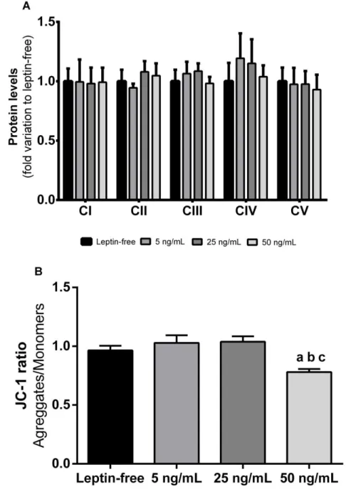
1. Leptin alters rat Sertoli cells metabolic activity but has no effect in their proliferation 43 2. Exposure of rat Sertoli cells to a supraphysiological concentration of leptin decreases mitochondrial membrane potential ... 44
3. Leptin does not alter the expression levels of genes involved in mitochondrial biogenesis
and copy number in rat Sertoli cells ... 46
4. Exposure of human Sertoli cells to a supraphysiological concentration of leptin increases SIRT1 mRNA levels... 48
5. Exposure of human Sertoli cells to leptin modulates mitochondrial complex II expression ... 49 V. Discussion ... 51 VI. Conclusion ... 59 VII. References ... 61 VIII. Annex I ... 83 IX. Annex II ... 85
List of Figures
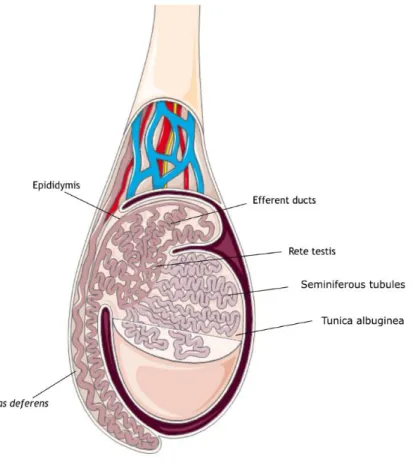
Figure 1: Schematic representation of a human testis. The testis is coated by tunica
albuginea, and divided in lobules. The seminiferous tubules are located inside these lobules, highly coiled and organized. The rete testis is responsible for transporting the spermatozoa from the seminiferous tubules into the efferent ducts. From this point forward, spermatozoa enter the epididymis where they go through several processes until they are ready to leave the male reproductive tract into the female reproductive tract. ... 2
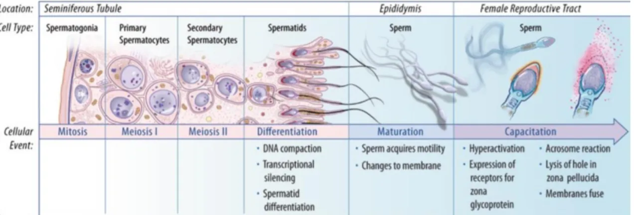
Figure 2: Schematic representation of major events in spermatogenesis. Spermatogonia go
through successive mitosis until they differentiate into primary spermatocytes. After the first meiotic division, secondary spermatocytes are formed. Meiosis II follows, which yields haploid spermatids. The spermatids then go through spermiogenesis (differentiation) followed by spermiation, in which they are released into the lumen of the seminiferous tubule. Afterwards, they go through the final steps of maturation in the epididymis, becoming motile. The final steps are achieved in the female reproductive tract, where they go through several biochemical changes, a process known as capacitation. Adapted from (Sharma and Agarwal 2011). ... 8
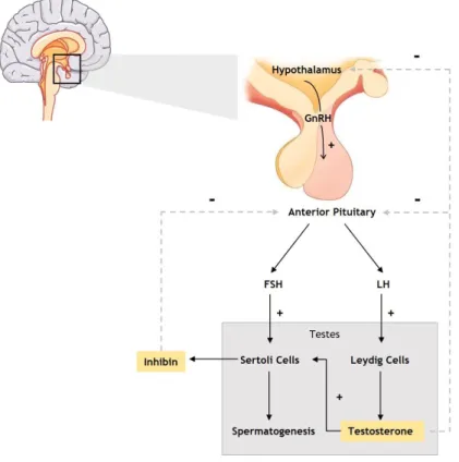
Figure 3: Hormonal regulation of male reproductive function. GnRH is synthetized in the
hypothalamus which in turn stimulates the anterior pituitary to produce LH and FSH. LH acts on Leydig cells stimulating testosterone production while FSH acts on Sertoli cells, stimulating spermatogenesis. High testosterone levels inhibit the release of GnRH by hypothalamus and LH by anterior pituitary. Inhibin production by Sertoli cells negatively regulates FSH production by anterior pituitary. Abbreviations: GnRH – gonadotropin releasing hormone, LH – luteinizing hormone, FSH – follicle-stimulating hormone. Legend: + stimulation; - inhibition. 10
Figure 4: Signaling pathways of leptin. Leptin binds to LepRb receptor causing a
conformational change in the receptor, activating JAK2, which phosphorylates other tyrosine residues located in the LepRb-JAK2 complex, triggering several downstream signaling pathways. Leptin signaling through LepRb also involves other signaling pathways, as depicted in the figure. Abbreviations: AgRP - agouti-related protein, JAK -Janus kinase, POMC – proopiomelanocortin, SOCS3 - suppressor of cytokine signaling 3, STAT - signal transducer and activator of transcription. Adapted from (Kwon, Kim et al. 2016). ... 18
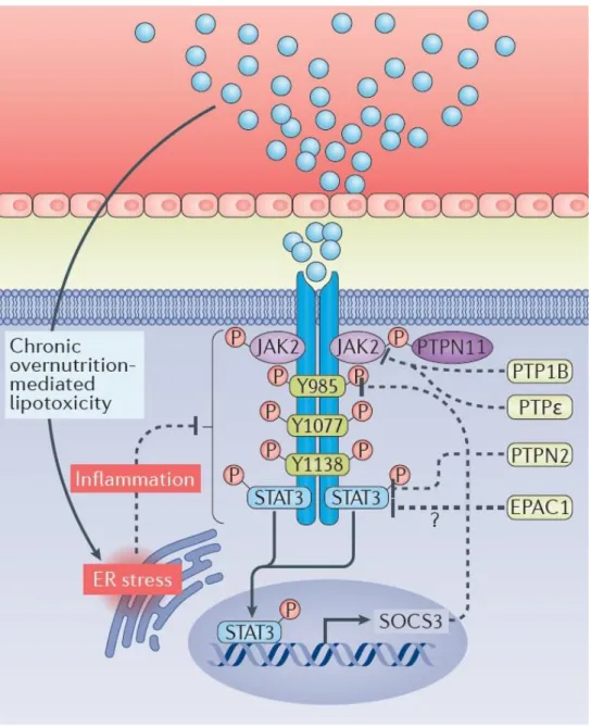
Figure 5: Leptin signaling and the proposed molecular mechanisms behind leptin resistance in obesity. In obese individuals, leptin is present at higher levels. However, a
feedback mechanisms, like SOCS3 leads to impaired leptin signaling. ER stress also triggers inflammatory responses, which could be involved in the diminished response to leptin that happens in obesity. Leptin signaling pathways are simplified in this image. Solid line – stimulates; Dashed line – inhibits. Abbreviations: SOCS3 – suppressor of cytokine signaling 3, ER – endoplasmic reticulum. Adapted from (Cui, Lopez et al. 2017). ... 21
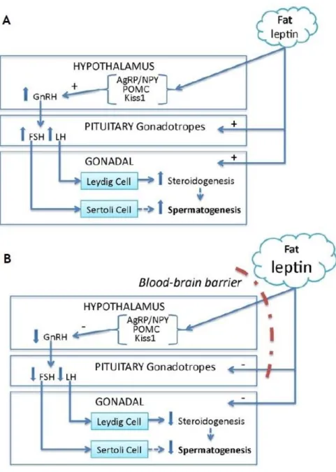
Figure 6: Leptin actions in the hypothalamic-pituitary-gonadal (HPG) axis under normal and pathological conditions. A) In an individual with a normal body mass index, leptin is
produced in the adipose tissue and travels to the hypothalamus, interacting with anorexigenic, oxiregenic and kisspeptin neurons leading to an increase in GnRH production. Leptin also stimulates pituitary gonadotropes secretory activity. This causes an increase in Leydig cell steroidogenesis through stimulation by LH together with a stimulation of Sertoli cells through FSH, further stimulating spermatogenesis. B) In an obese individual, leptin inability to stimulate hypothalamic neurons and pituitary gonadotropes leads to lower levels of LH and FSH in the testis, compromising steroidogenesis and spermatogenesis. Abbreviations: GnRH – gonadotropin releasing hormone, LH – luteinizing hormone, FSH – follicle-stimulating hormone. Adapted from (Landry, Cloutier et al. 2013). ... 27
Figure 7: Effect of leptin in rat Sertoli cells metabolic activity and proliferation. The figure
shows pooled data of independent experiments, indicating metabolic activity (panel A) and proliferation (panel B) of rat Sertoli cells cultured in the absence or presence (5, 25 and 50 ng/mL) of leptin. Results are expressed as mean ± SEM (n=6 for each condition). Significantly different results (P < 0.05) are indicated as: a – relative to leptin-free group. ... 44
Figure 8: Effect of leptin in rat Sertoli cells mitochondria. The figure shows pooled data of
independent experiments, indicating OXPHOS protein levels (panel A) and JC-1 ratio (panel B) of rat Sertoli cells cultured in the absence or presence (5, 25 and 50 ng/mL) of leptin. Results are expressed as mean ± SEM (n=6 for each condition). Significantly different results (P < 0.05) are indicated as: a – relative to leptin-free group; b – relative to 5 ng/mL group; c- relative to 25 ng/mL group. ... 45
Figure 9: Effect of leptin in mRNA levels of peroxisome proliferator-activated receptor γ coactivator 1α (PGC-1α), transcription factor A, mitochondrial (TFAM), nuclear respiratory factor 1 (NRF1), Sirtuin 1 (SIRT1) and mitochondrial copy number of rat Sertoli cells. The
figure shows pooled data of independent experiments, indicating mRNA levels of PGC-1α (panel A), TFAM (panel B), NRF1 (panel C), SIRT1 (panel D) and mitochondrial copy number (panel E) of rat Sertoli cells cultured in the absence or presence (5, 25 and 50 ng/mL) of leptin. Results are expressed as mean ± SEM (n=6 for each condition). ... 47
Figure 10: Effect of leptin in mRNA levels of peroxisome proliferator-activated receptor γ coactivator 1α (PGC-1α), nuclear respiratory factor 1 (NRF1), Sirtuin 1 (SIRT1) and mitochondrial copy number of human Sertoli cells (hSCs). The figure shows pooled data of
independent experiments, indicating mRNA levels of SIRT1 (panel A), PGC-1α (panel B), NRF1 (panel C) and mitochondrial copy number (panel D) of hSCs cells cultured in the absence or presence (5, 25 and 50 ng/mL) of leptin. Results are expressed as mean ± SEM (n=6 for each condition). Significantly different results (P < 0.05) are indicated as: a – relative to leptin-free group; b – relative to 5 ng/mL group; c- relative to 25 ng/mL group. ... 48
Figure 11: Effect of leptin in OXPHOS protein levels of human Sertoli cells (hSCs). The
figure shows pooled data of independent experiments, indicating OXPHOS protein levels of hSCs cells cultured in the absence or presence (5, 25 and 50 ng/mL) of leptin. Results are expressed as mean ± SEM (n=6 for each condition). Significantly different results (P < 0.05) are indicated as: a – relative to leptin-free group. ... 49
List of Tables
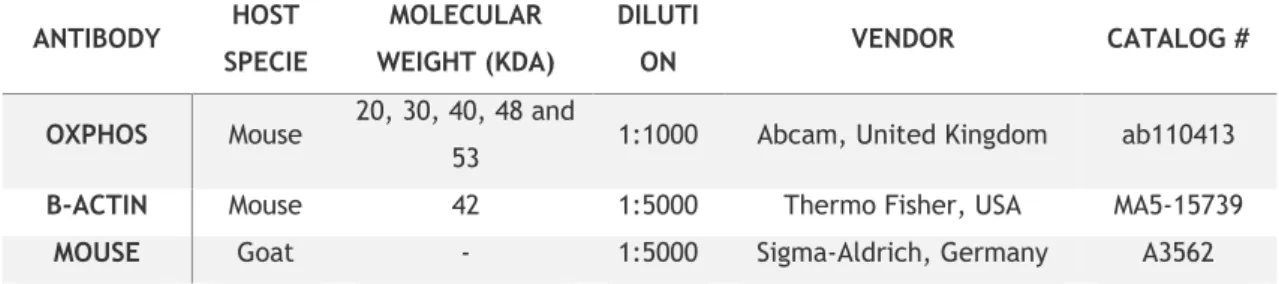
Table 1: List of the primary and secondary antibodies used in this study. ... 38
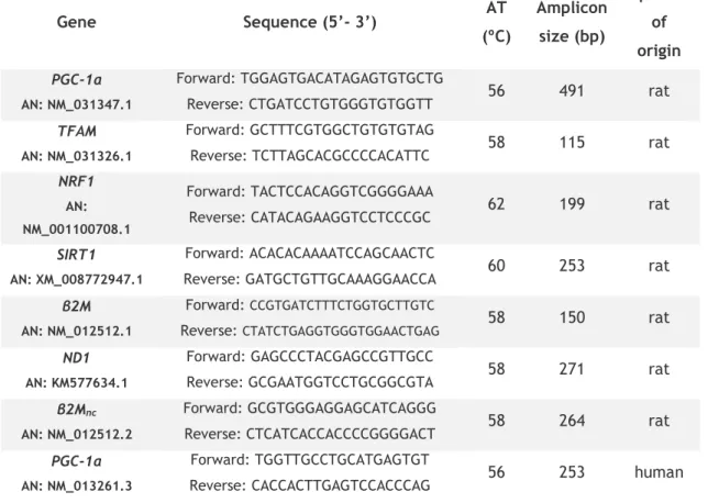
Table 2: Genes, oligonucleotide sequence and respective conditions for PCR amplification. 40
Table 3: Protein expression levels of mitochondrial complexes in rat Sertoli cells non-exposed
or exposed to 5, 25 and 50 ng/mL of leptin. ... 46
Table 4: Protein expression levels of mitochondrial complexes in human Sertoli cells
List of Abbreviations
18S 18S ribosomal RNA
AgRP Agouti-related peptide
ALCs Adult Leydig cells
AMPK AMP-activated protein kinase
ARC Arcuate nucleus
ATP Adenosine triphosphate
BBB Blood-brain barrier
BMI Body mass index
BSA Bovine serum albumin
BTB Blood-testis barrier
CART cDNA DIO
Cocaine- and amphetamine-regulated transcript Complementary DNA
Diet-induced obesity
DMEM:F12 Dulbecco’s Modified Eagle Medium Ham’s Nutrient Mixture F12
DMSO Dimethyl sulfoxide
DNA EDTA
Deoxyribonucleic acid
Ethylene diamine tetra acetic acid
ER Endoplasmic reticulum
ERK Extracellular signal-regulated kinase
FBS Fetal Bovine Serum
FDA Food and Drug Administration
FLCs Fetal Leydig cells
FSH Follicle-stimulating hormone
GnRH Gonadotropin releasing hormone
HIF-1α Hypoxia inducible factor 1α
HIFs Hypoxia inducible factors
HPG Hypothalamic-pituitary-gonadal
hSCs Human Sertoli cells
IGF-1 Insulin-like growth factor 1
ITS Insulin–transferrin–sodium selenite
JAK2 Janus kinase 2
JC-1 5,5′,6,6′-tetrachloro-1,1′,3,3′-tetraethylbenzimidazolylcarbocyanine iodide
LepR Leptin receptor
LH Luteinizing hormone
LHRH Luteinizing hormone-releasing hormone
MFN2 Mitofusin-2
M-PER Mammalian Protein Extraction Reagent
mRNA mtDNA
Messenger RNA Mitochondrial DNA
mTOR Mammalian target of rapamycin
MTT 3-(4,5-dimethylthiazol-2-yl)-2,5-diphenyltetrazolium bromide
ND1 Mitochondrially encoded NADH dehydrogenase 1
NPY Neuropeptide Y
NRF1 Nuclear respiratory factor 1
OXPHOS Oxidative phosphorylation
P450SCC Cytochrome P450 family 11 subfamily A member 1
PBS PCR PGC-1α
Phosphate buffered saline Polymerase chain reaction
Peroxisome proliferator-activated receptor γ coactivator 1α
PI3K Phosphatidylinositol-4,5-bisphosphate 3-kinase
PMCs Peritubular myoid cells
POMC Pro-opiomelanocortin
PPAR-γ Peroxisome proliferator-activated receptor-γ
qPCR RNA ROS
Quantitative PCR Ribonucleic acid
Reactive oxygen species
rSCs Rat Sertoli cells
RT-PCR s6k1
Reverse-transcriptase polymerase chain reaction S6 kinase-beta 1
SCs Sertoli Cells
SEM SHP2
Standard error mean
Protein tyrosine phosphatase 2
SIRT1 Sirtuin 1
SOCS3 Suppressor of cytokine signaling 3
SRB Sulforhodamine B
StAR Cholesterol transporter steroidogenic acute regulatory protein
STAT3 Signal transducer and activator of transcription 3
STAT5 Signal transducer and activator of transcription 5
TFAM Transcription factor A, mitochondrial
tRNA Total RNA
β2M β2-microglobulin
I. Introduction
1. Male reproductive system: an overview
The reproductive system is not essential for the survival of the individual; it is, however, required for the survival of the species. It is through the reproductive system that new individuals are born; the species are constantly repopulated and the genetic code is transmitted over generations. In humans, the sexual reproduction is the method used, which has several advantages, namely at the level of variability induced by the combination of progenitor’s genes. This variability ensures the evolution of the species throughout time. The reproductive system has some unique features. Unlike any other body systems, it is not fully functional at the time of birth, and it requires the action of sex hormones around the time of puberty to be fully active and ready to perform its purpose. In addition, the gender differences between the male and female reproductive system are clearly observed, a fact that does not occur in the other body systems (Van De Graaff 2001).
The male reproductive system has different structures that can be divided in primary and secondary sex organs. In males, the primary sex organs, also known as gonads, are the testes. They are responsible for the production of spermatozoa and secretion of sex hormones. The secretion of sex hormones is then responsible for the secondary sex organs development. Surrounding the testes is the scrotum, an outpouching of the abdominal wall that protects the testes. The secondary sex organs are structures responsible for the nourishment and storage or transport of the spermatozoa to the exterior or into the female reproductive tract. One of the organs responsible for this transport is the penis. The penis is the male organ used in sexual intercourse and can be divided into three structures: the root linked to the abdominal wall, the body of the penis that corresponds to the major portion of this organ and the glans, also referred as the head of the penis (Clark 2005). There are other secondary sex organs, such as the epididymis, vas deferens, ejaculatory ducts and urethra responsible for storage, maturation and transport of the spermatozoa and others responsible for secretion of fluids that are part of the ejaculate, such as seminal vesicles, prostate gland and bulbourethral glands (Figure 1). Sex hormones are also responsible for the development of the secondary sex characteristics, that appear during puberty, such as body hair, deep voice and Adam's apple development (VanPutte, Russo et al. 2010).
The testes are the male gonads, paired ovoid organs that are responsible for the production of spermatozoa and sex hormones. They are suspended in the scrotum by the spermatic cords. Each one is about 4-5 cm long and 2.5 cm in diameter and weighs between 14-18 g in humans (Johnson, Petty et al. 1984). Both testes are covered by two tunics. The
outer tunica is the tunica vaginalis and their visceral layer covers the surface of each testis, except where the testis attaches to the epididymis and spermatic cord. This tunica is a thin closed peritoneal sac that has origin on the peritoneum during testes descent. The parietal layer of the tunica vaginalis covers more tissue than the previous one, extending superiorly onto the distal part of the spermatic cord. The separation between the visceral and parietal layers is filled with fluid, allowing the movement of the testes in the scrotum (Van De Graaff 2001, Rizzo 2009, VanPutte, Russo et al. 2010). The testes also have a tough fibrous outer membrane called tunica albuginea. This tunica has characteristic extensions that move to the inside of each testis dividing it into testicular lobules. Each of these lobules contains long and highly coiled seminiferous tubules that are nearly 80 cm long, if uncoiled in humans. It is inside these tubules, considered the functional unit of the testis, that spermatogenesis occurs (Figure 1). Here, spermatozoa are produced at a rate of about thousands per second. Sertoli cells (SCs), which function as a physical support to germ cells and nourish their development into sperm, form the seminiferous tubules. Between these numerous tubules is the interstitial space, where Leydig cells are placed.
Figure 1: Schematic representation of a human testis. The testis is coated by tunica albuginea, and
divided in lobules. The seminiferous tubules are located inside these lobules, highly coiled and organized. The rete testis is responsible for transporting the spermatozoa from the seminiferous tubules into the efferent ducts. From this point forward, spermatozoa enter the epididymis where they go through several processes until they are ready to leave the male reproductive tract into the female reproductive tract.
1.1 Functional organization of the testes
The testes are responsible for the production of male gametes and male sex hormones, which are processes known as spermatogenesis and steroidogenesis, respectively. These two distinct processes take place in two morphologically and functionally different compartments: the seminiferous tubular compartment and the interstitial compartment. Despite being anatomically and functionally different, these two compartments function closely to one another and they are both required to achieve the correct parameters in sperm quality and production.
The tubular compartment is composed by the seminiferous tubules. It is responsible for 60 to 80% of the testicular volume and contains the germ cells, SCs and the peritubular myoid cells (PMCs). Sertoli and germ cells are organized in a highly-polarized system to efficiently support the spermatogenesis. Adjacent SCs form tight junctions with each other, providing an immune-privileged microenvironment suitable to germ cells development. Tight junctions formed between adjacent SCs establish the blood-testis barrier (BTB), responsible for controlling the movement of nutrients in the seminiferous tubules (Wong and Cheng 2005). However, there are other types of cooperation used between adjacent SCs to strengthen the BTB, such as ectoplasmic specializations and desmosomes (Mruk and Cheng 2004, Lie, Cheng et al. 2011). This barrier has the function of “gate”, preventing solutes and large molecules from reaching the germ cells and the function of “fence”, restricting the movement of proteins and lipids between adluminal and basal compartments. BTB also divides the seminiferous tubules in basal and adluminal compartments. Spermatogonia and pre-meiotic spermatocytes are present in the basal compartment whereas meiotic spermatocytes, spermatids and spermatozoa reside in the adluminal compartment, showing distinct polarity depending on their location (Pelletier 2011, Mruk and Cheng 2015). In addition, Sertoli cell nuclei and Golgi complexes are also found in the basal compartment where early phagosomes and early processes involved in spermatids development are all confined to the adluminal compartment. However, the most obvious form of cell polarity present in the testes is observed during the development of spermatids, where the heads of spermatids point towards the basal compartment while the tails point towards the adluminal compartment (Gao and Cheng 2016, Gao, Xiao et al. 2016). The testis is divided by a septum of connective tissue into about 250-300 lobules, each one containing 1-3 convoluted seminiferous tubules. Generally, there are about 600 seminiferous tubules present in the human testis and each one has an average length of 30-80 cm which varies according to whether they are uncoiled. The total length of the seminiferous tubules is, on average, 300 meters per testis and 600 meters per man (Xiao, Mruk et al. 2014, Griswold 2016).
Surrounding the seminiferous tubules, in the interstitial compartment, are the PMCs which have several functions. In humans, three or four layers of PMCs surround the seminiferous tubules while in mice, a single layer of PMCs is present (Gardner and Holyoke
1964). In adult testis, PMCs mediate the contraction of the seminiferous tubules and, during testis development and adulthood, they work together with SCs to deposit the basement membrane (composed by laminin, collagen IV and fibronectin) that surrounds the seminiferous tubules. This is a critical interaction to ensure a correct spermatogenesis and architecture of the seminiferous tubules (Skinner, Tung et al. 1985, Bichoualne, Thiébot et al. 1994, Maekawa, Kamimura et al. 1996, Verhoeven, Hoeben et al. 2000). There are other functions attributed to PMCs. In rats, these cells where shown to be an important part of the barrier function, restricting the entry of substances into the seminiferous tubules (Dym and Fawcett 1970). The interstitial compartment is located between the seminiferous tubules and is filled with Leydig cells, the major component of this compartment. In fact, there are two populations of Leydig cells, the fetal Leydig cells (FLCs) and the adult Leydig cells (ALCs) (O'Shaughnessy, Baker et al. 2006). After birth, FLCs start degenerating and it is not yet clear if they give rise to ALCs. On the other hand, ALCs derive from Leydig stem cells, capable of self-renewal. These cells develop into Leydig progenitor cells, which express a number of factors such as luteinizing hormone receptors and 3β-hydroxysteroid dehydrogenase (Haider 2004). Further differentiation occurs into adult cells that no longer proliferate. In the presence of luteinizing hormone (LH), these cells produce testosterone that is fundamental for the establishment and maintenance of the secondary sex characteristics and the continuation of spermatogenesis (Walker 2011, Smith and Walker 2014). There are other cell types present in the interstitium, namely the immune cells (macrophages, T-cells, dendritic cells), where they respond according to the stimuli received (Perez, Theas et al. 2013). From this group, macrophages are the most abundant in the interstitium, corresponding to approximately 25% of the interstitial cells present in the adult rodent testis (Niemi, Sharpe et al. 1986). Several studies have shown that there is cross-talk between immune cells and spermatogonia, where the number of spermatogonia declines after ablation of macrophages (DeFalco, Potter et al. 2015). These also establish cell junctions with Leydig cells, to facilitate an eventual response.
1.2 Sertoli cells
SCs are the somatic cells present in the seminiferous epithelium that support and nourish the developing germ cells. They are highly polarized cells that extend upwards from the basement membrane of the germinal epithelium to the lumen of the seminiferous tubules in a direct interaction with the developing germ cells (Mruk and Cheng 2004). SCs are irregularly shaped, columnar cells with a characteristic oval nucleus with a dark nucleolus. They have a large surface area which is correlated with the number of germ cells that they can support (Vogl, Vaid et al. 2008). This characteristic is also important for germ cell movement during spermatogenesis. First described in 1865 by Enrico Sertoli, who used the term “mother cells” to describe them, SCs are known as the “nurse cells” for their role in
providing structural and nutritional support for germ cells development (França, Hess et al. 2016). Additionally, SCs also have apoptosis functions, are involved in the establishment of the BTB, secrete the seminiferous tubular fluid that assists in the transport of mature spermatozoa into the epididymis and secrete factors such as inhibin that are involved in the hormonal control of spermatogenesis (França, Hess et al. 2016). The number of SCs is heavily tied with the fertility of an individual as each SC has a limit on the number of germ cells that can support. Since these cells become terminally differentiated during puberty, a lower number of SCs implicates a lower number of supported germ cells, which translates into lower levels of daily sperm production with direct consequences in male fertility (Orth, Gunsalus et al. 1988, Sharpe, McKinnell et al. 2003).
As discussed before, the tight junctions formed between adjacent SCs establish the BTB that controls the microenvironment inside the seminiferous tubules. BTB has two main functions: (1) act as a “gate” and a “fence” to restrict the paracellular flow of substances from the basal to the adluminal compartment. The blood vessels and lymphatic vessels are located in the interstitium between the seminiferous tubules which confers to SCs the task of regulating the entry of nutrients and important molecules, such as hormones, into the adluminal compartment where the later stages of spermatogenesis are occurring. However, the BTB also regulates the entry of harmful components that might be present in the blood vessels such as drugs and chemicals. This characteristic selectivity of BTB is crucial for the establishment of a safe microenvironment for the germ cells development. BTB also acts as a “fence”, restricting the movement of proteins and lipids between the basal and adluminal compartments (Wong and Cheng 2005, Cheng and Mruk 2012); (2) creating an immunological barrier, to prevent the immunological response of anti-sperm antibodies that would attack the developing germ cells, leading to male infertility. The BTB sequesters germ-cell specific antigens that surge during meiosis and spermiogenesis, preventing an immunological response. Furthermore, several studies have shown that SCs also play a role in the maintenance of the testis as an immune-privileged organ by secreting immunosuppressive molecules (Su, Mruk et al. 2011, Mruk and Cheng 2015).
SCs possess a Warburg-like metabolism, prioritizing glycolysis over oxidative metabolism which is a less effective pathway for adenosine triphosphate (ATP) production. Furthermore, SCs also sustain a high glycolytic flux, in a similar way to cancer cells, which is a consequence of the metabolic cooperation required between SCs and germ cells for a successful spermatogenesis (Oliveira, Martins et al. 2015). One of the end products of glycolysis, pyruvate, is then converted to lactate which is a crucial factor for germ cells development due to its anti-apoptotic effect in germ cells. Furthermore, lactate is also the major energy source of germ cells (Robinson and Fritz 1981). However, spermatogonia utilize glucose as the major energy substrate while germ cells in further stages of spermatogenesis such as spermatids use lactate for energy production (Mita and Hall 1982, Nakamura, Okinaga et al. 1984). Spermatozoa also utilize glucose/fructose as the major source of energy
illustrating that the substrate required for energy production changes at each stage of spermatogenesis (Bajpai, Gupta et al. 1998). The metabolism of SCs is also highly susceptible to hormones. Follicle-stimulating hormone (FSH) and insulin have well documented effects in the regulation of SCs metabolism, stimulating glucose metabolism and lactate production (Mita, Price et al. 1982, Oliveira, Alves et al. 2012). SCs metabolism and its regulation is one of the most important processes for a normal spermatogenesis and male fertility. However, a grey area of knowledge surrounding SCs metabolism still exists which when undisclosed could contribute to improve several infertility cases.
2. Spermatogenesis
Spermatogenesis is a multi-step process that produces spermatozoa in the seminiferous tubules determining male fertility (O'Shaughnessy 2014). Beginning at puberty and during the reproductive life of a fertile men, more than 40 million spermatozoa are produced every day (Cheng and Mruk 2013). This process is controlled by several endocrine and other regulatory factors and its duration depends on the species with a duration of 40 to 50 days in rodents and nearly 80 days in humans (Sharpe 1994). The final objective of spermatogenesis is the development of mature spermatozoa with half the number of chromosomes (haploid), from the immature germ cells, spermatogonia (diploid). Immature germ cells undergo several processes including mitosis, meiosis and differentiation to give rise to mature spermatozoa (Rato, Alves et al. 2012, Alves, Rato et al. 2013). This process occurs in the seminiferous tubules, the functional unities of the testis, through a close association of germ cells with the somatic cells, SCs (Walker and Cheng 2005, Rato, Alves et al. 2012). The tight junctions formed between adjacent SCs constitute the BTB that divides the seminiferous epithelium into two compartments: the basal compartment where spermatogonia and pre-meiotic spermatocytes are present and the adluminal compartment where pre-meiotic spermatocytes, spermatids and spermatozoa reside. (Cheng, Wong et al. 2010). SCs are also responsible for the germ cells movement towards the lumen where mature spermatozoa are released. Spermatogenesis can be divided in four different phases: mitosis, meiosis, spermiogenesis and spermiation (O'Shaughnessy 2014). These events are a cycle of cellular changes and can be further divided into stages, with different stages per specie. In humans, spermatogenesis occurs during the 6 stages of the seminiferous epithelial cycle while in mice it occurs during the 12 stages of the seminiferous epithelial cycle and in rats during the 14 stages of the seminiferous epithelial cycle (Cheng, Wong et al. 2010). During this process, germ cells are located from the periphery to the center of the seminiferous tubule, according to their degree of maturation (Alves, Rato et al. 2013).
Spermatogonia are the most immature cells in the testis. In the early stages of spermatogenesis, they go under successive mitosis renewing themselves and giving origin to a new cell type. At this stage, they are called spermatogonia type A (Lie, Cheng et al. 2009). These cells are located outside the BTB, continuing their process of self-renewal until death which is the reason why men, unlike women, are fertile throughout life. After several mitoses, spermatogonia type A has three options: it can divide and form another population of spermatogonia (self-renewal), it can undergo apoptosis or it can differentiate into the first committed stem cell type. Intermediate spermatogonia are the first population of cells committed to becoming spermatozoa and give origin to the spermatogonia type B (Mauch and Schoenwolf 2001). These cells are located in the basement membrane of the seminiferous tubules, surrounded by SCs and are the last cells of the line that go under mitosis. They migrate in direction to the lumen of the seminiferous tubule differentiating into the primary spermatocytes (Mauch and Schoenwolf 2001). The factors behind the decisions to follow the path of differentiation instead of self-renewal or to choose to divide meiotically instead of mitotically are still a matter of debate among the scientific community. Primary spermatocytes undergo a meiotic division, meiosis I, to yield a pair of secondary spermatocytes. These haploid cells then undergo another meiotic division, meiosis II, originating haploid spermatids (Figure 2) (O'Donnell, Robertson et al. 2001).
At last, spermiogenesis takes place, in which spermatids maturation into mature spermatozoa occurs. During spermiogenesis several morphological changes occur that involve development of the flagellum, chromosomal condensation, formation of the acrosome and elongation of the nucleus (Meistrich, Trostle-Weige et al. 1992, Rato, Alves et al. 2012). In humans, 12 spermatid maturation steps are described (Weinbauer, Luetjens et al. 2010). SCs are crucial in this phase, as the adhesive contacts and ectoplasmic junctional specializations between spermatids and SCs are shattered which culminates with the release of spermatozoa into the lumen of the seminiferous tubule, in a process called spermiation (Griswold 1998, Hai, Hou et al. 2014). At this point, spermatozoa are fully differentiated cells; however, they are not yet motile and capable of fertilizing the oocyte. The non-motile spermatozoa are transported to the epididymis in testicular fluid secreted by SCs, on which they acquire motility, in a final step of spermatozoa maturation. The ability to fertilize the egg is acquired during the last stages of spermatogenesis, along the epidydimal duct and during the transit inside the female reproductive tract. These series of biochemical changes in which spermatozoa become functional are referred as capacitation (Figure 2) (Dun, Aitken et al. 2012, Dacheux and Dacheux 2014).
Figure 2: Schematic representation of major events in spermatogenesis. Spermatogonia go through
successive mitosis until they differentiate into primary spermatocytes. After the first meiotic division, secondary spermatocytes are formed. Meiosis II follows, which yields haploid spermatids. The spermatids then go through spermiogenesis (differentiation) followed by spermiation, in which they are released into the lumen of the seminiferous tubule. Afterwards, they go through the final steps of maturation in the epididymis, becoming motile. The final steps are achieved in the female reproductive tract, where they go through several biochemical changes, a process known as capacitation. Adapted from (Sharma and Agarwal 2011).
2.1 Hormonal regulation of spermatogenesis
Spermatogenesis is a tightly regulated process due to the complexity and the number of the events involved. A single malfunction in one of the many steps that are part of spermatogenesis can compromise the entire process. Beyond the need to maintain proper growth and differentiation of germ cells, somatic cells proliferation and functionality is also required for a successful spermatogenesis. The hypothalamic-pituitary-gonadal (HPG) axis is responsible for this regulation, through the actions of two endocrine factors, LH and FSH (Ramaswamy and Weinbauer 2014). Within this axis, neurons of the hypothalamus produce gonadotropin releasing hormone (GnRH) in a characteristic pulsatile way (Ramaswamy and Weinbauer 2014). This pulse is susceptible to the metabolic status of body and in a fasting state, GnRH secretion is suppressed which decreases the levels of LH and testosterone compromising male reproductive function if longer periods of fasting occur frequently (Trumble, Brindle et al. 2010). GnRH travels to the anterior pituitary and stimulates the synthesis and release of LH and FSH in the gonadotrophs, the cells that express GnRH receptors. These endocrine factors then act at a testicular level to regulate spermatogenesis (Rato, Alves et al. 2012, Alves, Rato et al. 2013). LH binds to receptors on the surface of Leydig cells and stimulates testosterone production, a sex steroid hormone that diffuses into the seminiferous tubules. Within the seminiferous tubules, SCs are the only cells that possess receptors for testosterone and FSH (Walker and Cheng 2005). FSH is crucial to stimulate spermatogenesis since its involved in the maintenance of testicular size and in the regulation of spermatogonia proliferation and differentiation (Dym 1994, Walker and Cheng 2005). Synergistically, testosterone and FSH stimulate SCs to secrete paracrine agents, such as
androgen-binding protein, that modulate spermatogenesis (Ritzen, Hagenas et al. 1975, Hall, Conti et al. 1990). A complex interaction between the hypothalamus and the testes regulates gonadotropins synthesis and release (Figure 3). On one side, hypothalamus through GnRH production stimulates LH and FSH production; on the other the testes establish a negative feedback mechanism with the pituitary, through the production of sex steroid hormones and inhibin (Tilbrook and Clarke 2001). Several autocrine and paracrine factors are also reported to be involved in local mechanisms that modulate spermatogenesis (Le Magueresse, Pineau et al. 1988, Nehar, Mauduit et al. 1998, Huleihel and Lunenfeld 2002). Inhibin, secreted by SCs, can act on the anterior pituitary decreasing the FSH levels (O'Connor and De Kretser 2004) while its counterpart, activin is involved in SCs proliferation (Nicholls, Stanton et al. 2012). Testosterone, produced by Leydig cells, inhibits LH production through two distinct mechanisms: it acts on the hypothalamus diminishing the frequency of the GnRH pulsatile release which ultimately decreases the secretion of gonadotropins in the pituitary; or acts directly in the pituitary decreasing the secretion of LH (Figure 3) (Widmaier, Raff et al. 2011). Testosterone has a crucial role in male reproductive function, as its absence prevents germ cells to go beyond meiosis, prevents the release of mature spermatozoa and compromises the establishment of the BTB (Walker 2010). The disturbance of one of these steps compromises spermatogenesis and consequently male fertility.
Spermatogenesis regulation is not summed up to androgens and gonadotropins. In fact, estrogens, insulin-like growth factor 1 (IGF-1) and growth hormone have showed distinct functions with implications in spermatogenesis. Estrogens, usually referred as the female sex hormone, are reported to have an essential role regulating the HPG axis, indirectly modulating LH and testosterone equilibrium through a feedback loop (O'Donnell, Robertson et al. 2001). IGF-1, synthetized in the testes by SCs, has also shown to be capable of stimulate the proliferation of Leydig cell precursors and spermatid maturation (Itoh, Nanbu et al. 1994). Additionally, despite usually being referred as being involved in other processes, growth hormone also has an important role in reproductive function. Growth hormone plays a role in gonadal steroidogenesis and spermatogenesis either directly at a gonadal level or indirectly via IGF-1. In fact, growth hormone regulates Leydig cells steroidogenesis through regulation of IGF-1 production (Spiteri-Grech and Nieschlag 1992, Hull and Harvey 2000). Despite a lot of knowledge acquired in the last decades about the hormones and processes involved in spermatogenesis, there is still much to be done. The complex signaling and hormonal network behind the control of spermatogenesis still needs to be deciphered to better understand the mechanisms behind male fertility and to understand which part of this endless chain of biochemical events is comprised in infertility cases.
Figure 3: Hormonal regulation of male reproductive function. GnRH is synthetized in the
hypothalamus which in turn stimulates the anterior pituitary to produce LH and FSH. LH acts on Leydig cells stimulating testosterone production while FSH acts on Sertoli cells, stimulating spermatogenesis. High testosterone levels inhibit the release of GnRH by hypothalamus and LH by anterior pituitary. Inhibin production by Sertoli cells negatively regulates FSH production by anterior pituitary. Abbreviations: GnRH – gonadotropin releasing hormone, LH – luteinizing hormone, FSH – follicle-stimulating hormone. Legend: + stimulation; - inhibition.
3. Leptin: from discovery to energy control
Leptin was discovered in 1994 by Friedman et al., through positional cloning (Zhang, Proenca et al. 1994). Since then, the knowledge about this adipocyte-derived protein has consistently grown. In brief, leptin is a 16 kDa cytokine encoded by the LEP gene, located in chromosome 7q31.3 in humans consisting of three exons separated by two introns, and chromosome 6 in mice (He, Chen et al. 1995, Isse, Ogawa et al. 1995). The majority of its production occurs in adipocytes but it can also be locally produced in other tissues such as stomach, placenta, ovary, mammary gland, immune cells and skeletal muscle (Cioffi, Van Blerkom et al. 1997, Bado, Levasseur et al. 1998, Schubring, Prohaska et al. 1999, Wang, Wang et al. 2011, Wolsk, Mygind et al. 2012). In these tissues, leptin has been implicated in several functions such as regulation of immune responses, reproductive function, and fetal development (Lago, Gomez et al. 2008, Herrid, Palanisamy et al. 2014, Perez-Perez, Sanchez-Jimenez et al. 2015). These evidences indicate that leptin has pleotropic effects in several biological functions and more importantly, that leptin function is generally independent of its
action in the regulation of body weight. After being produced by white adipose tissue, leptin enters the bloodstream and circulates in a concentration that is associated with the amount of adipose tissue depots on an individual (Frederich, Hamann et al. 1995, Maffei, Halaas et al. 1995). Therefore, leptin levels indicate body fat reserves. This information is then communicated to specific areas in the brain that regulate energy homeostasis (Rosenbaum and Leibel 2014). Higher circulating leptin levels suggest a higher number of adipose tissue depots which triggers a response to reduce food intake and to promote energy consumption. In contrast, a decrease in leptin circulating levels triggers an opposite response, promoting food intake and reducing energy consumption (Hamann and Matthaei 1996, Kolaczynski, Ohannesian et al. 1996). This mechanism is often dysregulated in several metabolic diseases such as obesity. In those cases, leptin resistance occurs where higher levels of circulating leptin are present but do not trigger the expected response in order to reduce food intake and promote energy expenditure (Jung and Kim 2013, Sainz, Barrenetxe et al. 2015). Thus, since its discovery, leptin has emerged as a key control point for energy homeostasis and its relevance in health and disease has been in spotlight for more than two decades. In the last years, the effects of leptin in male reproductive function gained attention and novel molecular mechanisms showing how it acts through the male reproductive tract have been identified.
3.1 Leptin as a satiety hormone
Leptin exerts its physiological effects through specific receptors. First identified in 1995 (Tartaglia, Dembski et al. 1995), the leptin receptor (LepR) was isolated from mouse choroid plexus and due to strong similarities in the extracellular ligand-binding domain with the gp130 signal-transducing subunit is considered part of the large family of class I cytokine receptors. This receptor is encoded by the LEPR gene located in the chromosome 1 and chromosome 4 in humans and mice, respectively. Activated via ligand induced conformational changes, this receptor forms homodimers even when leptin is not present (Devos, Guisez et al. 1997). Several isoforms of this receptor are known (LepRa-f), all products of the LEPR gene, resulting of alternative messenger RNA (mRNA) splicing or post translational modifications (Lee, Proenca et al. 1996). These isoforms have identical extracellular domains consisting of over 800 amino acids; however, their intracellular domains terminate at different lengths that are characteristic of each isoform (Gorska, Popko et al. 2010).
Secreted isoforms are characterized by only possessing an extracellular domain unlike the other isoforms. This can result of alternative mRNA splicing or proteolytic cleavage products of membrane-bound isoforms of LepR. Discovered by Sinha et al., this isoform is soluble and functions as the main binding protein for leptin circulating in the bloodstream,
modulating its bioavailability and the cellular effects of leptin itself (Sinha, Opentanova et al. 1996, Lammert, Kiess et al. 2001). Unlike what happens in mice, the human secreted isoform (LepRe) is exclusively generated through proteolytic cleavage of membrane-bound isoforms of LepR, a process referred as shedding, thus being an excellent indicator of the number of membrane-anchored leptin receptors (Maamra, Bidlingmaier et al. 2001). In normal conditions this process occurs inherently, however several studies have shown that it can be induced by different kinds of stimuli such as lipotoxicity and apoptosis (Unger 2003, Schaab, Kausch et al. 2012). Soluble isoforms can have either antagonistic or agonistic effects although the former prevails (Di Marco, Gloaguen et al. 1997, Hardy, Owczarek et al. 2001, Chalaris, Garbers et al. 2011). LepRe effect is still a matter of debate, however it has been shown that an antagonistic effect is present (Schaab, Kausch et al. 2012). Higher concentrations of LepRe over leptin have a prevailing inhibitory effect on leptin signal transduction, nullifying a crucial step of leptin-mediated activation, the signal transducer and activator of transcription 3 (STAT3) activation thus neutralizing the anorexigenic effects of leptin in rats (Zhang and Scarpace 2009). Moreover, several other mechanisms through which LepRe promotes leptin resistance have been identified including inhibition of leptin transport through the blood-brain barrier (BBB) (Tu, Kastin et al. 2008). These antagonistic effects are also present in a state of negative energy balance, such as anorexia nervosa or malnutrition, where LepRe is found in higher concentrations when compared with leptin (Krizova, Papezova et al. 2002, Stein, Vasquez-Garibay et al. 2006).
Short isoforms (LepRa, LepRc, LepRd, LepRf) are very similar between them and their role is not yet very clear. They possess a similar extracellular domain and a similar transmembrane domain consisting of 34 amino acids. Their intracellular domain is also similar, sharing the first 29 amino acids, and consisting of 32-40 amino acids in total (Wada, Hirako et al. 2014). Due to lack of intrinsic tyrosine kinase activity, LepR binds to cytoplasmic kinases, specifically Janus kinase 2 (JAK2) (Ghilardi and Skoda 1997). The first 29 amino acids contain a constant box 1 motif and a JAK2. Although these isoforms are also able to recruit JAKs and activate certain signaling cascades, they lack the box 2 motif to be able to induce the pivotal STAT. More recently, studies highlight for their involvement in leptin signaling cascades through MAP kinase signaling pathways (Akasaka, Tsunoda et al. 2010). Other possibility is their involvement in leptin transport across the BBB and leptin clearance (Hileman, Tornoe et al. 2000, Hileman, Pierroz et al. 2002).
LepRb, also known as long isoform of LepR, is the fully functional isoform of leptin receptor. The major difference between LepRb and the short isoforms resides in the intracellular domain, formed by over 300 amino acids (Gorska, Popko et al. 2010). Similar to what happens in short isoforms, this domain contains a box 1 motif and a JAK2, however it also contains two box 2 motifs that are crucial to induce the STAT signaling pathway, characteristic of leptin signaling (Allison and Myers 2014). This isoform is essential to the
correct signaling of leptin as seen in animal models that lack this receptor, such as db/db mice. Homozygous for the LepR spontaneous mutation and thus lacking the LepRb isoform, phenotypically these animals are hyperleptinemic, hyperphagic, obese, leptin resistant and have reproductive dysfunction (Chen, Charlat et al. 1996). These animals also display a phenotype similar to db3J/db3J mice, a model that is null for all known isoforms of the LepR. Interestingly, expression in these mice of a LepRb transgene induces a depletion of body weight, hyperphagia, glucose intolerance and interestingly, it restored male fertility (Kowalski, Liu et al. 2001). However, mutations in the LEPR and LEP gene are extremely rare in humans. In male reproductive tract, LepR has already been identified in the testes, epididymis and seminiferous tubules in several species (Rago, Aquila et al. 2009). In humans, LepR was already identified in somatic cells, germ cells and spermatozoa although its function and involvement in reproductive processes such as spermatogenesis and steroidogenesis is not yet enlightened (Jope, Lammert et al. 2003, Ishikawa, Fujioka et al. 2007, Martins, Moreira et al. 2015). However, several findings point towards a significant role of leptin in these cells underpinning the importance of further scientific research in this specific field.
3.1.1 Leptin and LepR expression in testis
The first demonstrations of LepR expression in murine testis, specifically in sperm and Leydig cells, appeared soon after leptin itself was discovered (Hoggard, Mercer et al. 1997). After these findings, several studies have shown that LepR expression in testis is a shared trait among different species (Rago, Aquila et al. 2009). Interestingly, LepR isoforms present different expressions levels per tissue. For example, LepRb, the fully functional signaling isoform of LepR, is found mainly in the hypothalamus, while LepRa, a short isoform with a role that is not yet clear, is found in peripheral tissues of most species studied, including the testis (Margetic, Gazzola et al. 2002). In fact, some studies report that LepRb is not found at all in testis while others report that LepR in testis is functional and JAK/STAT pathways are involved in leptin signaling (Hoggard, Mercer et al. 1997, Yuan, Huang et al. 2014). This could be due to the fact that LepR activation involves cell-specific signal transduction pathways. Moreover, LepR short isoforms still have signaling potential and are still able to recruit JAKs and activate certain signaling cascades which could explain leptin signaling occurring without the functional isoform of LepR. However, there is very little information about LepR short isoforms and the signaling cascades involved which is an obstacle when trying to understand the possible leptin signaling implications in the testis related to these isoforms.
LepR connection with Leydig cells started to be in the spotlight as soon as the inhibitory effect of leptin in testosterone production was established. LepR has been already identified in Leydig cells in several species including humans (Caprio, Isidori et al. 1999,