UNIVERSIDADE DA BEIRA INTERIOR
Ciências da Saúde
Phenotypic and Functional Aspects of Lymphocyte
Populations in Patients with Blood Dyscrasias
Undergoing Phlebotomy
Ana Raquel Costa Brito
Dissertação para obtenção do Grau de Mestre em
Ciências Biomédicas
(2º ciclo de estudos)
Orientador: Prof. Doutor Fernando Arosa
Coorientadora: Profª. Doutora Elsa Cardoso
ii
Agradecimentos
Em primeiro lugar gostava de agradecer ao meu orientador, Professor Doutor Fernando Aguilar Arosa, pela oportunidade de poder trabalhar nesta área pela qual nutro grande interesse, pela sua orientação científica e dedicação e por nunca me deixar desistir e me fazer sempre ver o lado positivo de todas as situações. Gostava também de agradecer à minha coorientadora, Professora Doutora Elsa Maria Cardoso por toda a ajuda laboratorial e por estar sempre disponível para ajudar independentemente da distância. A ambos, por todos os conhecimentos que me transmitiram e pelas sugestões e contribuições ao longo deste último ano de trabalho.
Ao Centro de Sangue e da Transplantação de Coimbra, principalmente à Dra. Cristina Caeiro, pelo fornecimento de buffy coats de dadores de sangue indispensáveis para a realização deste trabalho, bem como ao Centro Hospitalar Cova da Beira, em especial ao Dr. Jorge Martinez, pela fundamental colaboração neste projecto desde a seleção de doentes ao fornecimento das flebotomias. A todos os funcionários do Serviço de Imunohemoterapia do Centro Hospitalar Cova da Beira pela amabilidade com que sempre fui tratada e por toda a disponibilidade que sempre demonstraram.
Um especial obrigada à Dra. Patrícia Amantegui Ibarzabal e à Dra. Andreia Monteiro, do Serviço de Patologia Clínica do Centro Hospitalar Cova da Beira, por me terem permito o acesso ao citómetro de fluxo deste serviço e em função disso alterado as suas rotinas diárias. Muito obrigada pela disponibilidade, simpatia e importante ajuda de ambas, sem tal, a conclusão deste trabalho teria sido impossível.
Aos meus colegas do CICS-UBI muito obrigada por todo o companheirismo, pelas conversas e por toda a ajuda prestada quando mais necessitei.
Finalmente aos meus pais, Anabela e Joaquim, por me terem proporcionado a oportunidade de seguir os meus sonhos e a toda a minha família pela confiança que depositaram em mim e pelos incentivos nas horas mais difíceis. Ao Zé Pedro por estar sempre disponível para ouvir os meus desabafos, pela sinceridade, por todo o apoio e carinho, por tudo. À Nádia pelas conversas, pela companhia e por estar sempre presente. Aos meus amigos pela paciência e por acreditarem sempre em mim.
iii
Resumo
As discrasias sanguíneas são patologias associadas a alterações funcionais ou estruturais de um qualquer constituinte do sangue. A Policitemia Vera (PV) e a Policitemia Secundária (SP), inseridas neste grupo, são doenças associadas com o aumento da quantidade de eritrócitos (eritrocitose) na corrente sanguínea. O aumento da quantidade de glóbulos vermelhos (RBC) na PV parece estar maioritariamente associado à presença de uma mutação na cinase Janus 2 (JAK2) que culmina na ativação constitutiva desta cinase nas células hematopoiéticas e consequente produção excessiva de RBC. Contrariamente à PV, a SP resulta de mecanismos que não envolvem alterações nas células progenitoras da medula óssea, estando maioritariamente associada à presença de outras patologias. Por sua vez, a Hemocromatose Hereditária (HH) e a Hemocromatose Secundária (SH) são doenças associadas ao aumento da quantidade de ferro e também elas são inseridas no grupo das discrasias sanguíneas. Enquanto a HH é uma doença associada a mutações no gene HFE ou a alterações nos mediadores envolvidos na absorção e transporte de ferro, a SH é uma patologia que surge maioritariamente como consequência da necessidade de transfusões sanguíneas recorrentes verificada em algumas doenças. Atualmente, e tendo em conta as evidências experimentais, pensa-se que os RBC estão envolvidos em várias funções para além do transporte de gases metabólicos e nutrientes para os tecidos. De facto, a presença de RBC em culturas com linfócitos humanos isolados do sangue periférico e estimulados com mitogénios é responsável pelo aumento da proliferação e sobrevivência de linfócitos T. Nos poucos estudos prévios onde foram estudadas populações de linfócitos em doentes com PV e SP, foi descrito um aumento do rácio CD4+/CD8+, assim como um aumento da percentagem e da capacidade
supressora de linfócitos T CD4+ reguladores (CD4+CD25+FoxP3+) no sangue periférico de
doentes com PV. A nível da HH e da SH, o panorama é distinto, tendo sido realizados estudos imunológicos com maior frequência, sendo que a maioria reporta anomalias a nível das populações de linfócitos T CD8+, incluindo a diminuição da população T CD8+ e o aumento da
percentagem de células CD8+CD28-.
O objetivo primordial deste trabalho foi avaliar se doentes com anomalias no número de RBC apresentavam alterações fenotípicas e funcionais a nível das diferentes populações de linfócitos e se estas alterações poderiam ser influenciadas pelo tratamento com flebotomias. Para isso, foi realizado o estudo fenotípico das populações de linfócitos em doentes com PV, SP, HH e SH, assim como num grupo controlo constituído por dadores regulares de sangue. Para além disso, foi também avaliada a influência da presença de RBC de doentes e controlos na proliferação e sobrevivência de linfócitos T, recorrendo à realização de culturas com células mononucleares do sangue periférico (PBMC). As PBMC usadas neste estudo foram isoladas a partir de amostras de sangue periférico de dezanove dadores saudáveis, dezassete doentes com HH, três doentes com SH, três doentes com PV e dez doentes com SP. As
iv percentagens de populações de linfócitos, assim como a percentagem de proliferação e sobrevivência das células em cultura foram determinadas através da análise por citómetria de fluxo. Várias condições de cultura foram incluídas neste estudo, mais especificamente a estimulação das células com um mitogénio de linfócitos T ou a presença de RBC autólogos ou heterólogos.
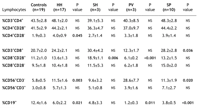
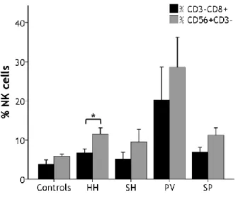
Quando as percentagens das diferentes populações de linfócitos foram analisadas, foi possível observar que a percentagem de linfócitos NK se encontrava elevada em todos os grupos de doentes em comparação ao grupo de indivíduos saudáveis, no entanto este aumento só era estatisticamente significativo nos grupos de doentes com HH e SP. Em contraste, a percentagem de linfócitos B encontrava-se significativamente diminuída nos doentes com HH, PV e SP em relação ao grupo controlo. Outras diferenças, nomeadamente o aumento das populações de linfócito T CD8+ e CD8+CD28+ nos doentes com SH e SP, assim como a
diminuição destas populações em doentes com PV, foram também observadas. Em relação à monotorização das percentagens de linfócitos dos doentes ao longo do tratamento com flebotomias, foi verificada uma tendência para a diminuição da percentagem de linfócitos NK em cinco dos sete doentes seguidos. Em relação à influência dos RBC nas culturas de PBMC, apenas ligeiras diferenças em termos de proliferação e sobrevivência foram observadas, com a excepção das culturas com PBMC de doentes com PV onde, de modo geral, se registou uma menor percentagem de proliferação em todas as condições de cultura em comparação com os restantes grupos de doentes e com o grupo controlo, provavelmente devido ao aumento da população de linfócitos T CD4+ reguladores no sangue periférico descrito recentemente em
doentes com PV. No entanto, e talvez uma das mais inesperadas observações relativa às experiências de proliferação, foi o facto de ser constantemente verificado que os próprios RBC, principalmente os RBC isolados de dadores saudáveis, eram capazes de induzir proliferação de uma população de linfócitos T na ausência de estímulo e sem a observação do aumento de tamanho e complexidade desta população (blastos), independentemente da origem dos PBMC onde eram adicionados.
Os resultados obtidos neste estudo sugerem que o excesso de RBC, bem como o excesso de ferro em circulação, podem estar associados com alterações a nível dos linfócitos NK e B. Futuramente, é necessário realizar mais estudos com vista a determinar se para além das alterações a nível da quantidade de linfócitos NK e B associadas às discrasias sanguíneas, também se encontram anomalias noutros parâmetros destas populações, como por exemplo na expressão de recetores superficiais. Existe também a necessidade de alargar o período de monotorização dos doentes tratados com flebotomias, assim como a necessidade de aumentar o número de indivíduos estudados, tendo em conta que este foi apenas um estudo de carácter preliminar. É ainda imprescindível realizar estudos exaustivos sobre as implicações que a idade e o tempo de armazenamento dos RBC podem ter na sobrevivência e proliferação de células T, provavelmente responsáveis pelos inesperados resultados obtidos neste estudo.
v
Palavras-chave
vi
Abstract
Blood dyscrasias are pathological conditions in which any of the constituents of the blood are structuraly or functionally abnormal. Polycythemia Vera (PV) and Secondary Polycythemia (SP) are two disorders characterized by the increase of the amount of red blood cells (RBC) while hereditary hemochromatosis (HH) and secondary hemochromatosis (SH) are associated with iron overload. It is currently believed that RBC play an important role in T cells growth and survival, which may suggest that anomalies in red blood cells could be associated with lymphocyte alterations. The few immunological studies conducted in erythrocytosis patients reported an increase of CD4+/CD8+ ratio as well as an increase in the percentage of T
CD4+CD25+FoxP3+ cells in patients with PV. A number of earlier studies reported anomalies in
CD8+ T populations in patients with HH. The main objective of this work was to ascertain if
patients with anomalies in RBC numbers have alterations in lymphocyte populations and whether therapeutic phlebotomy influences these populations. In order to do that, blood samples from nineteen healthy donors, seventeen HH patients, three SH patients, three PV patients and ten SP patients were used as a source of peripheral blood mononuclear cells and RBC. The percentages of lymphocyte populations as well as the extent of T cell proliferation and survival were determined by flow cytometry. It was observed that the percentage of NK cells was higher in all groups of patients when compared to controls. However, this increase was statistically significant only in HH and SP patients. In contrasts, B cells were significantly decreased in HH, PV and SP in relation to controls. When the influence of RBC on T cell proliferation and survival was ascertained, it was possible to note that cells from PV patients tended to proliferate less than the cells from the other groups of patients and controls. Unexpectedly, it was also observed that RBC by themselves were able to induce T cell proliferation in the absence of any mitogen stimulus. This effect was most striking with RBC obtained from buffy coats of regular blood donors. These results suggest that an excess of circulating RBC and/or iron are associated with alterations in NK and B populations. Further studies are needed in order to ascertain if the anomalies in NK and B cells include alterations of other parameters, like the expression of surface receptors. More importantly, based on the evidences of the effect of the addition of RBC from controls to cultures, it is necessary to perform extensive studies to elucidate the implications of RBC storage and aging on T cell proliferation and survival.
vii
Keywords
viii
Table of contents
I. Introduction 1 1. Blood dyscrasias 1 1.1. Polycythemia Vera 1 1.2. Secondary polycythemia 3 1.3. Hereditary Hemochromatosis 4 1.4. Secondary Hemochromatosis 6 1.5. Therapeutic phlebotomy: implications for RBC anomalies 6 2. Cross-talk between RBC and T cells 7 2.1. RBC and erythropoiesis 7 2.2. RBC functions and transfusion-related immunomodulation 7 2.3. RBC and T cell growth and survival 9 3. Lymphocyte anomalies associated with blood dyscrasias: A role for RBC? 10II. Aims of the study 13
III. Materials and methods 15
1. Subjects 15
2. Reagents and antibodies 15
3. Cells 16
4. Culture conditions 17
5. CFSE labeling 18
6. Lymphocyte subsets and phenotyping and flow cytometry 18 7. Determination of T cell proliferation 18
8. Statistical analysis 19
IV. Results 21
1. Clinical data 21
2. Peripheral blood lymphocytes phenotypic characterization 22 3. Correlations between lymphocyte subsets and clinical data 26
4. Follow-up studies 28
5. Effect of RBC in T cell proliferation and survival 30
V. Discussion 38
VI. Conclusions 44
VII. Future perspectives 46
ix
List of figures
Figure 1. Erythropoietin receptor signaling 3
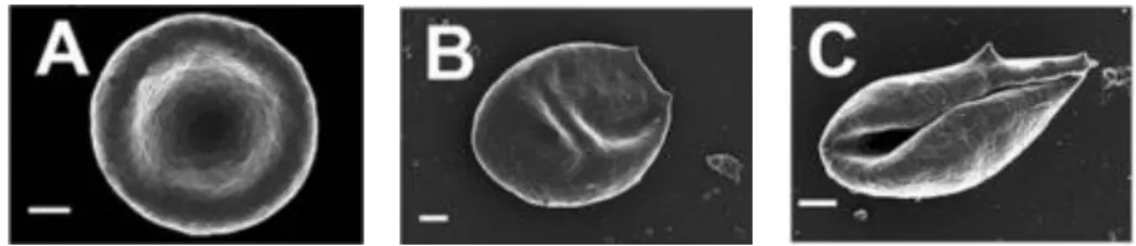
Figure 2. RBC morphological alterations related with iron overload disorders 5
Figure 3. Scheme of PBMC and RBC isolation from peripheral blood samples from
patients and controls
17
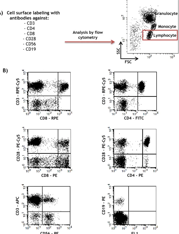
Figure 4. Schematic representation of the flow cytometry analysis of the different
lymphocyte populations
23
Figure 5. Percentage of lymphocyte populations among patients and controls 25
Figure 6. Comparison between the percentage of CD3-CD8+ and NK cells among
patients and controls
26
Figure 7. Correlations between clinical data and lymphocyte populations from
patients
27
Figure 8. Correlations between clinical data and lymphocyte populations in
erythrocytosis patients
28
Figure 9. Lymphocyte populations follow-ups in two patients with HH 29
Figure 10. Lymphocyte populations follow-ups in two patients with PV 29
Figure 11. Lymphocyte populations follow-ups in three patients with SP 30
Figure 12. Analysis of culture proliferation and survival by flow cytometry 31
Figure 13. PI labeling of activated T cells 32
Figure 14. Percentages of proliferation and survival of cultures with PBMC 33
Figure 15. RBC are able to induce cell proliferation in unstimulated cultures of PBMC 34
Figure 16. Proliferating cells in cultures of unstimulated PBMC in the presence of
RBC are T cells
35
Figure 17. Comparison between the addition of autologous and heterologous RBC to
cultures with cells from patients
35
Figure 18. Comparison of the addition of autologous and heterologous to cultures
with cells from controls
x
List of tables
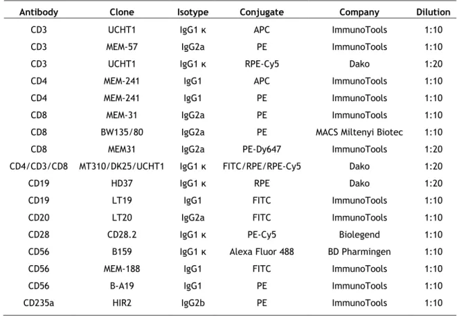
Table 1. Description of the mouse fluorochrome-conjugated anti-human monoclonal antibodies used in the study
17
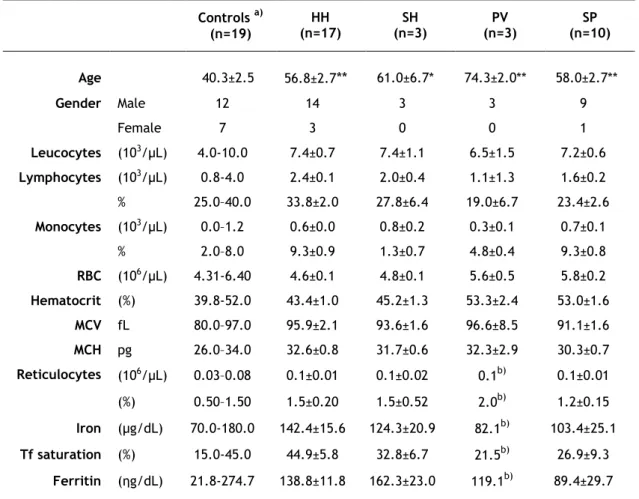
Table 2. Relevant clinical data of patients and controls 22
Table 3. Differences of the relevant clinical data among iron overload disorders and erythrocytosis
23
Table 4. Total percentages of lymphocyte populations 25
xi
Abbreviations
BSA Bovine serum albumin
CFSE 5-(and -6)-Carboxyfluorescein diacetate succinimidyl ester COPD Chronic obstructive pulmonary disease
DNA Deoxyribonucleic acid EPO Erythropoietin
FBS Fetal bovine serum FSC Forward scatter
HH Hereditary hemochromatosis HLA Human leukocyte antigen
IL Interleukin JAK2 Janus kinase
KIR Killer immunoglobulin receptor MAP Mitogen-activated protein MCH Mean corpuscular hemoglobin MCV Mean corpuscular volume
MHC Major histocompatibility complex NK Natural killer
NO Nitric oxide
PBMC Peripheral blood mononuclear cells PBS Phosphate buffered saline solution PHA Phytohemagglutinine
PI Propidium iodide
PI3K Phosphatidylinositol 3 kinase PV Polycythemia Vera
RBC Red blood cells
RBCA Autologous red blood cells RBCH Heterologous red blood cells
SH Secondary hemochromatosis SP Secondary Polycythemia SSC Side scatter
STAT5 Signal transducer and activator of transcription protein 5 Treg T regulator lymphocytes
1
I. Introduction
1. Blood dyscrasias
Blood dyscrasias are pathological conditions in which any of the constituents of the blood are structuraly or functionally abnormal. Sereveral hematological diseases may be included in this group like leukemias, iron-related disorders and myeloproliferative neoplasms. Iron-related disorders are characterized by alterations in iron-related parameters, which may lead to the development of iron overload, as seen in hereditary hemochromatosis (HH) or secondary hemochromatosis (SH) [1], or to the development of iron deficiencies [2]. In contrast, myeloproliferative neoplasms are a group of diseases that include Polycythemia Vera (PV), Essential Thrombocythemia and primary myelofibrosis and are characterized by clonal expansion of an abnormal hematopoietic stem cell [3].
1.1. Polycythemia Vera
Polycythemia Vera is a bone marrow disorder characterized by an excessive production of blood cells in the marrow, including red blood cells (RBC), granulocytes and megakaryocytes (responsible for the production of platelets) [4]. Hemoglobin values above 18.5 g/100 mL in men or 16.5 g/100 mL in women [5], or hematocrit values over 52% in men or 48% in women [6] are indicative of erythrocytosis. Nonetheless, PV individual manifestations may differ greatly between subjects. Some subjects present a moderate elevation in the amount of erythrocytes with an extreme degree of thrombocytosis, while in others the leukocytes count may be at or close to leukemia levels, with only a slight increase in RBC and platelets [4]. Polycythemia Vera has an estimated incidence of about 2.3 per 100,000 every year with a median age of presentation of approximately 60 years old [7], with rare juvenile cases [8]. The incidence of PV is slightly higher in men than women (2.8 versus 1.3 cases/100,000 per year), and is highest for men aged 70–79 years (24 cases/100,000 persons per year)[9]. Common symptoms associated to this disease include headache, vertigo, visual disturbance, vascular disturbances of the extremities, hemorrhage after minor trauma, venous and arterial thrombosis [4], weight loss, pruritus after a warm bath and arthritis [10]. PV is associated with significant morbidity and mortality [7]. This disease may evolve into myelofibrosis, anemia, or less commonly into acute leukemia [7], but the primary causes of morbidity and mortality are thrombotic complications and bleeding [11].
In 2005, the discovery of an acquired Janus kinase 2 (JAK2) mutation was considered as the cause of classical myeloproliferative neoplasms [4,12,13]. Patients with these disorders had a homozygous GT transversion that cause a phenylalanine-to-valine substitution at the position 617 of JAK2 (V617F)[12]. The JAK2V617F mutation is present in more than 95% of PV
2 patients [14,15]. PV patients without the JAK2V617F mutation virtually all have a JAK2 exon 12 mutation [14], another JAK2 mutation associated with this disease [9]. However, some studies suggest that the JAK2V617F mutation may not entirely account for the development of PV [16]. The homozygous JAK2V617F mutation is the result of two steps. The first step is an acquired point mutation which results in a heterozygous state. Then, loss of heterozigosity occurs as the result of mitotic recombination between homologous chromosomes 9p [12,14]. Homozygous JAK2V617 patients (about half of PV patients) tend to have a longer duration of the disease, higher hemoglobin levels, increased incidence of pruritus and are more likeliness to progress to myelofibrosis [17].
The JAK2V617F somatic mutation results in constitutive activation of JAK2 leading to cytokine hypersensitivity and erythrocytosis [7]. This mutation is located in the JAK2 auto-inhibitory domain that results in the autonomous activation of the JAK2 downstream signaling pathways including signal transducer and activator of transcription protein 5 (STAT5), phosphatidylinositol 3 kinase (PI3K), Akt serine threonine kinase and mitogen-activated protein (MAP) kinase (Figure 1)[5,8]. Under normal conditions JAK2 signaling is initiated by the biding of erythropoietin (EPO), a glycoprotein hormone produced predominantly in the kidney that controls erythropoiesis, to its receptor [5]. Experiments performed in murine cell lines showed that the mutated JAK2V617F was constitutively active and able to activate the EPO receptor-dependent signaling pathways in the absence of EPO [8]. Genetic evidences and in vitro functional studies suggest that V617F gives hematopoietic precursors proliferative and survival advantages [12]. The dominance exerted by the JAK2V617F erythroid clone over polyclonal erythroid precursors could be due, in part, to its ability to complete differentiation more efficiently in a low EPO milieu [18]. This dominance may also involve transforming growth factors produced by PV patients mononuclear cells and the hypersensitivity of polycythemia erythroid progenitor cells to interleukin (IL)-3, granulocyte macrophage–colony-stimulating factor, stem cell factor, and insulin like growth factor [4,9,18].
The JAK2V617F mutation is present in granulocytes, erythroblast and in all the EPO-independent erythroid colonies. Initially it was thought that the JAK2V617F mutation was not present in B and T cells [19], however, Ishi et al. demonstrated that the JAK2V617F mutation is also present in B and T lymphocytes in a subpopulation of patients with PV [20]. Two possible mechanisms of the JAK2V617F mutation are suggested. In the first JAK2V617F mutation alone or in combination with one or more preexisting somatic mutations may cause the myeoloproliferative disorder [12]. The second model suggests that the JAK2V617F mutation is a secondary genetic event that occurs after the onset of the myeloproliferative phenotype, which may be caused by mutation(s) in a gene or genes that remain unknown [12,21]. In any case, the detailed mechanism involved in the abnormal activation of JAK2 and its transforming activity remain to be elucidated [13].
3
Figure 1. Erythropoietin receptor signaling. Binding of Epo to its receptor results in receptor
homodimerization and autophosphorylation of the receptor-associated JAK2. Activated JAK2 in turn mediates the phosphorylation of key tyrosine residues on the distal cytoplasmic region of EpoR, which then serve as docking sites for downstream effectors, including STAT5 and PI3K. Activated STAT5 translocates to the nucleus to affect gene transcription. Adapted from [5].
Besides the alterations in the amount of RBC, other RBC anomalies have been associated with PV. The removal of excessive RBC from the circulation in PV patients as therapy may imply changes in RBC morphology. The reduction of iron stores in PV due to the continuous need of this metal for hemoglobin production, may lead to the production of smaller and hypochromic RBC [4]. Alterations in the adhesion properties of RBC from PV patients have also been reported [8]. In a study conducted by Wautier et al. RBC from PV individuals were abnormally adherent to resting human umbilical vein endothelial cells under static and flow conditions [22]. A later study from the same group confirmed that the increased adhesion levels were due to a constitutive phosphorylation of an adhesion molecule that correlated with the presence of JAK2V617F mutation [23].
1.2. Secondary Polycythemia
Secondary Polycythemia (SP), previously designated as polyglobulia, is a condition that comprises a group of diseases characterized by an increase in RBC mass or RBC amount like PV, but that results from a mechanism other than alterations in bone marrow progenitors [6]. SP is differentiated from PV by the absence of JAK2 mutations and by raised EPO levels
4 [10,24,25]. SP can be classified as congenital, which are associated with genetic abnormalities, or acquired, usually related to a secondary disorder or external factors that lead to an increase of erythrocyte production [6,9].
Congenital SP are associated with genetic abnormalities which include mutations in genes from the oxygen-sensing pathway that culminate in the increase of EPO levels (e.g. mutations in the von Hippel Lindau gene), heterozygous mutations in the prolyl hydroxylase 2 gene or mutations in the hypoxia inducible factor [5,6,24,26]. High oxygen-affinity hemoglobin and rare enzyme deficiencies, such as bisphosphoglycerate mutase deficiency, are other congenital causes that can culminate in SP [5,24].
The causes of acquired SP belong to three main categories: hypoxia, pathological EPO production and exogenous erythropoietin. The hypoxic process may be associated with conditions that limit oxygen availability in the arterial circulation, such as chronic obstructive pulmonary disease (COPD), cyanotic heart disease or obstructive sleep apnea [27,28]. High altitude habitat is another hypoxia-related factor that may lead to the development of SP [5,24,28]. Pathological EPO production is mainly a consequence of renal alterations like local renal hypoxia or tumors. The presence of exogenous EPO may also drive to the over production of RBC and is generally associated with the administration of androgens or corticosteroids or with the use of EPO to increase the red blood cell mass in athletes [24,28].
1.3. Hereditary Hemochromatosis
Hereditary Hemochromatosis (HH) is an inherited autosomal recessive disorder [29] characterized by the progressive accumulation of iron [30] that is caused by dysregulated intestinal iron absorption [31]. Some of the symptoms associated with the disorder include fatigue, depression, weight loss, joint pain, decreased libido, hair loss and abdominal discomfort [32]. The iron deposition in parenchymal organs that takes place in this disease can be responsible for serious organ dysfunctions leading to liver failure and cirrhosis, hepatocellular carcinoma, atherosclerosis, arthritis, various endocrinopathies including diabetes, hypermelanotic pigmentation of the skin, or compromised immune defense [29]. Hereditary Hemochromatosis is considered a disorder with slow progression with typical age of presentation for HH related symptoms usually occurring between 40 and 60 years old for males and after menopause in females [32]. Men tend to develop symptoms and signs of organ injury at a rate of 5 to 10 times higher than women, a particularity that is likely related with the recurrent blood loss in women due to menses and childbirth and varying dietary habits among men and women [31].
The most common type of HH is associated with mutations in the HFE gene [29]. The HFE gene was first identified in 1996 as a major histocompatibility complex (MHC) class I-like gene in which homozygosity for a missense mutation that results in cystein-to-tyrosine substitution
5 at amino acid 282 of human HFE protein (C282Y), was responsible for most cases of HH [33]. C282Y is the most prevalent mutation in Western populations affecting approximately one out of 250 individuals [34,35] being much less common in Hispanic, Asian American, Pacific Islander, and black persons [36].
The HFE protein interacts with transferrin (Tf) receptors 1 and 2, senses iron body status, and activates downstream signaling pathways that regulate iron homeostasis [37]. HFE mutations result in low levels of hepdicin, a hormone produced in the liver that plays a major role in the regulation of systemic iron homeostasis, acting as a negative regulator of iron absorption from the gut and iron egress from macrophages [31]. The C282Y mutation disrupts a disulphide bond apparently required for proper folding of the HFE molecule, preventing its association with β2-microglobulin as well as its surface expression [38]. Approximately 80% of HH patients are homozygous for C282Y while only a few cases of patients are heterozygotes for a C282Y and H63D mutation, a second mutation in the HFE gene associated with HH [29]. Other factors that affect iron absorption, transport, and mobilization include hepdicin, hemojuvelin, Tf receptor-1 and -2, ferroportin [30], and CD8+ T cells [39]. Mutations in some
of these genes (e.g. haemojuvelin gene, hepdicin gene and Tf receptor-2 gene) have been associated with non-HFE-related forms of HH [1], namely aggressive juvenile subtypes [2]. These mutations are much less frequent than HFE mutations. However, in contrast to HFE mutations, they are spread in different parts of the world and are not linked to a specific race [35].
Morphological alterations of RBC have been described in hemochromatosis patients and individuals with high serum ferritin levels relative to those from normal controls [40]. RBC from these individuals presented a significantly elongated shape than RBC from controls (Figure 2). Alterations in fibrin network morphology from RBC were also observed. These differences could largely be reversed in the presence of some iron chelating agents [40].
1.4. Secondary iron overload
Figure 2. RBC morphological alterations related with iron overload disorders. RBC scanning electron
microscope images from a normal healthy individual (A), a HH patient (B), and an individual with high ferritin levels (C). Figure adapted from [40].
6
1.4. Secondary Hemochromatosis
Secondary Hemochromatosis (SH), also known as secondary iron overload, is mainly associated with recurrent blood transfusions in patients with hereditary or acquired anemia subtypes that arise from mutations in genes that lead to insufficient or malfunctioning erythrocytes, such as myelodysplastic syndrome, thalassemia or sickle cell disease [2]. Although blood transfusions may be able to correct the anemia state in patients with myelodysplastic syndrome, thalassemia, and sickle disease, chronic transfusion therapy remains one of the most important causes of SH [1,41–43].
Other causes of secondary iron overload include liver diseases [44] and ingestion of excessive dietary amounts of iron [42]. The release of iron from injured hepatocytes is one of the main causes of iron overload in chronic liver diseases like hepatitis C, alcoholic liver disease and non-alcoholic fatty liver disease [1]. Hepdicin dysregulation and HFE mutations may also contribute to iron overload in these particular diseases, however further studies are necessary to confirm this association [1].
1.5. Therapeutic phlebotomy: implications for RBC anomalies
Phlebotomy, also known as bloodletting or venesection, is a major therapeutic procedure that has been performed in various civilizations since antiquity up to the present [10]. Currently, therapeutic phlebotomy is one of the main treatments in PV, SP, HH and SH. Classical phlebotomy is a simple, safe, and low-cost method [45] that allows reduction of RBC mass and depletion of iron stores. The amount of blood that is removed at each session as well as the phlebotomy frequency depend of the hematocrit values in PV and SP patients and serum ferritin levels in patients with iron overload, but in the majority of the cases approximately 450 to 500 mL of blood are removed at each session [4,10,30].
As most therapeutic approaches, phlebotomy is associated with positive and negative effects. Several studies demonstrated that hepatic fibrosis in HH patients can be reversed by phlebotomy therapy [30]. Also, phlebotomy has been associated with a reduced risk of the development of leukemia, an increased risk of vascular events and a better overall median survival in PV patients when compared to other treatment strategies [9,14]. Some negative aspects referenced by patients undergoing phlebotomy include venous access problems, time wasted (for travel, waiting, and the procedure) and the fact that in many cases the blood is not used for donation [30]. The lack of long term follow-up studies of patients with erythrocytosis that are treated exclusively with therapeutic phlebotomy makes it difficult to assess the clinical benefits of this treatment.
The need of recurrent venesections as the main treatment of iron overload disorders and erythrocytosis, and the fact that this blood cannot be reused for other purposes like blood
7 donation, makes phlebotomy samples an ideal source for the study of lymphocyte populations.
2. Cross-talk between RBC and T cells
In the past decades evidence has been accumulated supporting the view that RBC are more than just mere carriers of gases and nutrients. Marked alterations in the immune system after transfusions of RBC suggest that these cells may be associated with immunomodulation [46,47]. Previous studies conducted by our group also indicate that RBC play an important role in T cell growth and survival [48,49]. The characteristics of RBC, as well as their functions are further discussed in the subsequent sections of this chapter.
2.1. RBC and erythropoiesis
Erythrocytes represent the most common cell type in adult blood. Human blood contains approximately 5x10⁶ erythrocytes per microliter (normal range 4.7x10⁶ to 6.1x10⁶ for males
and 4.2x10⁶ to 5.4x10⁶ for females) and have an average life span of 120 days [50]. In normal
adults, approximately 200 billion of the oldest erythrocytes (about 1% of the total number) are replaced every day by an equal number of newly formed erythrocytes [51]. The small size of RBC, usually 6 to 8 µm of diameter, and its biconcave shape creates a large surface area for gas exchange and allows their entry into the micro capillaries of the tissues [50]. Mature RBC in the bloodstream lack the nucleus and cytoplasmic organelles, including mitochondria and ribosomes. As RBC age, their surface area and volume, but not hemoglobin, are progressively lost [52].
Erythropoiesis is a tightly regulated process by which the hematopoietic tissue of the bone marrow produces RBC. It consists of several developmental stages: hematopoietic stem cell, burst-forming unit-erythroid, colony-forming unit-erythroid, proerythroblast, basophilic erythroblast, polychromatic erythroblast, orthochromatic erythroblast, reticulocyte and ultimately to mature RBC [25,51–53]. In humans, the new red cells enter the circulation as reticulocytes that need approximately one week to complete the maturation process [50], which include the complete elimination of traces of DNA, mitochondria, endoplasmic reticulum, and ribosomes [54]. EPO is the main regulator of erythropoiesis [51]. Alterations in this process, like mutations in the hematopoietic cells or the increase of EPO levels, have been associated with the development of classical myeloproliferative neoplasms and SP, respectively, as described previously.
2.2. RBC functions and transfusion-related immunomodulation
Red blood cells have been considered almost exclusively as transporters of metabolic gases and nutrients for the tissues. Currently, a number of clinical and experimental evidence support the notion that these are not the only functions played by erythrocytes. RBC are one of the major components of blood antioxidant capacity and one of the cells more resistant to
8 oxidative stress [55]. Within inflamed areas, RBC can contribute to detoxify reactive oxygen and nitrogen species and thus to rescue or partially “protect” cells under intense oxidative stress. In some physiological conditions, for instance when RBC become targets of xenobiotics or parasites, or when they pass through tissues where an intense production of reactive oxygen or nitrogen species occurs, RBC can also act as sources of reactive oxygen species [55].
Red blood cells may also play a pivotal role in the regulation of the local vascular processes matching oxygen supply and demand in active skeletal muscle in humans [56–58]. It is believed that RBC contribute to these local regulatory processes releasing vasoactive substances into the vascular lumen in response to several metabolic and mechanical stimuli [57]. Indeed, several lines of evidence have implicated RBC as modulators of nitric oxide (NO) signaling by effecting both NO formation and inhibition of NO signaling [59]. Under hypoxic conditions it has been demonstrated that RBC induce NO-dependent vasodilatation [60]. It is thought that nitrite is reduced to NO in a manner that is regulated by the fractional saturation of hemoglobin and hence regulated by mechanisms that control oxygen affinity [61]. In addition, Srihirun et al. reported that nitrite in the presence of RBC had an inhibitory effect on platelet aggregation, but nitrite alone at physiological concentrations did not have the same effect [62]. This inhibitory effect was promoted by deoxygenated hemoglobin [62]. Despite these evidences, the mechanisms of release and potential sources of NO in RBC are still a matter of debate [60], since RBC can act both as NO scavengers and NO producers. Most of the current knowledge on the influence of RBC in the immune system is the result of the investigation of the consequences of RBC transfusions
.
Immune mediated reactions to transfusion include hemolytic reactions, febrile non-hemolytic reactions, allergic reactions, transfusion related acute lung injury, bacterial contamination, transfusion related immunomodulation, and transfusion associated graft versus host disease [46]. Transfusion-related immunomodulation consists of both immunostimulatory and immunosuppressive phenomena. Proinflammatory responses, like systemic inflammatory responses and multiple organ failure, are often seen after massive transfusions, whereas the immunosuppressive effects are seen in lower-volume transfusion [47]. Transfusion-related immunomodulation persists despite leukoreduction (removal of the contaminating donor white cells and platelets in red cell concentrates) [63], which implicates RBC in the production of mediators during storage that may be responsible for transfusion related outcomes [64].Allogeneic (but not autologous) transfusion has been associated with down regulation of cellular immunity and dysregulation of inflammatory innate immunity. Experimental evidence support the hypothesis that immune deviation towards type 2 immune responses and corresponding inhibition of type 1 responses may explain post-operative infection and cancer recurrence after transfusion [64]. Additionally, increases in T regulatory (Treg) cells have
9 been demonstrated after allogeneic exposure to stored RBC [65] which may explain, in part, the immunosuppression outcomes observed sometimes after RBC transfusion [66].
Recent studies suggest that the consequences of RBC transfusion are dependent of the time of storage of the RBC units [67]. Most storage lesion effects appear to worsen with increased storage duration. Inflammatory response, mediators of oxidative injury and risk of hyper-coagulation appear to increase when RBC with a long period of storage are compared with fresh RBC [68]. The excessive activation of the innate immune system by danger associated molecular pattern molecules generated throughout RBC storage may explain some of the adverse outcomes of transfusion [46,69].
In a study conducted by Bernard et al. it was reported that banked human RBC suppressed mitogen-stimulated human and antigen-stimulated mouse T-cell proliferation by mechanisms independent of arginine depletion (associated with the generation of suppressor cells) or cell death [70]. More recently, the same group demonstrated that B cell proliferation was also suppressed by blood bank RBC. They also concluded that T cells and B cells suppression was abrogated when fresh RBC were used [47]. The authors suggested that the lack of suppression observed with fresh RBC could be related with alterations of RBC properties during the isolation procedure [47].
2.3. RBC and T cell growth and survival
Previous studies performed by our group have demonstrated that the presence of RBC in cultures of mitogen-stimulated human peripheral blood lymphocytes was able to increase T cell proliferation and survival [48]. Moreover, the presence of RBC in cultures without serum allowed T cells to proliferate to levels very similar to those observed in cultures with serum, literally rescuing the activated T cells from cell death [71]. The increase in T cell survival was associated with the inhibition of activation-induced T cell death by RBC. This finding was correlated with a decrease in oxidative stress within the activated T cells and the inhibition of the formation of acrolein, a lipid-derived product that function as a marker of oxidative stress. In this case, the RBC protection-effect required the presence of intact RBC and RBC/T cells contact and was independent of monocytes [48]. It is also believed that RBC may have a different impact on CD4+ and CD8+ T cells. Indeed, some studies suggest that RBC support the
preferential expansion of CD8+ T cells in cultures [48,72].
Several attempts to identify the molecular mechanisms responsible for the protective effect exerted by RBC on T cells have been performed [73]. It is currently known that RBC are capable to augment the percentage of T cells that enter cell division and also the percentage of cell survival both after T cell receptor-dependent and T cell receptor-independent stimuli [71]. However, many aspects related with the molecular mechanisms associated with the cross-talk between RBC and T cells remain to be elucidated.
10
3. Lymphocyte anomalies associated with blood dyscrasias: a
role for RBC?
Few immunological studies have been conducted in SP and PV. In 1987 Laurence and collaborators reported that CD4+/CD8+ T lymphocyte ratios were significantly increased in
patients with PV. The finding of reduced CD8+ T lymphocytes, and hence increased CD4+/CD8+
ratios in PV, seemed to be a persistent feature of the condition, remaining uncorrected by previous treatment. They also demonstrated that these alterations were not verified in patients with SP [74].
More recently, alterations in T cell subpopulations have been associated with PV. Zhao et al. showed that the percentage of T CD4+ regulator cells (CD4+CD25+FoxP3+) in peripheral blood
of PV patients was significantly higher compared to healthy controls. Moreover, when co-cultured with activated CD4+CD25– T cells, the CD4+CD25+ Treg cells showed enhanced
suppressive function in PV [7]. In a more recent study, an increase in CD4+ Treg cells was also
demonstrated in PV patients undergoing long term treatment with interferon alpha. However, this increase was not observed in untreated patients [75]. T cells from PV patients also seem to produce growth factors that contribute to endogenous erythroid and megakaryocyte colony formation [16]. Decrease in natural killer (NK) cells activity in patients with PV was also reported [76].
The immunological studies performed in hereditary and secondary hemochromatosis patients are much more abundant and mostly describe alterations in T cell subsets. Interestingly, like in PV, high CD4+/CD8+ T cell ratios were also observed in some HH patients. In HH, high
CD4+/CD8+ T cell ratios were associated with high levels of iron overload [77]. Other T cell
anomalies reported in HH patients include expansion of CD8+CD28- T cells linked with the
HLA-A3 haplotype, an increase of the relative percentage of CD8+HLA-DR+ cells (a marker of
recent activation) and a decrease in the cytotoxic activity of CD8+ T lymphocytes [78]. In
another study, Fabio et al. described a decrease on total lymphocyte counts of several lymphocyte populations, including CD3+CD4+, CD4+, CD28+, CD8+CD28+ and NK cells in HH.
They also reported a major increase in the production of IL-4, IL-5 and IL-10 by the CD3+CD8+
T cell subset [79]. More recently, the low numbers of CD8+ lymphocytes observed in HH were
associated with a decrease of the most mature CD8+ effector memory T cells [80].
Besides phenotypic alterations, functional abnormalities in CD8+ T lymphocytes were also
described in HH patients. The level of autophosphorylation of the CD8-associated p56lck, a protein kinase that plays a key role in T cell activation, as well as its phosphotransferase activity was significantly reduced up to three-fold in HH patients relative to healthy donors [81]. Interestingly, a recent study performed using transient HFE transfection assays in a model of antigen-presenting cells demonstrate that wild type HFE inhibits T lymphocyte
11 activation independently of the nature of the peptide being presented by MHC-I molecules and of MHC-I/peptide complex stability when compared to C282Y-mutated HFE [37].
The lymphocyte and RBC alterations found in PV, SP, HH and SH may suggest the existence of a cross-talk between RBC and T cells that may impact the development and survival of lymphocyte subsets. In this context, the study of phenotypic and functional lymphocyte alterations in patients with blood dyscrasias may provide further evidences that can lead to a better understanding of the cross-talk between RBC and T cells as well as the implications of RBC alterations in this interaction.
12
13
II. Aims of the study
The main objective of this work was to ascertain if patients with anomalies in RBC numbers and/or cell mass have alterations in the four major lymphocyte populations (T, B, NK and NKT) and whether therapeutic phlebotomy influences these populations. For that purpose, patients with PV, SP, HH and SH, along with a group of healthy blood donors were studied. In this context, the specific aims of this work were:
1. To analyze lymphocyte populations in patients and controls.
14
15
III. Materials and methods
1. Subjects
Seventeen HH patients (14 males and 3 females; mean age 57 years; range 30-77 years), three SH patients (3 males; mean age 61 years; range 48-70 years), three PV patients (3 males; mean age 74 years; range 71-78 years) and ten SP patients (9 males and 1 female; mean age 58 years; range 38-72 years) were selected to this study. Nineteen healthy regular blood donors (12 males and 7 females; mean age 40 years; range 21-55 years) from Centro de Sangue e da Transplantação de Coimbra were used as controls. All buffy coats included in this study were between 2-3 days old.
HH patients were diagnosed taking into account abnormal elevated values of serum Tf saturation (>50% to 55% in men and >45% in women) [31] and serum ferritin (>300 µg/L in men and >200 µg/L in women) [30,31] and the presence of HFE mutations. SH patients also presented elevated levels of Tf saturation and serum ferritin, similarly to HH patients. However, the presence of HFE mutations was excluded in these patients and the raised iron levels were associated in most of the cases with the presence of liver disorders, recurrent blood transfusions and the ingestion of excessive amounts of dietary iron.
The diagnosis of PV was established according to the World Health Organization diagnosis criteria. These include high hematocrit values (>52% in men and >48% in women) or raised RBC mass (>25% above predicted) and the presence of JAK2 mutations. In JAK2 mutations negative PV patients minor criteria like bone marrow biopsy changes, serum EPO below the normal range or in vitro endogenous erythroid colony formation are also required [6]. Hematocrit values and RBC mass were also raised in SP patients. However, SP diagnosis was established by the absence of JAK2 mutations and generally by raised serum EPO. In most cases, SP were secondary pathologies associated with pulmonary disorders, like COPD and emphysema, or with smoking habits.
This study was approved by the Ethics Committee of Centro Hospitalar Cova da Beira and is part of a joint research project between CICS-UBI and the Immunohemotherapy Service from Centro Hospitalar Cova da Beira (projectnº105/2013). Informed consent was obtained from all subjects following the guidelines of local institutions.
2. Reagents and antibodies
Phytohemagglutinin (PHA, from Phaseolus vulgaris), bovine serum albumin (BSA), trizma base, ammonium chloride, RPMI-1640 medium, propidium iodide (PI) and the antibiotic-antimycotic solution were obtained from Sigma-Aldrich (Madrid, Spain). Biocoll (density 1.10 g/mL) and fetal bovine serum (FBS) were from Biochrom (Berlin, Germany). Lymphoprep was from
16 STEMCELL Technologies (Genobre, France). Sodium azide was obtained from Amresco (Solon, USA). CellTrace Carboxyfluorescein diacetate succinimidyl ester (CFSE) Cell Proliferation Kit was from Molecular Probes (Amesterdan, The Netherlands). The mouse fluorochrome-conjugated anti-human monoclonal antibodies used in this work and its characteristics are described in Table 1.
Table 1. Description of the mouse fluorochrome-conjugated anti-human monoclonal antibodies used in the study.
Antibody Clone Isotype Conjugate Company Dilution
CD3 UCHT1 IgG1 κ APC ImmunoTools 1:10
CD3 MEM-57 IgG2a PE ImmunoTools 1:10
CD3 UCHT1 IgG1 κ RPE-Cy5 Dako 1:20
CD4 MEM-241 IgG1 APC ImmunoTools 1:10
CD4 MEM-241 IgG1 PE ImmunoTools 1:10
CD8 MEM-31 IgG2a PE ImmunoTools 1:10
CD8 BW135/80 IgG2a PE MACS Miltenyi Biotec 1:10
CD8 MEM31 IgG2a PE-Dy647 ImmunoTools 1:20
CD4/CD3/CD8 MT310/DK25/UCHT1 IgG1 κ FITC/RPE/RPE-Cy5 Dako 1:20
CD19 HD37 IgG1 κ RPE Dako 1:20
CD19 LT19 IgG1 FITC ImmunoTools 1:10
CD20 LT20 IgG2a FITC ImmunoTools 1:10
CD28 CD28.2 IgG1 κ PE-Cy5 Biolegend 1:10
CD56 B159 IgG1 κ Alexa Fluor 488 BD Pharmingen 1:10
CD56 MEM-188 IgG1 FITC ImmunoTools 1:10
CD56 B-A19 IgG1 PE ImmunoTools 1:10
CD235a HIR2 IgG2b PE ImmunoTools 1:10
3. Cells
Buffy coats were obtained from the patients fresh collected peripheral blood bags (therapeutic phlebotomies) after a centrifugation at 2200xg for 15 minutes (Figure 3). Peripheral blood mononuclear cells (PBMC) were obtained from the buffy coats of healthy donors and from the buffy coats of patients by performing a gradient centrifugation at 1000xg for 30 minutes over Lymphoprep or Biocoll (Figure 3). PBMC were washed with 1x phosphate buffered saline solution
(
PBS) followed by centrifugation at 700xg for 10 minutes. Contaminating RBC were lysed in pre-heated lysis solution (10 mM Tizma base, 150 mM Ammonium Chloride, pH 7.2), for 10 minutes at 37 °C. After RBC lysis, PBMC were washed once again with PBS and centrifuged at 500xg for 10 minutes.17 RBC were obtained from the pellet region after Lymphoprep/Biocoll centrifugation or from the pellet region after centrifugation of total blood at 2200xg for 15 minutes and diluted 1:10 in RPMI-1640 (Figure 3). The RBC solution was stored until use for a maximum of one week at 4 °C.
4. Culture conditions
PBMC (1.5x10⁶) were cultured in 6-well plates in RPMI-1640 supplemented with 1%
heat-inactivated FBS and 1% antibiotic-antimycotic solution in a final volume of 5 mL. Cultured cells were left in an incubator at 37 °C, 5% CO2 and 95% humidity, between 6 to 7 days. PBMC
were either left unstimulated or stimulated with 5 µg/mL of PHA, a T cell mitogen, in the absence or presence of autologous RBC. In some experiments PBMC from controls and patients were cultured in the presence of heterologous RBC. All the cultures with RBC were performed at a PBMC:RBC ratio of 1:10. Unstimulated PBMC with no RBC were used as a negative control. Samples (PBMC/RBC) obtained from different phlebotomies of the same patient were considered as independent proliferation experiments.
Figure 3. Scheme of PBMC and RBC isolation from peripheral blood samples from patients and controls. The figure shows the process of PBMC and RBC isolation from samples from patients (A) and
from healthy donors (B). Buffy coats from patients were obtained after whole blood (phlebotomy samples) centrifugation at 2200xg for 15 minutes. In the case of healthy donors, the blood samples received from Centro do Sangue e da Transplantação de Coimbra were already in the form of buffy coats. PBMC from patients and controls were obtained after a gradient centrifugation of buffy coats at 1000xg for 30 minutes over Lymphoprep or Biocoll. RBC were collected from the pellet region after centrifugation of whole blood or after Lymphoprep/Biocoll centrifugation.
B) Controls
2200xg 15 minutes Buffy coats PBMC 1000xg 30 minutes RBC RBC Lymphoprep/Biocoll Phlebotomy bag PBMC RBC Lymphoprep/Biocoll Buffy coatsA) Patients
1000xg 30 minutes18
5. CFSE labeling
CFSE is a fluorescent dye that passively diffuses into cells and forms dye-protein adducts which are retained by the cells until they enter mitosis, where the dye-protein adducts are splitted between daughter cells. For CFSE labelling PBMC were resuspended in PBS at a concentration of 10x106 cells/mL and incubated with 5 µM of CFSE for 10 minutes at 37 °C,
with occasional mixing. After labeling, PBMC were washed twice with PBS/20% heat inactivated FBS and resuspended in culture media. CFSE labeling efficiency was analyzed by flow cytometry immediately after label procedure and further confirmed by the analysis of fluorescence from unstimulated PBMC at the end of cultures.
6. Lymphocyte subsets phenotyping and flow cytometry
PBMC or total leukocytes were used for lymphocyte phenotyping. When total leukocytes were used, peripheral blood samples were first treated with RBC lysis solution (10 mM Tizma base, 150 mM Ammonium Chloride, pH 7.2), for 10 minutes at 37 °C. Approximately 0.5x10⁶ PBMC or
2.5x10⁶ total leukocytes (in case of whole blood staining) were placed per well (in 96-wells
round bottom plates) and washed twice with staining solution (PBS 1x, 0.2% BSA, 0.1% Sodium azide). Cells were then incubated on ice for 30 minutes, in the dark, with 50 µL of the appropriated diluted anti-human monoclonal antibodies on staining solution. Cells incubated with 50 µL of staining solution (no antibody added) were used as negative control. Afterwards, PBMC or total leukocytes were washed again twice with staining solution and once with PBS. Cells were resuspendend in 150 µL of PBS and placed in FACS tubes at a final volume of 500 µL. After labeling, cells were then acquired in a FACSCalibur (Becton Dickison S.A. Madrid). For each sample 10,000 viable lymphocytes were acquired, whenever possible, using FSC/SSC characteristics and analyzed with CellQuest Pro or CellQuest Software (Becton Dickison S.A. Madrid).
Different combinations of monoclonal antibodies were used in order to discriminate distinct lymphocyte populations (see Table 1). Lymphocyte subsets were defined as follow: CD8+ T
cells (CD3+CD8+), CD4+ T cells (CD3+CD4+), NK (CD3-CD56+), NKT (CD3+CD56+) and B cells (CD19+
or CD20+). The expression of co-stimulatory molecule CD28+ was also investigated among CD8+
and CD4+ T cells. Simultaneous staining with CD3 and CD8 demonstrated that the percentage
of CD8bright T cells (CD28+ plus CD28-) approximated the percentage of CD3+CD8+ T cells. In
order to avoid discrepancies between populations, only CD8bright cells were considered as
CD3+CD8+ T cells.
7. Determination of T cell proliferation
T cell activation and division were studied by two methods: (1) determination of cell size and complexity according to FCS/SSC parameters (blasts) and (2) sequential halving of CFSE-fluorescence intensity. T cell survival was also investigated by the determination of cell size
19 and complexity according to FCS/SSC parameters and PI labeling (5 µg/mL). At the end of the culture period, PBMC cultured with RBC were treated with lysis solution (10 mM Tizma base, 150 mM Ammonium Chloride, pH 7.2) for 10 minutes at 37 °C to remove RBC. This treatment did not significantly alter the activation parameters studied when compared with non-treated activated T cells [48]. Before FACSCalibur acquisition, all cells were washed with PBS and centrifuged at 500xg for 10 minutes. T cell death was determined by PI staining in a couple of experiments.
In some experiments, after the treatment with lysis solution, cells were labeled with fluorochrome-conjugated monoclonal antibodies against CD3 and CD235a (glycophorin A). Glycophorin A is a major protein expressed in the human RBC membrane and it was used in this study to investigate the presence of RBC proteins in the lymphocyte region. For each sample 10,000 events were acquired in the gate encompassing both the “small cells” and “blast cells” regions using FSC/SSC characteristics by flow cytometry and analyzed using CellQuest or CellQuest Pro software.
8. Statistical analysis
Statistical analysis was performed using IBM SPSS Statistics version 21 (SPSS Inc. USA) software. Student’s independent samples T-test was used to determine the significance of the differences between group means. Mann-Whitney test was used to determine the significance of the differences between group medians when non parametric variables were compared, namely ferritin. Pearson’s correlation was used to assess the correlation between the percentages of lymphocyte populations and patients’ clinical data. Statistical significance was defined as p<0.05.
20
21
IV. Results
1. Clinical data
Relevant clinical data of patients and controls included in this study are summarized in Table 2. Regarding the control population, only age and gender were available. It was possible to observe that the mean age of the four groups of patients was significantly higher than the mean age of the control group. The mean age of PV patients in relation to HH patients was also statistically significant higher. Interestingly, both the percentage of lymphocytes and the total number of lymphocytes, were decreased in PV and SP patients, when compared to patients with HH and SH (p<0.001).
Table 2. Relevant clinical data of patients and controls†.
†
Values represent the mean±SEM. Differences between controls’ age and the different groups of patients (Student’s T-test) are indicated (**p<0.001, *p<0.05). a) Clinical data from controls was not available, but reference values from healthy male populations are shown, since the majority of patients were men. b) SEM is not applicable since this data was only available in one of the patients. MCV, mean corpuscular volume; MCH, mean corpuscular hemoglobin; n, number of subjects; Tf, transferrin.
Controls a) (n=19) HH (n=17) SH (n=3) PV (n=3) SP (n=10) Age 40.3±2.5 56.8±2.7** 61.0±6.7* 74.3±2.0** 58.0±2.7** Gender Male 12 14 3 3 9 Female 7 3 0 0 1 Leucocytes (103/µL) 4.0-10.0 7.4±0.7 7.4±1.1 6.5±1.5 7.2±0.6 Lymphocytes (103/µL) 0.8-4.0 2.4±0.1 2.0±0.4 1.1±1.3 1.6±0.2 % 25.0–40.0 33.8±2.0 27.8±6.4 19.0±6.7 23.4±2.6 Monocytes (103/µL) 0.0–1.2 0.6±0.0 0.8±0.2 0.3±0.1 0.7±0.1 % 2.0–8.0 9.3±0.9 1.3±0.7 4.8±0.4 9.3±0.8 RBC (106/µL) 4.31-6.40 4.6±0.1 4.8±0.1 5.6±0.5 5.8±0.2 Hematocrit (%) 39.8-52.0 43.4±1.0 45.2±1.3 53.3±2.4 53.0±1.6 MCV fL 80.0–97.0 95.9±2.1 93.6±1.6 96.6±8.5 91.1±1.6 MCH pg 26.0–34.0 32.6±0.8 31.7±0.6 32.3±2.9 30.3±0.7 Reticulocytes (106/µL) 0.03–0.08 0.1±0.01 0.1±0.02 0.1b) 0.1±0.01 (%) 0.50–1.50 1.5±0.20 1.5±0.52 2.0b) 1.2±0.15 Iron (µg/dL) 70.0-180.0 142.4±15.6 124.3±20.9 82.1b) 103.4±25.1 Tf saturation (%) 15.0-45.0 44.9±5.8 32.8±6.7 21.5b) 26.9±9.3 Ferritin (ηg/dL) 21.8-274.7 138.8±11.8 162.3±23.0 119.1b) 89.4±29.7
22 Other differences between the groups included higher hematocrit and RBC amount and lower levels of iron, Tf saturation and ferritin in patients with PV and SP when compared with HH and SH patients (Table 2). These differences were confirmed when the four groups of patients were paired in two groups, iron overload (patients with HH and SH) and erythrocytosis (patients with PV and SP). As shown in Table 3, lymphocytes, both total and in percentage, were statistically significant lower in the erythrocytosis group in relation to the iron overload group (1.5±0.1x10³/µL versus 2.3±0.1x10³/µL, and 22.4±2.4% versus 32.9±1.9%, respectively).
In contrast, hematocrit values as well as RBC amount were statistically significant higher in the erythrocytosis group in relation to the iron overload group (53.0±1.3% versus 43.7±0.8% and 5.8±0.2x10⁶/µL versus 4.6± 0.1x10⁶/µL). Although the values of iron, Tf saturation and
ferritin were higher in the iron overload group, no statistically significant differences were observed among the two groups.
Table 3. Differences of the relevant clinical data among iron overload disorders and erythrocytosis†.
† Values represent the mean±SEM. Iron overload group includes patients with HH and SH; and
erythrocytosis group includes patients with PV and SP. P values of the comparison between the two groups are shown (Student’s T-test and Mann-Whitney test in the case of ferritin). NS, not significant; n, number of subjects.
2. Phenotypic characterization of peripheral blood lymphocytes
In order to investigate if peripheral blood lymphocyte populations were different among healthy individuals and patients with blood dyscrasias, PBMC or leukocytes from all individuals were labeled with antibodies against selected surface receptors and analyzed by flow
Iron Overload (n=20) Erythrocytosis (n=13) P value Age 57.5±2.5 61.8±2.9 NS Leucocytes (103/µL) 7.4±0.6 7.0±0.6 NS Lymphocytes (103/µL) 2.3±0.1 1.5±0.1 <0.001 % 32.9±1.9 22.4±2.4 0.002 Monocytes (103/µL) 0.7±0.03 0.6±0.1 NS % 9.4±0.7 8.3±0.8 NS RBC (106/µL) 4.6±0.1 5.8±0.2 <0.001 Hematocrit (%) 43.7±0.8 53.0±1.3 <0.001 MCV fL 95.5±1.8 92.4±2.2 NS MCH pg 33.9±0.2 33.3±0.3 NS Reticulocytes (106/µL) 0.1±0.01 0.1±0.01 NS (%) 1.5±0.2 1.3±0.2 NS Iron (µg/dL) 139.5±13.5 99.8±20.8 NS Tf saturation (%) 43.0±5.0 26.0±7.7 NS Ferritin (ηg/dL) 142.5±10.5 94.4±24.8 NS
23 cytometry, as described in material and methods. The expression of the surface markers was then analyzed in the “lymphocyte” region according to the FSC/SSC characteristics, as shown in the scheme of Figure 4.
Cell surface labeling with antibodies against: - CD3 - CD4 - CD8 - CD28 - CD56 - CD19 Monocyte Granulocyte Lymphocyte FSC SSC CD8 - RPE CD8 - PE CD3 – RP E -Cy5 CD28 – PE -Cy5
Figure 4. Schematic representation of the flow cytometry analysis of the different lymphocyte populations. As mentioned in materials and methods section, PBMC or leukocytes were labeled with
combinations of different fluorochrome-conjugated antibodies against cell surface receptors and analyzed by flow cytometry. The expression of the surface markers was analyzed in the “lymphocyte” region, according to the FSC/SSC characteristics (A). A representative phenotypical experiment based in the expression of the different receptors studied in this work is shown in the dot plots (B).