UNIVERSIDADE DA BEIRA INTERIOR
Ciências
The effect of plant extracts in human prostate
cells: implications in cancer
Sara Isabel Hilário Gonçalves
Thesis for obtaining the Master Degree in
Biochemistry
(2
rdcycle of studies)
Supervisor: Sílvia Cristina da Cruz Marques Socorro, PhD
Co-Supervisor: Ana Paula Coelho Duarte, PhD
“For artists, scientists, inventors, schoolchildren, and rest of us, intrinsic motivation
- the drive to do something because it is interesting, challenging and absorbing - is
essential for high levels of creativity “
- Dan Pink
Agradecimentos
Manifesto a minha gratidão e o meu maior reconhecimento:
À Professora Doutora Sílvia Socorro, minha orientadora, pelo incansável apoio, paciência e confiança com que acolheu e proporcionou este estudo, pelos ensinamentos, orientação científica, revisão crítica, sem os quais teria sido impossível concretizar este trabalho.
À Professora Doutora Ana Paula Duarte, minha co-orientadora, pela confiança com que acolheu e proporcionou este projeto, pela orientação científica e apoio, determinantes para a realização deste projeto.
Doutra Cátia Vaz, pelo incansável apoio, paciência e ensinamentos sem os quais teria sido impossível concretizar este trabalho.
A todos os colegas envolvidos no Centro de Investigação em Ciências da Saúde da Universidade da Beira Interior, não deixando de destacar o meu agradecimento à Mariana Feijó, pela amizade e apoio incondicional.
À minha família e amigos que sempre acreditaram em mim e me ajudaram.
Aos meus sobrinhos, pela energia positiva e pelo amor incondicional.
Ao Diogo Rodrigues, pelo apoio, pela motivação e ajuda na revisão ortográfica e linguística deste trabalho.
E por último, de forma especial, à minha mãe, meu verdadeiro ídolo, que de forma incondicional sempre me apoiou, confortou, ajudou e acreditou que conseguiria finalizar esta etapa da minha vida.
Resumo
O cancro da próstata é uma das doenças neoplásicas mais frequentes no sexo masculino e a segunda causa de morte oncológica, nos países ocidentais. Apesar dos desenvolvimentos ocorridos nas últimas décadas, ainda são necessárias novas abordagens terapêuticas para fazer face ao desenvolvimento e progressão desta patologia. Desde a antiguidade que os produtos naturais são utilizados para fins terapêuticos. E ao longo dos anos tem havido crescentes evidências de que as plantas medicinais desempenham um papel crucial na prevenção e mitigação de diferentes doenças humanas. Os compostos fenólicos constituem um dos grupos mais extensos de produtos químicos no reino vegetal, aos quais têm sido atribuídas propriedades medicinais. Estes compostos apresentam efeitos antiproliferativos e antioxidantes, aos quais têm estimulado um grande interesse na investigação da sua aplicabilidade no controlo do crescimento de células cancerígenas. Contudo, são ainda muito pouco conhecidos os efeitos dos compostos fenólicos no controlo de outras propriedades das células cancerígenas, como por exemplo, sobre a sua capacidade de contrariar o metabolismo híper-glicolítico e a reprogramação do metabolismo. Crataegus monogyna e Arbutus unedo são exemplos de plantas ricas em compostos fenólicos. O C. monogyna, “pirliteiro”, é uma espécie da classe Mognoliopsida, ordem Rosales, família Rosaceae, que se encontra amplamente distribuído na Península Ibérica, Europa Ocidental e Central e Norte da África. É uma planta com interesse farmacológico, devido aos seus diversos tipos de benefícios, tais como no tratamento da hipertensão, distúrbios cardíacos e distúrbios digestivos. O A. unedo, “medronheiro”, é uma espécie da classe Mognoliopsida, ordem Ericales, da família Ericaceae. É uma planta que cresce espontaneamente ao redor da bacia do Mediterrâneo, bem como noutras regiões com verões quentes e invernos amenos e chuvosos. Esta planta tem vindo a ser utilizada desde há muito tempo na medicina popular, e a sua aplicação surge de diversas características promotoras da saúde, tais como no tratamento de problemas gastrointestinais e urológicos, hipertensão, doenças cardíacas, diabetes, assim como agente anti-inflamatório, entre outras. O presente estudo teve como objetivo investigar a atividade dos extratos de C.
monogyna e A. unedo no controlo do crescimento e metabolismo de células da próstata
humana não neoplásicas (PNT1A) e neoplásicas (LNCaP e PC3). Extratos metanólicos das folhas secas de ambas as plantas foram obtidos através de uma extração assistida por ultra-som. As células PNT1A, LNCaP e PC3 foram tratadas com diferentes concentrações dos extratos purificados (0, 10, 50, 100, 200, 500 e 1000 µg / ml) durante 24, 48 e 72 h. A viabilidade celular nas diferentes condições experimentais foi determinada pelo ensaio MTT. Integrando os resultados de viabilidade celular obtidos para as três linhas celulares de próstata em estudo na presença das diferentes concentrações de ambos os extratos, foi seleccionada a concentração de 200 μg / ml e o tempo de incubação de 72 h para análise posterior da apoptose e do metabolismo glicolítico. As alterações na expressão proteica de
reguladores chave da apoptose e do metabolismo foram analisadas pela técnica do Western Blot. O consumo de glicose, a produção de lactato, a atividade do lactato desidrogenase (LDH) e a atividade da Caspase-3 foram avaliados por ensaios bioquímicos usando kits específicos com posterior análise espectrofotométrica. Diferentes respostas foram observadas nas células prostáticas não neoplásicas (PNT1A) e neoplásicas (LNCap e PC3) sob o efeito dos extractos de C. monogyna e A. unedo. A diminuição significativa da viabilidade das células LNCaP e PC3 foi observada a partir da presença de 100 e 200 µg / ml de extratos de plantas durante 72 h de tratamento. Os resultados da proliferação das células PNT1A foram diferentes, demonstrando uma diminuição significativa da proliferação apenas a partir da concentração de 500 µg / ml. No geral, os resultados obtidos demonstraram que ambos os extratos diminuíram significativamente a proliferação das células da próstata. Considerando a morte celular, apenas a atividade dos extratos de A. unedo aumentou a taxa apoptótica nas células PNT1A, esta por sua vez foi sustentada pela expressão alterada de reguladores chave da apoptose. Nas células neoplásicas LNCaP e PC3 não se observou qualquer efeito na atividade apoptótica na presença dos extratos. Em relação ao metabolismo glicolítico, as células LNCaP tratadas por extratos de C. monogyna apresentaram uma supressão do metabolismo glicolítico com diminuição da atividade da LDH e produção de lactato, o que foi acompanhado pela expressão/atividade alterada dos transportadores glicolíticos e enzimas. Curiosamente, nas células PC3, no global, os extractos de C. monogyna e A. unedo demonstraram regular negativamente tanto a atividade apoptótica como o metabolismo glicolítico, apesar da diminuição da proliferação. Os presentes resultados indicam que os extratos de C. monogyna e A. unedo podem ter efeitos benéficos sobre as células neoplásticas e não neoplásticas da próstata, diminuindo a proliferação e controlando as vias apoptóticas e glicolíticas. Estes efeitos foram mais pronunciados no caso das células sensíveis aos androgénios, as células LNCaP.
Palavras-chave
Cancro da Próstata, Apoptose, Metabolismo Glicolítico, Compostos Fenólicos, Crataegus
Resumo Alargado
O cancro da próstata é uma das doenças neoplásicas mais frequentes no sexo masculino e a segunda causa de morte oncológica, nos países ocidentais. Apesar dos desenvolvimentos ocorridos nas últimas décadas, ainda são necessárias novas abordagens terapêuticas para fazer face ao desenvolvimento e progressão desta patologia. O envelhecimento geral da população, entre outros factores de risco, contribuem para um aumento anual da incidência desta doença.
Assim, estratégias preventivas, que visem diminuir a proliferação dos tecidos prostáticos durante o seu envelhecimento, evitando a progressão de condições como a hiperplasia prostática benigna e posteriormente cancro da próstata, têm um grande interesse para a diminuição da prevalência desta doença de difícil tratamento.
Desde a antiguidade que os produtos naturais são utilizados para fins terapêuticos. E ao longo dos anos tem havido crescentes evidências de que as plantas medicinais desempenham um papel crucial na prevenção e mitigação de diferentes doenças humanas.
Os compostos fenólicos constituem um dos grupos mais extensos de produtos químicos no reino vegetal, aos quais têm sido atribuídas propriedades medicinais. Estes compostos apresentam efeitos antiproliferativos e antioxidantes, os quais têm estimulado grande interesse na investigação da sua aplicabilidade no controlo do crescimento de células cancerígenas. Contudo, são ainda muito pouco conhecidos os efeitos dos compostos fenólicos no controlo de outras propriedades das células cancerígenas. Uma vez que o metabolismo energético tumoral apresenta um elevado consumo de glicose e libertação de lactato, mesmo na presença de oxigénio (Efeito Warburg) e altera outras vias metabólicas, processo conhecido como reprogramação metabólica, é pertinente descobrir se há compostos com capacidade de contrariar o metabolismo híper-glicolítico e a reprogramação do metabolismo. Sendo assim é de grande interesse científico identificar novos agentes anticancerígenos a partir de fontes naturais e descobrir se os compostos fenólicos poderão ter acção ao nível do metabolismo. As espécies Crataegus monogyna e Arbutus unedo são exemplos de plantas ricas em compostos fenólicos.
O C. monogyna, “pirliteiro”, é uma espécie da classe Mognoliopsida, ordem Rosales, família
Rosaceae, que se encontra amplamente distribuída na Península Ibérica, Europa Ocidental e
Central e Norte da África. É uma planta com interesse farmacológico, devido aos seus diversos tipos de benefícios tais como no tratamento da hipertensão, distúrbios cardíacos e distúrbios digestivos. Já foi demonstrado o seu efeito antiproliferativo em células do cancro do pulmão, NCI-H460 74, e também em células do cancro da mama, MCF-7.
O A. unedo, “medronheiro”, é uma espécie da classe Mognoliopsida, ordem Ericales, da família Ericaceae. É uma planta que cresce espontaneamente ao redor da bacia do Mediterrâneo, bem como noutras regiões com verões quentes e invernos amenos e chuvosos. Esta planta tem vindo a ser utilizada desde há muito tempo na medicina popular, e a sua aplicação surge de diversas características promotoras da saúde, tais como no tratamento de problemas gastrointestinais e urológicos, hipertensão, doenças cardíacas, diabetes, assim como agente anti-inflamatório, entre outras. O seu efeito antiproliferativo já foi constatado em células do cancro do cólon, HCT 116.
O presente estudo teve como objetivo investigar a atividade dos extratos de C. monogyna e
A. unedo no controlo do crescimento e metabolismo de células da próstata humana não
neoplásicas (PNT1A) e neoplásicas (LNCaP e PC3).
Extratos metanólicos das folhas secas de ambas as plantas foram obtidos através de uma extração assistida por ultra-som. As células PNT1A, LNCaP e PC3 foram tratadas com diferentes concentrações dos extratos purificados (0, 10, 50, 100, 200, 500 e 1000 µg / ml) durante 24, 48 e 72 h. A viabilidade celular nas diferentes condições experimentais foi determinada pelo ensaio MTT. Integrando os resultados de viabilidade celular obtidos para as três linhas celulares de próstata em estudo na presença das diferentes concentrações de ambos os extratos, foi seleccionada a concentração de 200 μg / ml e o tempo de incubação de 72 h para análise posterior da apoptose e do metabolismo glicolítico. As alterações na expressão proteica de reguladores chave da apoptose e do metabolismo foram analisadas pela técnica do Western Blot. O consumo de glicose, a produção de lactato, a atividade do lactato desidrogenase (LDH) e a atividade da Caspase-3 foram avaliados por ensaios bioquímicos usando kits específicos com posterior análise espectrofotométrica.
Diferentes respostas foram observadas nas células prostáticas não neoplásicas (PNT1A) e neoplásicas (LNCap e PC3) sob o efeito dos extractos de C. monogyna e A. unedo. A diminuição significativa da viabilidade das células LNCaP e PC3 foi observada a partir da presença de 100 e 200 µg / ml de extratos de plantas durante 72 h de tratamento. Os resultados da proliferação das células PNT1A foram diferentes, demonstrando uma diminuição significativa da proliferação apenas a partir da concentração de 500 µg / ml. No geral, os resultados obtidos demonstraram que ambos os extratos diminuíram significativamente a proliferação das células da próstata. Considerando a morte celular, apenas a atividade dos extratos de A. unedo aumentou a taxa apoptótica nas células PNT1A, esta por sua vez foi sustentado pela expressão alterada de reguladores chave da apoptose. Nas células neoplásicas LNCaP e PC3 não se observou qualquer efeito na atividade apoptótica na presença dos extratos. Em relação ao metabolismo glicolítico, as células LNCaP tratadas por extratos de C.
monogyna apresentaram uma supressão do metabolismo glicolítico com diminuição da
atividade da LDH e produção de lactato, o que foi acompanhado pela expressão/atividade alterada dos transportadores glicolíticos e enzimas. Curiosamente, nas células PC3, no global,
os extractos de C. monogyna e A. unedo demonstraram regular negativamente tanto a atividade apoptótica como o metabolismo glicolítico, apesar da diminuição da proliferação. Os presentes resultados indicam que os extratos de C. monogyna e A. unedo podem ter efeitos benéficos sobre as células neoplásticas e não neoplásticas da próstata, diminuindo a proliferação e controlando as vias apoptóticas e glicolíticas. Estes efeitos foram mais pronunciados no caso das células sensíveis aos androgénios, as células LNCaP.
Abstract
Prostate cancer is one of the most frequent malignancies diagnosed in men and the second cause of cancer-related deaths in Western countries. Despite several developments in the last decades, better approaches are still needed for the treatment and management of this pathology. Natural products have been used as a therapeutic resource since ancient times. Over the years there has been increasing evidence that medicinal plants play a crucial role in the prevention and mitigation of different human diseases. Polyphenols are a large class of chemicals found in plants that have attracted much attention in the last decades due to their antiproliferative properties and antioxidant effects, which has increased the interest on the investigation of its applicability controlling the growth of cancer cells. Much less is known considering the effects of polyphenols controlling other specific features of cancer cells, as their ability to suppression of highly glycolytic metabolism and the reprogram metabolism.
Crataegus monogyna and Arbutus unedo are examples of plants rich in phenolic compounds.
The C. monogyna, “pirliteiro”, is a species of the class Mognoliopsida, order Rosales, family
Rosaceae, widely distributed in the Iberian Peninsula, Western and Central Europe and North
of Africa. This plant has been studied due to its pharmacological interest, having various benefits in hypertension, heart and digestive disorders. The A. unedo, “medronheiro”, is a species of the class Mognoliopsida, order Ericales and family Rosaceae. It grows spontaneously around the Mediterranean basin, as well as, in other regions with hot summers and mild rainy winters. This plant has been long used in folk medicine, on traditional remedies, due to its several health-promoting characteristics, for treatment of gastrointestinal and urological problems, hypertension, cardiac diseases, and diabetes and as antiinflammatory agent, among other interesting properties. The present study investigated the activity of C. monogyna and A. unedo extracts controlling human non-neoplastic (PNT1A) and neoplastic (LNCaP and PC3) prostate cells growth. Methanolic extracts of dry leaves were obtained by ultrasound-assisted extraction. PNT1A, LNCaP and PC3 cells were treated with different concentrations of the purified extracts (0, 10, 50, 100, 200, 500 and 1000 µg/ml) for 24, 48, and 72 h. Cell viability at different experimental conditions was determined by the MTT assay. Integrating the results of cell viability obtained for the three prostate cell lines under study in the presence of both plant extracts, the 200 μg/ml concentration and the incubation time of 72 h were selected for the subsequent analysis of apoptosis and metabolism. The alteration on the protein expression of key apoptosis and metabolic regulators was analyzed using the Western Blot. The glucose consumption, lactate production, activity of lactate dehydrogenase (LDH) and activity of Caspase-3 were evaluated by biochemical assays using specific kits and subsequent spectrophotometric analysis. Distinct responses were observed in non-neoplastic (PNT1A) and neoplastic (LNCap and PC3) prostate cells upon exposure to C. monogyna and A. unedo extracts. For both extracts, the viability of
LNCaP and PC3 cells started to decrease from 100 and 200 µg/ml on, for 72 h of treatment. PNT1A cells proliferation was significantly different, starting only at a concentration of 500 µg/ml. Nevertheless, the administration of both plants extracts decreased prostate cell proliferation. Considering cell-death, A. unedo extracts increased the apoptotic rate of PNT1A cells, which was underpinned by the altered expression of key apoptosis regulators. An effect not observed in the apoptotic rate of the neoplastic LNCaP and PC3 cells. LNCaP cells treated by C. monogyna extracts markedly displayed a suppression of glycolytic metabolism with decreased LDH activity and lactate production, which was accompanied by altered expression/activity of glycolytic transporters and enzymes. Curiously, PC3 cells had an overall down-regulation of apoptotic and metabolic activity in the presence of C. monogyna and A.
unedo extracts, despite the decreased proliferation. The present findings indicate that C. monogyna and A. unedo extracts may have beneficial effects on neoplastic and
non-neoplastic prostate cells, diminishing proliferation and controlling the apoptotic and glycolytic pathways. These effects were more pronounced in the case of the androgen-sensitive LNCaP cells.
Keywords
Prostate cancer, Apoptosis, Glycolytic Metabolism, Proliferation, Phenolic compounds,
List of contents
Chapter I - General Introduction ... 1
1. Brief overview of prostate anatomy and physiology ... 1
2. Epidemiological notes on prostate cancer ... 3
3. Prostate cancer diagnosis and treatment: the basis ... 5
4. Development and progression of prostate cancer ... 5
5. Molecular basis of apoptotic cell death ... 7
6. Metabolism of cancer cells: the glycolytic pathway ... 9
7. Plant extracts, polyphenols and cancer ... 12
8. Characterization of Crataegus monogyna ... 13
9. Characterization of Arbutus unedo ... 14
Chapter II - Aim ... 17
Chapter III - Material and Methods ... 19
1. Preparation of plants extracts ... 19
2. Cell Culture and treatment ... 19
3. Cell viability assay (MTT) ... 19
4. Protein quantification ... 20
5. Quantification of glucose and lactate ... 20
6. LDH enzymatic activity ... 21
7. Caspase-3-like colorimetric activity assay ... 21
8. Western Blot ... 22
9. Statistical Analysis ... 23
Chapter IV - Results ... 24
1. Crataegus monogyna and Arbutus unedo extracts decrease the viability of human prostate cells ... 24
2. Effect of Crataegus monogyna and Arbutus unedo extracts modulating apoptosis of human prostate cells ... 27
2.1 Differential expression of apoptosis regulators in the non-neoplastic PNT1A cell in the presence of Crataegus monogyna and Arbutus unedo extracts ... 27
2.2 Expression of pro-apoptotic genes and the activity of caspase-3 are unaltered in LNCaP cells by exposure to Crataegus monogyna extract and decreased after exposure to Arbutus unedo ... 29
2.3 Expression of pro-apoptotic genes and activity of caspase-3 are unaltered or down-regulated in PC3 cells after exposure to Arbutus unedo extract ... 31
3. Analysis of human prostate cells glycolytic metabolism in response to Crataegus monogyna and Arbutus unedo extracts ... 34
3.1 The glycolytic metabolism of non-neoplastic PNT1A cell is suppressed in the presence of Arbutus unedo extracts ... 34
3.2 Both Crataegus monogyna and Arbutus unedo extracts stimulate glucose consumption in the neoplastic LNCaP cells differentially affecting lactate ... 38
3.3 Crataegus monogyna extract enhanced glucose consumption and lactate export in neoplastic PC3 cells ... 41
Chapter V – Discussion ... 47
Chapter VI – Conclusions and future perspectives... 55
List of Figures
Figure 1 – Sagittal view showing the male reproductive structures………1
Figure 2 - The zonal anatomy of the prostate gland………2
Figure 3 – Development of prostate cancer in prostate Epithelium………7
Figure 4 - The intrinsic and extrinsic apoptotic pathways………8
Figure 5 - Glycolysis and the two main possible pathways of pyruvate………12
Figure 6 – Antioxidant compounds present in the Arbutus unedo tress………16
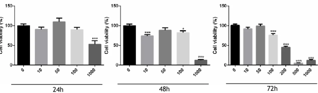
Figure 7 - Percentage of viable non-neoplastic human prostate epithelial PNT1A cells after exposure to different concentrations of C. monogyna extracts (10, 50, 100, 200, 500 and 1000 μg/mL) for 24, 48 and 72 h determined by the MTT assay………24
Figure 8 - Percentage of viable neoplastic human prostate LNCaP cells after exposure to different concentrations of C. monogyna extracts (10, 50, 100, 200, 500 and 1000 μg/mL) for 24, 48 and 72 h determined by the MTT assay. ………25
Figure 9 - Percentage of viable neoplastic human prostate PC3 cells after exposure to different concentrations of C. monogyna extracts (10, 50, 100, 200, 500 and 1000 μg/mL) for 24, 48 and 72 h determined by the MTT assay………25
Figure 10 - Percentage of viable non-neoplastic human prostate epithelial PNT1A cells after exposure to different concentrations of A. unedo extracts (10, 50, 100, 200, 500 and 1000 μg/mL) for 24, 48 and 72 h determined by the MTT assay. ………26
Figure 11 - Percentage of viable neoplastic human prostate LNCaP cells after exposure to different concentrations of A. unedo extracts (10, 50, 100, 200, 500 and 1000 μg/mL) for 24, 48 and 72 h determined by the MTT assay. ………26
Figure 12 - Percentage of viable neoplastic human prostate PC3 cells after exposure to different concentrations of A. unedo extracts (10, 50, 100, 200, 500 and 1000 μg/mL) for 24, 48 and 72 h determined by the MTT assay………27
Figure 13 - Expression of the apoptosis regulator, Bax, in non-neoplastic human prostate epithelial PNT1A cells after treatment with 200 μg/ml of C. monogyna extracts for 72 h, determined by Western blot analysis after normalization with β-actin………28
Figure 14 - Activity of Caspase-3 in non-neoplastic human prostate epithelial PNT1A cells after treatment with 200 μg/ml of C. monogyna extracts for 72 h, determined by a colorimetric assay………28
Figure 15 - Expression of the apoptosis regulators (A-B) in non-neoplastic human prostate epithelial PNT1A cells after treatment with 200 μg/ml of A. unedo extracts for 72 h, determined by Western blot analysis after normalization with β-actin………29
Figure 16 - Activity of Caspase-3 in non-neoplastic human prostate epithelial PNT1A cells after treatment with 200 μg/ml of A. unedo extracts for 72 h, determined by a colorimetric assay………29
Figure 17 - Expression of the apoptosis regulators (A-B) in neoplastic human prostate epithelial LNCaP cells after treatment with to 200 μg/ml of C. monogyna extracts for 72 h, determined by Western blot analysis after normalization with β-actin………30
Figure 18 - Activity of Caspase-3 in neoplastic human prostate epithelial LNCaP cells after treatment with 200 μg/ml of C. monogyna extracts for 72 h, determined by a colorimetric assay………30
Figure 19 - Expression of apoptosis regulators (A-B) in neoplastic human prostate epithelial LNCaP cells after treatment with 200 μg/ml of A. unedo extracts for 72 h, determined by Western blot analysis after normalization with β-actin………31
Figure 20 - Activity of Caspase-3 in neoplastic human prostate epithelial LNCaP cells after treatment with 200 μg/ml of A. unedo extracts for 72 h, determined by a colorimetric assay………31
Figure 21 - Expression of the apoptosis regulator, Bax (A), in neoplastic human prostate epithelial PC3 cells after treatment with 200 μg/ml of C. monogyna extracts for 72 h, determined by Western blot analysis after normalization with β-actin………32
Figure 22 - Activity of Caspase-3 in neoplastic human prostate epithelial PC3 cells after treatment with 200 μg/ml of C. monogyna extracts for 72 h, determined by a colorimetric assay………32 Figure 23 - Expression of the apoptosis regulators (A-B) in neoplastic human prostate epithelial PC3 cells after treatment with 200 μg/ml of A. unedo extracts for 72 h, determined by Western blot analysis after normalization with β-actin.……… …………33 Figure 24 - Activity of Caspase-3 in neoplastic human prostate epithelial PC3 cells after treatment with 200 μg/ml of A. unedo extracts for 72 h, determined by a colorimetric assay. ………33 Figure 25 - Glucose consumption (A) and lactate production (B) in non-neoplastic human prostate epithelial PNT1A cells in response to 200 μg/ml treatment of C. monogyna extracts for 72 h.………34 Figure 26 – Expression of metabolism-associated proteins (A-E) in non-neoplastic human prostate epithelial PNT1A cells in response to 200 μg/ml treatment of C. monogyna extracts for 72 h, determined by Western blot analysis after normalization with β-actin. ………35 Figure 27 – LDH enzymatic activity in non-neoplastic human prostate epithelial PNT1A cells in response to 200 μg/ml treatment of C. monogyna extracts for 72 h, determined by spectrophotometric assays. ………36 Figure 28 - Glucose consumption (A) and lactate production (B) in non-neoplastic human prostate epithelial PNT1A cells in response to 200 μg/ml treatment of A. unedo extracts for 72 h. ………36 Figure 29 – Expression of metabolism-associated proteins (A-E) in neoplastic PNT1A cells in response to 200 μg/ml treatment of A. unedo extracts for 72 h, determined by Western blot analysis after normalization with β-actin. ………37 Figure 30 – LDH enzymatic activity in non-neoplastic human prostate epithelial PNT1A cells in response to 200 μg/ml treatment of A. unedo extracts for 72 h, determined by spectrophotometric assays. ………38 Figure 31 - Glucose consumption (A) and lactate production (B) in neoplastic human prostate LNCaP cells in response to 200 μg/ml treatment of C. monogyna extracts for 72 h. ………38 Figure 32 – Expression of metabolism-associated proteins (A-E) in neoplastic human prostate LNCaP cells in response to 200 μg/ml treatment of C. monogyna extracts for 72 h, determined by Western blot analysis after normalization with β-actin. ………39 Figure 33 – LDH enzymatic activity in neoplastic human prostate LNCaP cells in response to 200 μg/ml treatment of C. monogyna extracts for 72 h, determined by spectrophotometric assays. ………40 Figure 34 - Glucose consumption (A) and lactate production (B) in neoplastic human prostate LNCaP cells in response to 200 μg/ml treatment of A. unedo extracts for 72 h. ………40 Figure 35 – Expression of metabolism-associated proteins (A-E) in neoplastic human prostate LNCaP cells in response to 200 μg/ml treatment of A. unedo extracts for 72 h, determined by Western blot analysis after normalization with β-actin. ………41 Figure 36 – LDH enzymatic activity in neoplastic human prostate LNCaP cells in response to 200 μg/ml treatment of A. unedo extracts for 72 h, determined by spectrophotometric assays. ………41 Figure 37 - Glucose consumption (A) and lactate production (B) in neoplastic human prostate PC3 cells in response to 200 μg/ml treatment of C. monogyna extracts for 72 h. ………42 Figure 38 – Expression of metabolism-associated proteins (A-E) in neoplastic human prostate PC3 cells in response to 200 μg/ml treatment of C. monogyna extracts for 72 h, determined by Western blot analysis after normalization with β-actin. ………43 Figure 39 – LDH enzymatic activity in neoplastic human prostate PC3 cells in response to 200 μg/ml treatment of C. monogyna extracts for 72 h, determined by spectrophotometric assays. ………44 Figure 40 - Glucose consumption (A) and lactate production (B) in neoplastic human prostate PC3 cells in response to 200 μg/ml treatment of A. unedo extracts for 72 h. ………44
Figure 41 – Expression of metabolism-associated proteins (A-E) in neoplastic human prostate PC3 cells in response to 200 μg/ml treatment of A. unedo extracts for 72 h, determined by Western blot analysis after normalization with β-actin. ………45 Figure 42 – LDH enzymatic activity in neoplastic human prostate PC3 cells in response to 200 μg/ml treatment of A. unedo extracts for 72 h, determined by spectrophotometric assays. ………45
List of Tables
Table 1 - Synthesis of the effects of C. monogyna extracts in proliferation, apoptosis and glycolytic metabolism of non-neoplastic (PNT1A) and neoplastic (LNCaP and PC3) prostate cells. ………53 Table 2 - Synthesis of the effects of A. Unedo extracts in proliferation, apoptosis and glycolytic metabolism of non-neoplastic (PNT1A) and neoplastic (LNCaP and PC3) prostate cells. ……… 53
List of Abbreviations
1,3BPG 1,3-Bisphosphoglyceric Acid2PG 2-Phosphoglycerate
3PG 3-Phosphoglyceric acid
ADP Adenosine diphosphate
ADT androgen deprivation therapy
AP Acid phosphatase
APAF-1 Apoptotic protease activating factor 1
AR Androgen Receptor
ATP Adenosine triphosphate
BAK-1 Bcl-2 Homologous Antagonist Killer
Bax Apoptosis Regulator Bax
Bcl-2 B cell lymphoma 2
BID Bcl-2 Homologous Antagonist Killer BPH Benign Prostatic Hyperplasia
Ca2+ Cálcio
CGA Chlorogenic Acids
CK Cytokeratin
CRPC Castration-resistant prostate cancer DHAP Dihydroxyacetone Phosphate
DHT Dihydrotestosterone
DNA Deoxyribonucleic Acid
FADD Fas-Associated Protein with Death Domain FAS Ligand acts as a prototypic death factor Fructose-1,6-p Fructose- 1,6-Phosphate
Fructose-6-p Fructose 6-Phosphate GADP Glyceraldehyde 3-Phosphate
GADPH Glyceraldehyde 3-Phosphate Dehydrogenase GLUT Glucose transporter
H2O Água
HOXB13 Gene associate with an increased risk for autosomal dominant prostate cancer
IFN-γ Interferon-γ
LNCaP Human cell line of prostate cancer derived from lymph node metastasis MCT Monocarboxylate transporter
MDM2 Mouse double minute 2
MTT 3-(4,5-Dimethylthiazol-2-Yl)-2,5-Diphenyltetrazolium Bromide
NOXA Proapoptotic protein
nsSNPs Non-synonymous single nucleotide polymorphisms p21 Cyclin-dependent kinase inhibitor
p53 Tumour Suppressor p53
P63 Tumor protein
PAP Prostatic acid phosphatase
PC3 Human cell line of prostate cancer derived from bone metastasis
PCa Prostate Cancer
PCA3 Prostate Cancer Gene 3 PFK-1 Phosphofructokinase-1
PGI Enzyme phosphoglucose isomerase PGK Mono Phosphoglycerate Kinase
PGM Phosphoglycerate mutase
PNT1A Human cell line of prostate PSA Prostate specific antigen
PUMA p53 Upregulated Modulator of Apoptosis
RNA Ribonucleic acid
SNP Single nucleotide polymorphism
tBID truncated Bid
TCA Tricarboxylic acid
TNF Tumor necroses factor
TRAIL TNF-related Apoptosis-inducing Ligand
Chapter I - General Introduction
1. Brief overview of prostate anatomy and physiology
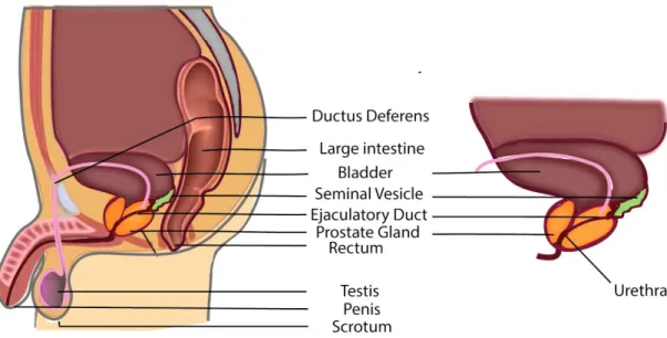
The prostate is a compound tubule alveolar exocrine gland that is part of the male reproductive system (figure 1). The prostate gland develops after puberty as a result of the testosterone surge. It reaches a size of three centimeters in length, four centimeters in width and two centimeters in depth, in the average adult. It’s the largest gland of the male reproductive system and it weighs about twenty grams, being detectable via a rectal examination [1].
Figure 1. Sagittal view showing the male reproductive structures.
This gland is located just below the bladder and it lies above the urogenital diaphragm between the rectum and the symphysis pubis. It surrounds the ejaculatory ducts, and the beginning of the urethra, which goes right through the center of the prostate and is known as the prostatic urethra [1][2]. A simplified representation of prostate position in the male reproductive structures is observable in Figure 1. The prostate is composed by three kinds of cells: glandular cells, which secrete the prostatic fluid part of the semen; muscle cells, which regulate urine flow and ejaculation; and fibrous cells, which support the gland. The glandular cells of the prostate comprise the luminal cells, basal cells and neuroendocrine cells [3]. The prostate lies in a hammock of nerves, which can be divided into three zones: proximal neurovascular plate, predominant neurovascular bundles, and accessory distal neural pathways [1].
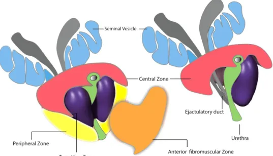
In the twentieth century, several researched claimed the division of prostate gland into lobes, by analogy with laboratory animals, a concept that became popular even though no distinct lobes can be seen in the human prostate. The current and most widely accepted structural division of prostate encompasses four distinct zones drained by specific ducts and with unique distinguishable features, namely (Figure 2) [4]: the central zone, which occupies about 25 percent of the prostate’s volume and is traversed by the ejaculatory ducts; the transition zone, which comprises only 5–10 percent of the glandular tissue; the peripheral zone, corresponding to 70 percent of the prostate volume at the back and to the sides of the gland; and the anterior fibromuscular zone or stroma, the entire anterior surface of the prostate, composed by fibromuscular tissue contiguous with the bladder, without glandular tissue. The susceptibility of these different zones to diseases affecting the prostate are not equal: the peripheral zone is very susceptible to cancer, as it is affected in approximately 85–90 percent of cases; the transition zone is very prone to benign prostatic hyperplasia (BPH); and the central zone is very resistant to BPH and prostate cancer [1] [4] [5].
Figure 2. The zonal anatomy of the prostate gland. Using the prostatic urethra and ejaculatory ducts
as reference, the prostate comprises three principal zones and an anterior fibromuscular layer. The central zone (pink) located at the top of prostate near the bladder and the ejaculatory ducts pass through the central zone to the urethra. The transition zone (purple) surrounds the portion of the urethra closest to the bladder. The peripheral zone (yellow) is located at the posterior side of the prostate closest to the rectum. Peripheral and central zones are collectively referred to as outer prostate, whereas the transition zone and anterior fibromuscular layer (orange) are termed inner prostate.
The prostate is an exocrine gland that produces alkaline secretions righ in sugars and proteins, contributing to the seminal fluid (about 30 percent in volume). These are thought to be important in aiding fertilization in several ways: by maintaining the alkaline pH (that protects the sperm from the acidic environment of the female reproductive system), by helping to nourish the sperm in the semen and by affecting the degree of viscosity of the semen, promoting the viability of sperm after ejaculation [1]. Secretion products of the prostate include zinc, citric acid, spermine, prostaglandins, cholesterol, clotting enzyme,
acid phosphatase (AP), prostatic acid phosphatase (PAP), and prostatic specific antigen (PSA). The AP and PAP are both enzymes that have been used for a long time to monitor the course of prostatic cancer disease in advanced stages. The PSA, is only detected in the epithelial cells of the prostatic ductal elements and is used for detection and monitoring of prostate cancer (PCa) [6].
The prostate also has the ability of converting testosterone into 5α-dihydrotestosterone (DHT), by the activity of 5 α–reductase. DHT is the most important prostatic androgen with a higher affinity for the androgen receptor (AR) than testosterone. DHT binding to AR activates the AR that in the nucleus activates transcription, resulting in the production of messenger RNAs, and protein synthesis increasing cellular growth and prostate volume [7].
2. Epidemiological notes on prostate cancer
The PCa is the most common cancer affecting men concerning the number of new cases diagnosed, and represents one of the major causes of cancer death in men worldwide [8]. For example, in America, about 1 man in 7 will be diagnosed with PCa during his lifetime and about 1 man in 39 will die of PCa [9]. The American Cancer Society estimates that for 2018 there will be about 164,690 new cases of PCa and 29,430 deaths by PCa in the United States [8]. In Portugal, PCa incidence has been increasing since 1998 (1.8%/year), with the exception of the North Region, with a decrease since 2006 (−3.2%/year). If these trends are maintained, 8600 incident cases and 1700 deaths are estimated for 2020 [10].
PCa is mostly common in North America, northwestern Europe, Australia, and the Caribbean islands and less common in Asia, Africa, Central America, and South America [1]. PCa in African American men exhibits a relatively high incidence and mortality (incidence is 1.6-fold higher than in other populations). The risk factors that drive this are unknown and potentially consist of social, environmental and genetic influences. The biological basis for this disparity is unclear in PCa [11] [12].
PCa develops mainly in older men: about 6 cases in 10 are diagnosed in men aged 65 or older and it is rare before the age of 40, so the average age at the time of diagnosis is about 66 [13]. Apart from age, geography and race/ethnicity, the hereditary can be another risk factor for PCa. This human malignancy has the highest degree of genetic transmission. In some families, the hereditary pattern is so strong that mimics an autosomal dominance trait [14]. Single nucleotide polymorphisms (SNPs) are considered to be the primary genetic cause for hereditary PCa and the study of functional non-synonymous single nucleotide polymorphisms (nsSNPs) would give an insight into the exact cause underlying the onset of hereditary PCa and possible methodologies for the cure or early management of the disease. So far it has been proven that the nsSNPs (missense) have a profound damaging effect on the homeobox domain of the HOXB13 gene which might lead to the altered binding patterns of HOXB13
in the normal prostate development and is also associated with the increased risk of inherited PCa. Subsequent advances in genetic studies have proved that HOXB13 has a significant role in PCa susceptibility, but the exact mechanism behind it remains undiscovered. However, the exact mechanism and pathology of those predicted nsSNPs should further be validated by in
vivo experiments and population based studies [15].
The lifestyle and diet also have been indicated as risk factors for PCa [16]. Accordingly, several generally widely recommended healthy lifestyle habits such as not smoking, maintaining body weight within limits, and practicing regular vigorous physical exercise appear to counteract PCa development and progression [17]. Concerning obesity, the body’s fat content affects the handling of testosterone and this was suggested as the link between fat and PCa. Recently, it was shown that obesity favors the migration of PCa cells and dissemination of disease by an action promoted directly by the periprostatic adipocyte cells over prostate tumor cells [16]. Several dietary factors, flavonoids and other similar antioxidants are important protective agents because oxidation damages complex biological materials, including DNA, and may be one factor causing cancer. Vitamin E, vitamin A, vitamin C, food rich in lycopene (e.g. tomato), cruciferous vegetables, healthy sources of vegetable fats, and coffee, may also have a role in reducing risk of PCa progression [16] [17]. For this reason, vegetarians are said to be at a much lower risk of developing PCa, perhaps by as much as 50 per cent [16].
Others risk factors for PCa have been pointed out, though with less clear effects. This includes industrial exposure, for example to cadmium, which can occur in men working in copper smelting [18]; chemical exposure to agents such as the Agent Orange, that was a commercially manufactured defoliate sprayed extensively during the Vietnam War [19]; sexual behavior, as some analysis reports demonstrated slight increases of risk associated with the number of sexual partners and history of sexually transmitted infection [20]; vasectomy, as some studies have suggested that men who have a vasectomy have a slightly increased risk for PCa; and inflammation of the prostate, hading a specific attention for your study due to inflammation’s role in multiple stages of PCa development. The inflammasome associated with PCa remains uncharacterized and studies characterizing the role of inflammation in PCa are at the beginning stage. Nevertheless, there is evidence that the inflammasomes regulate inflammatory cytokines, thereby modulating the course of inflammation. Inflammasomes activation is linked with infection, stress, or danger signals, which are common events within the prostate gland. Since the pro-inflammatory cytokines may serve as fuel for the developing neoplastic cells in the prostate, the tumor promoting effect of pro-inflammatory cytokines/chemokines may be reduced significantly by understanding the mechanistic regulation of inflammasomes [21].
3. Prostate cancer diagnosis and treatment: the basis
The tumor marker detection is of great significance to early clinical diagnosis and monitoring of disease recurrence. Despite the limitation it has, PSA still is the currently used diagnostic biomarker for PCa [22]. To date, a series of techniques have been applied for the quantitative detection of PSA, such the enzyme-linked immunosorbent assay, surface plasmon resonance, luminescence energy transfer, electrochemiluminescence, and electrochemistry methods. Among the various detection techniques, photoelectrochemical immunoassay has aroused increasing attention owing to its good stability, acceptable specificity and favorable sensitivity compared with conventional optical and electrochemical methods. When PSA concentration is higher than 4.0 ng/mL, the patient’s health is endangered, but PSA levels should be evaluated in line with other diagnostic techniques when managing a patient with PCa. Not all low or high PSA levels will necessarily indicate that a patient has or does not have PCa, as PSA levels are organ specific and not cancer specific. So PCa screening is done in part through the use of the PSA blood tests and is often combined with a digital rectal examination. Making a definitive diagnosis of PCa generally includes blood count and biochemical profile with PSA quantification (% of free/total PSA), transrectal ultrasound, echography, magnetic resonance imaging, PCA3 test and biopsy [22] [23].
Treatment choice in PCa is based on the well-established prognostic factors, namely, initial PSA level, clinical tumor stage and Gleason score, along with general considerations such as baseline urinary function, comorbidities and patient’s age [24]. The most important primary treatment for locally advanced or metastatic PCa is the androgen deprivation therapy (ADT). The progression of PCa is dependent on androgens, and treatments reducing the production of androgens, or antagonizing the function of AR, have been the first line option to treat advanced PCa. Other treatments available include radical prostatectomy, prostate brachytherapy, external beam radiation therapy and active surveillance [24] [25]. So far, basal treatment of PCa relies on surgery, chemotherapies and ionizing radiation, with a low efficacy and high toxicity to healthy tissues. New therapeutic approaches of decreasing the PCa associated mortality and minimizing the nonspecific and undesirable side effects are needed [26].
4. Development and progression of prostate cancer
In tumor development and progression, the interaction between cancer cells and the microenvironment is crucial. The development of PCa is dependent on the relationship between the levels of cell proliferation and the rate of apoptosis in prostate cells. Androgens are the main regulators of this relationship, stimulating cell proliferation and inhibiting apoptosis. Almost all prostate cancers are adenocarcinomas, cancers which develop from the glandular cells. There are other types of PCa but they are rare, as sarcomas, small cell carcinomas, neuroendocrine tumors and the transitional cell carcinomas. In PCa, both luminal
cells and basal cells have the potential to give rise to tumors with different phenotypic changes [2][3] [27].
As has been shown previously, the prostate is surrounded by a capsule consisting of a glandular epithelium embedded in a fibromuscular stroma and its epithelium consists of two histologically distinct layers: basal layer and luminal layer [1]. It is believed that the basal layer is the proliferative compartment of the prostate and consists of cuboidal epithelial cells that adhere to the basal membrane. Basal cells express proliferation markers like CK5, CK14 and p63. In turn, the luminal cells express high levels of AR, and are androgen-dependent and secretory cells responsible for production and secretion of PSA. The stem cells, present in the basal layer, have unlimited proliferative capacity, are able to self-renew and yet differentiate into cells of the glandular epithelium with more limited proliferative capacity. When disturbances occur in the cellular environment, the process of differentiation of stem cells can be affected and is believed to contribute, in addition to other factors, to the malignant phenotype of prostate [3] [28].
Overall, it has been suggested that the damage of the prostate epithelium, potentially inflicted by diverse environmental exposures, such as infections and trauma, and/or microenvironment stimuli, such as oxidative stress and hypoxia, triggers pre-malignant inflammatory processes to promote tumor development and progression. In this milieu, the damaged epithelium may generate proliferative inflammatory atrophy (PIA) lesions, which may progress through prostatic intraepithelial neoplasia (PIN) to PCa (Figure 3). High-grade prostatic intraepithelial neoplasia preferentially develops in the peripheral zone of the prostate, which is the site of origin for most adenocarcinomas [2] [27] [29].The AR hyperactivity leads to PCa initiation and progression to advanced disease, which can occur by AR overexpression resulting in a transition from an androgen-sensitive disease to the androgen-resistant cancer, the so-called castration-resistant prostate cancer (CRPC). Therefore, the constant activation of distinct growth signaling pathways in PCa cells, including the activation of AR, leads to the development of local and invasive carcinoma and ultimately to the metastization of different organs, including liver, lungs and bones [30].
Figure 3. Development of epithelial prostate cancer. The pathophysiology of the prostate comprises
multiple steps and starts with proliferative inflammatory atrophy (PIA), the appearance of pre-malignant lesions and transformation of prostate epithelial cells by processes like oxidative stress, inflammation, luminal and basal proliferation. In the next step, prostatic intraepithedial neoplasia (PIN), the luminal cell hyperproliferation, telemore shortening and stromal reactivity take place. Finally and consequently the stage of PCa is reached, occuring the luminal cell hyperproliferation, loss of basal epithelia, basement membrane breakdown, immune cell infiltration and stroma reactivity.
5. Molecular basis of apoptotic cell death
Apoptosis represents a controlled mechanism of cell death that is altered in cancer cells. Reactivation of apoptosis has been envisaged as a therapeutic tool to eliminate cancer cells. Unfortunately, advanced prostate tumors eventually progress to androgen-insensitive stages, which are resistant to current therapeutic approaches that act by triggering apoptosis [30] [31].
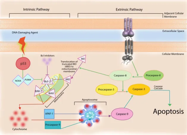
Apoptosis is triggered by signaling events involving a diverse array of protein networks, cellular organelles and macromolecular complexes that converge to the activation of caspases. Apoptotic characteristics include [31]: i) cell shrinkage, ii) membrane blebbing, iii) chromatin condensation, iv) DNA fragmentation caused by internucleosomal DNA cleavage, and v) finally ending with the engulfment by macrophages or neighboring cells, thereby avoiding an inflammatory response in surrounding tissues. Apoptosis execution occurs via two distinct signaling pathways: the intrinsic or mitochondrial-mediated pathway and the extrinsic or death receptor-mediated pathway (Figure 4) [31] [32].
Figure 4. The intrinsic and extrinsic apoptotic pathways. The intrinsic pathway is usually activated by
p53 that controls the activity of several mediators culminating in the activation of caspases 9 and 3, resulting in apoptosis. External death stimuli bind death receptors at cell membrane (e.g FAS) triggering caspase-8 and -3 activation resulting in apoptosis (Extrinsic pathway).
The intrinsic pathway is usually activated in response to many different damaging influences, for example, DNA damage, oxidative stress, hypoxia, or chemotherapeutic drugs, or by the absence of growth factor signals [32]. In conditions like these, p53 causes cell cycle arrest, primarily by activating the transcription of a cyclin-dependent kinase inhibitor, p21/waf1, and induces apoptosis via transcriptional activation of the pro-apoptotic PUMA, NOXA, and B-cell lymphoma 2 (Bcl-2) family genes (Figure 4). This pathway is mainly regulated by proteins of the Bcl-2 family, which control the release of pro-apoptotic factors from the mitochondrial intermembrane space. More than 20 members of this family have been identified to date in humans, including suppressors (Bcl-2, Bcl-xL, Mcl-1, Bfl-1/A1, Bcl-W, and Bcl-G) and promoters (Bax, Bak, Bok, Bad, Bid, Bik, Bim, Bcl-Xs, Krk, Mtd, Nip3, Nix, Nora, and Bcl-B) of apoptosis. These apoptosis regulators, especially Bax and BAK, will change their conformation, oligomerize, and be attracted to the mitochondria inducing the creation of apoptotic pores in the mitochondrial membrane. This will release one of the designated main “killing factors” of the cell, the enzyme cytochrome-c, alongside with another pro-apoptotic protein, Smac/DIABLO, which will aid in caspase activation. When cytochrome c is released into the cytoplasm the apoptotic protease-1 (APAF-1) and procaspase-9 are recruited, which in turn activates a series of nuclear and cytoskeletal proteins, and signaling molecules. These
proteins form a functional apoptosome that activates a caspase-9 and -3, resulting in programmed cell destruction [30] [32] [33].
An alternative and complementary signaling pathway that leads to programmed cell death includes the extrinsic death receptor pathway (Figure 4). The extrinsic pathway is initiated by the binding of apoptosis-inducing ligands such as FAS or TRAIL, tumor necrosis factor (TNF) receptors. Activation of death receptors by binding their natural ligands induces receptor clustering and formation of a death-inducing signaling complex. The complex recruits procaspase-8 via the adaptor molecule Fas-associated death domain protein (FADD), resulting in the activation of caspase-8.Procaspase-8 an inactive ‘initiator’ caspase is proteolytically cleaved becoming activated, and further activating downstream effectors’ proteins such as caspases-8 and -3, which induces the degradation of cytosolic, cytoskeletal, nuclear proteins, and DNA, causing inevitable apoptosis [30] [32].
The extrinsic and intrinsic apoptosis pathways are connected by the caspase-8-mediated cleavage of the pro-apoptotic Bcl-2 family member, BID (Figure 4). Therefore, the caspase-8 is also responsible for cleaving BID, a pro-apoptotic member of the Bcl-2 family that forms the principal link between the extrinsic and intrinsic pathways by stimulating the release of cytochrome c from the mitochondrion, followed by induction of apoptosis [33].
More than 50% of human cancers, including PCa, exhibit loss of normal p53 functions and/or defects in the p53 signaling pathway and these molecular alterations are associated with resistance to apoptosis [34]. The p53, tumor suppressor gene product, is a transcription factor that enhances the transcription of several genes known to play a critical role in transducing signals from DNA damage. Its expression and activity are elevated in response to ionizing radiation, UV light, or certain genotoxic chemicals and mediate DNA repair, cell cycle arrest, and apoptosis. The functional activity of p53 is regulated through transcription, translation, protein turnover, cellular compartmentalization as well as it is association with other proteins, such as MDM2 [35] [36].
For the maintenance of prostate growth, the complex equilibrium between cell growth, proliferation factors, and apoptosis-inducing factors is essential. Fluctuations in this balance cause overexpression of factors causing cell survival, cell proliferation and loss of apoptosis leading to tumorigenesis and cancer. The deregulation of cell growth in PCa is notable by apoptotic evasion, loss of differentiation, and uncontrolled proliferation [32].
6. Metabolism of cancer cells: the glycolytic pathway
Cancer cells display unique and exquisite features from which the most emblematic is their preference on glycolytic metabolism for obtaining energy. Glycolysis is the cytoplasmic pathway that converts glucose into two molecules of pyruvate, with only a modest releasing of energy is being captured in two substrate-level phosphorylations and one oxidation
reaction. In normal conditions, with functional mitochondria and oxygen supply, glycolysis is aerobic. If either mitochondria or oxygen is lacking, glycolysis may occur anaerobically although some of the available energy is lost, which results in the conversion of pyruvate to lactate [37].
In healthy cells the glycolytic process is tightly regulated and consists of three stages. Firstly, glucose is uptake from the extracellular space via glucose transporters (GLUTs) at cell membrane. These families of proteins allow the energy independent transport of glucose across the hydrophobic cell membrane, down its concentration gradient [38] [39]. GLUT1 and GLUT3 isoforms in particular are present across all mammalian cells and are responsible for a considerable amount of the basal glucose uptake [40]. Once inside the cell, glucose is converted into pyruvate through a chain of reactions. The first stage is phosphorylation of glucose into glucose-6-phosphate, consuming one molecule of ATP, through the action of hexokinase. Glucose-6-phosphate is then converted, in a reversible reaction, into fructose-6-phosphate through the action of the phosphoglucose isomerase (PGI). Fructose-6-fructose-6-phosphate in turn is phosphorylated into fructose 1,6-biphosphatase through the activity of phosphofructokinase-1 (PFK-1). The PFK-1 is a key enzyme in this process since it catalyzes the first irreversible reaction of glycolysis, being a rate-limiting step in glucose metabolism [39] [40].
The second stage of glycolytic process involves the cleavage of the six-carbon fructose in the fructose 1,6-biphosphatase molecule in order to generate two separate molecules. This is achieved by the enzyme aldolase, and it generates D-glyceraldehyde 3-phosphate (GADP) and dihydroxyacetone phosphate (DHAP). The triose phosphate isomerase will then convert the DHAP molecule into a second GADP molecule, thus meaning the final glycolytic stage will occur twice [40].
In the third and final glycolytic step, one GADP molecule will be oxidized by the enzyme glyceraldehyde phosphate dehydrogenase (GAPDH) into D-1,3-bisphosphoglycerate (1,3BPG). One of the phosphate groups from 1,3BPG will then be transferred to an ADP molecule by the enzyme phosphoglycerate kinase (PGK) in order to produce the process first molecule of ATP and one molecule of 3-phosphoglycerate (3PG). The 3PG molecule is isomerized by the enzyme phosphoglycerate mutase (PGM) into 2-phosphoglycerate (2PG). This molecule will then be dehydrated into phosphophenolpyruvate which will, in the last step of the glycolytic process, be converted to pyruvate, with one final ATP molecule, by the action of the enzyme pyruvate kinase [40].
In normal cells and aerobic glycolysis pyruvate will be converted into acetyl-CoA to its complete oxidation, through the mitochondrion-localized tricarboxylic acid (TCA) cycle and oxidative phosphorylation to CO2 and H2O, which generates 38 ATP molecules per molecule of
aborted at either of two steps. First, aerobic glycolysis in tumor cells implies conversion of glucose into pyruvate, which generates only two ATP molecules per molecule of glucose, and subsequently into the lactate by lactate dehydrogenase (LDH). Second, in tumor cells, acetyl-CoA tends to be introduced into a truncated TCA cycle, with the result that acetyl-acetyl-CoA is exported into the cytosol and serves as a building block for cell growth and proliferation [41]. A simplified representation model is observable in Figure 5.
Therefore under anaerobic conditions, or as an alternative used by cancer cells, the end-product of glycolysis, pyruvate, is metabolized by the LDH enzyme, which catalyzes the interconversion of pyruvate to lactate. The lactate produced is then exported to the extracellular medium by specific monocarboxylate transporters, namely by the MCT4, contributing to the acidification of tumor microenvironment. The lactate in the tumor microenvironment, favors cell migration and invasion, also suppressing the anticancer immune defenses [39]. So it has been reported that most cancer cells express high levels of MCTs to ensure the rapid efflux of the lactate produced. During PCa progression, the expression profile of MCT isoforms changes. MCT1 is expressed in normal and malignant prostate, whereas MCT2 expression increases from normal gland to PIN and in situ carcinoma [42]. On the other hand, MCT4 is only expressed in malignancy and highly in more advanced stages of disease [43]. While MCT1 and MCT2 transport a wider range of substrates, MCT4 is specifically associated with the export of lactate in cells with a high glycolytic rate [42]. Furthermore, it has been shown that lactate may enter in stroma or cancer cells following oxidative phosphorylation or alternatively regenerating pyruvate that, in turn, may be used as energetic substrate by tumor cells. This process generates complementary metabolic pathways, which lead to survival and progression of cancer cells. Moreover, MCTs have been shown to be overexpressed in different human cancers and for this reason identified as attractive targets for cancer therapy [42] [43].
Figure 5. Glycolysis and the two main possible routes of pyruvate. In normal cells under aerobiosis
pyruvate is converted to Ac-CoA and enters the d tricarboxylic acid (TCA) cycle. In anaerobiosis, or typically as a feature of cancer cells, pyruvate is converted to lactate by the activity of lactate dehydrogenase (LDH).
As previously described, progression of PCa is associated with tumor enrichment in androgen-insensitive proliferating cells, as well as the emergence of new cell phenotypes, leading to a disease stage that frequently displays a fatal behavior. Moreover, PCa has been considered to have unique metabolic features, related with the high levels of citrate, alanine and lactate [44] [45]. Also, the increased glucose consumption seems to be required for rapid proliferation of androgen-insensitive PCa cells [44], which raises the curiosity about the identification of factors that could hamper the glycolytic metabolism of cancer cells.
7. Plant extracts, polyphenols and cancer
Polyphenols are a large class of chemicals which are found in plants, have attracted much attention in the last decades due to their properties and the hope that they will show beneficial health effects. Phenolic compounds constitute one of the most extensive groups of chemicals in the plant kingdom. It is estimated that more than 8000 compounds have been isolated and described [46]. Polyphenols occur primarily in conjugated form, linked to sugars moieties, but also to other compounds, such as carboxylic and organic acids, amines, lipids and even to other polyphenols [47]. They can be subdivided in five main subclasses: flavonoids, phenolic acids, stilbenoids, lignans and ellagic acids [48]. Flavonoids, represent the most common and widely distributed group of plant phenolics, and can be further divided into classes including flavones, flavonols, flavanones, anthocyanins and isoflavonoids [49]. They are characterized as containing two or more aromatic rings, each bearing one or more
phenolic hydroxyl groups and connected by a carbon bridge. Phenolic acids are usually divided in two main groups derive from benzoic acids, containing seven carbon atoms, or from cinnamic acids, comprising nine carbon atoms. The smaller subclass of stilbenoids comprises polyhydroxylated stilbenes, the main representative being resveratrol. So, all these polyphenols are found in plants, etherified with glucose and other carbohydrates (glycosides) or as free aglycones [46].
The interest of plant polyphenols derives from the evidence from several studies of their hight antioxidant activity. They are able to scavenge free radicals and their wide range of pharmacologic effects including inflammatory, allergic, proliferative, anti-tumour, apoptosis-inducing, antioxidant and antibacterial activities, provide important health benefits related to metabolic syndrome, cancer, brain health and immune system [47] [49]. The antioxidant properties of phenolic acids and flavonoids have been related to their redox properties and chemical structures, which allow them to act as reducing agents, hydrogen donors and singlet oxygen quenchers. In addition, some of them display a metal chelation activity, which hinders transition metals from acting as oxidation promoters [49].The antioxidant capacity in human plasma results in increased antioxidant protection of lipids and proteins [46].
Crataegus monogyna and Arbutus unedo are plants found in the region of Beira Interior and
their composition in phenolic compounds is widely described. This point and the fact the PCa is the second most common cancer, hence the interest in studying this plants extracts effects on non-neoplastic (PNT1A) and neoplastic (LNCaP and PC3) prostate cells.
8.
Characterization of Crataegus monogyna
The genus Crataegus is of the class Mognoliopsida, order Rosales and family Rosaceae. This genus has approximately 280 species [50], being one of the most common C. monogyna, hawthorn [51]. These plants grow spontaneously in temperate zones, in Europe, Africa and Asia. In the Iberian Peninsula, it is widely distributed along the whole territory, Majorca and Minorca islands. The species C. monogyna is the most abundant species in Portugal and is popularly known as a “Pirliteiro” [52]. It is a prickly shrub / small tree 5-10 m tall that produces white flowers in spring, red fruits about 1 cm in size in autumn [53] [54]. Flowers, leaves and fruits are used as dried products for infusions or included as plant extracts in capsules because of their wide pharmacological effects and low toxicity, especially for cardiac and nervous system symptoms as well as for their antioxidant activities [55]. C.
monogyna was mentioned for the first time in the first century by Dioscorides as a
‘cardiotonic’ remedy. The use of hawthorn in therapeutics was also documented in America since the nineteenth century to treat different heart diseases, angina pectoris, to support the effect of Digitalis, or in cases of irregular heartbeat when Digitalis was not tolerated. Nowadays, traditional use in the European Union recognizes C. monogyna for two different


![Figure 6. Antioxidant compounds present in the Arbutus unedo trees [72].](https://thumb-eu.123doks.com/thumbv2/123dok_br/19173209.942089/40.892.110.761.94.410/figure-antioxidant-compounds-present-arbutus-unedo-trees.webp)