Safety, beneficial and technological properties of
Enterococcus faecium
isolated
from Brazilian cheeses
Karina Maria Olbrich dos Santos
1,2, Antônio Diogo Silva Vieira
2,3,
Hévila Oliveira Salles
2, Jacqueline da Silva Oliveira
2, Cíntia Renata Costa Rocha
4,
Maria de Fátima Borges
5, Laura Maria Bruno
5,
Bernadette Dora Gombossy de Melo Franco
3, Svetoslav Dimitrov Todorov
3 1Empresa Brazileira de Pesquisa Agropecuária, Agroindústria de Alimentos, Rio de Janeiro, RJ, Brazil. 2
Empresa Brazileira de Pesquisa Agropecuária, Caprinos e Ovinos, Sobral, CE, Brazil. 3
Laboratório de Microbiologia de Alimentos, Faculdade de Ciências Farmacêuticas, Universidade de São Paulo, São Paulo, SP, Brazil.
4
Departamento de Bioquímica, Laboratorio de Imunopatologia Keizo Asami, Universidade Federal de Pernambuco, Recife, PE, Brazil.
5
Empresa Brazileira de Pesquisa Agropecuária, Agroindústria Tropical, Fortaleza, CE, Brazil.
Submitted: November 21, 2013; Approved: June 6, 2014.
Abstract
This study aimed to characterize the safety and technological properties ofEnterococcus faecium
strains isolated from Brazilian Coalho cheeses. High levels of co-aggregation were observed between
Enterococcus faecium strains EM485 and EM925 and both Escherichia coli and Clostridium perfringens. Both strains presented low levels of hydrophobicity.E. faeciumEM485 and EM925 were both able to grow in the presence of 0.5% of the sodium salts of taurocholic acid (TC), taurodeoxycholic acid (TDC), glycocholic acid (GC), and glycodeoxycholic acid (GDC), although they showed the ability to deconjugate only GDC and TDC. Both strains showed good survival when exposed to conditions simulating the gastro intestinal tract (GIT). When tested for the presence of vir-ulence genes, only tyrosine decarboxylase and vancomycin B generated positive PCR results.
Key words:Enterococcus faecium, probiotic properties, technological properties, virulence factors, antibiotic resistance.
Introduction
Enterococcusspp. presents very high survival in the presence of salts and variable pH and is adapted to several food systems. In the Mediterranean region,Enterococcus
spp. have played important roles in the preparation of vari-ous fermented milk and meat products for centuries, and they are essential for the ripening of cheese products and to the development of their aroma (Giraffa, 2002; Foule-quié-Morenoet al., 2006; Franzet al., 2011) due to proteol-ysis, lipolproteol-ysis, and the production of diacetyl (Giraffa, 2003). In addition, various studies have shown that bacte-riocinogenic lactic acid bacteria (LAB), including
Enterococcusspp., are commonly isolated from Brazilian
dairy products (Gomeset al., 2008; Frazzonet al., 2010; Moraeset al., 2010; Ortolaniet al., 2010).
Numerous studies in the last decade have demon-strated the safe application of enterococci in foods (Franzet al., 2011; Ogier and Serror, 2008; Martin-Platero et al., 2009), and certain enterococci have been investigated with regard to their potential as probiotics (Franzet al., 2003; Foulquié-Morenoet al., 2006; Boteset al., 2008; Todorov and Dicks, 2008). The application of Enterococci as a starter culture or a probiotic has been increasingly investi-gated, andEnterococcus faeciumSF68®(NCIMB 10415, Cerbios-Pharma SA, Barbengo, Switzerland) and
E. faecalis Symbioflor 1 (SymbioPharm, Herborn,
Ger-DOI: http://dx.doi.org/10.1590/S1517-838246120131245
Send correspondence to S.D. Todorov. Laboratório de Microbiologia de Alimentos, Faculdade de Ciências Farmacêuticas, Universidade de São Paulo, São Paulo, SP, Brazil. E-mail: [email protected].
many) have been successfully applied for the treatment of diarrhea in dogs and cats (Bybeeet al., 2011). In addition,
E. duransM4-5 was found to produce butyrate, which in-duces significant anti-inflammatory effects and helps pre-serve the epithelial integrity of the intestine (Razet al., 2007; Avram-Hananelet al., 2010). Enterococci probiotics are also used to prevent or treat diarrhea in pigs, poultry, cattle, and pets (Franzet al., 2011). However, their role as probiotics is still controversial because of their increased association with nosocomial infections and because they harbor multiple antibiotic-resistant genes, which are trans-missible by conjugation to pathogenic microorganisms (Dickset al., 2011; Franzet al., 2011; Montalban-Lopezet al., 2011). Several putative virulence factors have been de-scribed in enterococci, such as aggregation substance pro-tein, gelatinase, cytolysin, enterococcal surface proteins, hyaluronidase, accessory colonization factors and endo-carditis antigens (Vankerckhovenet al., 2004; Martin-Pla-teroet al., 2009).
In this study, bacteriocin-producing strains isolated from artisanal Coalho cheese produced in Ceará state, Brazil (dos Santoset al., 2014) were evaluated regarding their beneficial and technological potential. The strains have been identified to beE. faecium(dos Santos et al., 2014). Moreover, their safety and technological properties were determined. To our knowledge, this is the first report on the characterization of beneficialE. faeciumstrains iso-lated from Coalho cheese with potential technological ap-plications.
Materials and Methods
Strains
E. faeciumEM485 andE. faeciumEM925 have been previously isolated from Coalho cheese and identified based on biochemical, physiological and genetic properties (dos Santos et al., 2014). Bacterial cultures were main-tained in the presence of 20% glycerol at -80 °C.
Safety evaluation
Antimicrobial susceptibility
Antimicrobial Susceptibility Test Discs (Oxoid, Basingstoke, UK) were employed to assess the susceptibil-ity of selected enterococcus strains to antimicrobials classi-fied as inhibitors of cell envelope synthesis (penicillin G, ampicillin and vancomycin), protein synthesis inhibitors (gentamicin, streptomycin, tetracycline, chloramphenicol, and eritromycin), and inhibitors of nucleic acid synthesis (co-trimoxazole, rifampicin and metronidazole). MRS agar (Difco) plates containing 106-107 cfu/mL of E. faecium
EM485 orE. faeciumEM925, respectively, were prepared after strain cultivation in MRS broth at 37 °C for 48 h. Discs impregnated with the antimicrobials were applied to the plates, which were subsequently incubated at 37 °C for 24 h. Inhibition zones around the discs were measured, and
the strain was considered resistant to the antimicrobial agent according to the size of the inhibition zone (Charteris
et al., 1998). The test was performed in triplicate.
In addition, the MIC (minimum inhibition concentra-tion) was determined by using MICE Test strips (Oxoid, Basingstoke, UK). MRS agar plates containing 106-107 cfu/mL were prepared after strain cultivation in MRS broth at 37 °C for 48 h. Antibiotic strips impregnated with a gradient of antimicrobials were applied to the plates, which were subsequently incubated at 37 °C for 24 h. Inhi-bition zones around the strips were recorded. The test was performed in triplicate.
Characterization of virulence potential
E. faecium EM485 and E. faecium EM925 were tested for the following virulence genes:gelE (gelatinase),
hyl (hyaluronidase), asa1 (aggregation substance), esp
(enterococcal surface protein), cylA (cytolysin), efaA (endocarditis antigen), ace (adhesion of collagen), vanA andvanB (both related to vancomycin resistance), and for genes for amino acid decarboxylases:hdc1 andhdc2 (both related to histidine decarboxylase),tdc(tyrosine decarbox-ylase), andodc(ornithine decarboxylase) using PCR proto-cols from Martin-Plateroet al.(2009), Rivaset al.(2005) and Vankerckhovenet al.(2004). The amplified products were separated by electrophoresis on 0.8 to 2.0% (w/v) agarose gels in 0.5x TAE buffer. The gels were stained in TAE buffer containing 0.5 mg/mL ethidium bromide (Sigma-Aldrich Co.). The primers are detailed in Table 1.
Hydrophobicity
To evaluate cell surface hydrophobicity, overnight stationary-phase cultures of E. faecium EM485 and
E. faeciumEM925 were centrifuged at 7000 xgfor 5 min at 4 °C (Centrifuge 5810R, Eppendorf, Hamburg, Germany), washed twice with phosphate buffer (50 mM K2HPO4/KH2PO4, pH 6.5), and re-suspended in the same buffer until A560values (A0) near 1.0 were obtained.N- he-xadecane was then added to the cell suspension (1:5) and the mixture was vortexed for 120 s. After a period of 1 h at 37 °C, the A560value (A) of the aqueous layer was mea-sured. Cell surface hydrophobicity was calculated accord-ing to the equation: %H = [(Ao - A)/Ao] x 100, where Ao and A are the absorbance values before and after extraction with the organic solvent, respectively. The assay was per-formed in sextuplicate.
Beneficial and technological properties
Viability in milk acidified with lactic acid
The growth in milk was evaluated as described by Vinderola et al. (2008) with some modifications.
were re-suspended to 10 mL with reconstituted skim milk (10%, w/v) and incubated at 35 °C. Changes in milk pH were recorded after 6, 24 and 48 h of incubation.
Each cell suspension (1.5%, v/v) was transferred to skim milk (10%, w/v) previously acidified with lactic acid to pH 4.0 or 5.0. The control was skim milk without added lactic acid. Cultures were incubated up to 30 days at 5 °C. Strains viability was tested by plating on MRS agar and counting colonies on days 0 and 30 as described by Vin-derolaet al.(2002).
Effect of simulated gastric and intestinal conditions on the viability of selected strains
To compare the survival ofE. faeciumEM485 and
E. faeciumEM925 throughout a simulated gastric and
in-testinal passage, anin vitromodel was employed, adapted from Pintoet al.(2006). MRS broth was inoculated with approximately 2 x 10⁸cfu/mL of an overnight culture, and an aliquot of 1 mL was serially diluted in peptone water, pour-plated onto acidified MRS agar (pH 5.4), and incu-bated anaerobically at 37 °C for 72 h to determine the cfu/mL at time 0. To simulate gastric conditions, 6 mL of the cell suspension was diluted in 10 mL of an artificial gas-tric fluid consisting of a sterile electrolyte solution (6.2 g/L NaCl, 2.2 g/L KCl, 0.22 g/L CaCl2 and 1.2 g/L NaHCO3, pH adjusted to 2.5) with 0.3% pepsin (Sigma-Aldrich, St. Louis, USA) and incubated 1 h at 37 °C under agitation (150 rpm; Dubnoff Bath, Tecnal, Piracicaba, Brazil). After this period, a 1 mL aliquot was removed, seri-Table 1- Primers sequences utilized in the investigation of presence/absence for virulence factors, vancomycin resistance and biogenic amine produc-tion.
Enterococcus faecium EM485
Enterococcus faeciumEM925
Primers (5’ - 3’) Reference
Virulence genes*
gelE - - TATGACAATGCTTTTTGGGAT Vankerckhovenet al., 2004
AGATGCACCCGAAATAATATA
Hyl - - ACAGAAGAGCTGCAGGAAATG Vankerckhovenet al., 2004
GACTGACGTCCAAGTTTCCAA
asa1 - - GCACGCTATTACGAACTATGA Vankerckhovenet al., 2004
TAAGAAAGAACATCACCACGA
Esp - - AGATTTCATCTTTGATTCTTG Vankerckhovenet al., 2004
AATTGATTCTTTAGCATCTGG
cylA - - ACTCGGGGATTGATAGGC Vankerckhovenet al., 2004
GCTGCTAAAGCTGCGCTT
efaA - - GCCAATTGGGACAGACCCTC Martin-Plateroet al., 2009
CGCCTTCTGTTCCTTCTTTGGC
Ace - - GAATTGAGCAAAAGTTCAATCG Martin-Plateroet al., 2009
GTCTGTCTTTTCACTTGTTTC Antibiotic resistance
VanA - - TCTGCAATAGAGATAGCCGC Martin-Plateroet al., 2009
GGAGTAGCTATCCCAGCATT
VanB + + GCTCCGCAGCCTGCATGGACA Martin-Plateroet al., 2009
ACGATGCCGCCATCCTCCTGC Biogenic amine
hdc1 - - AGATGGTATTGTTTCTTATG Rivaset al., 2005
AGACCATACACCATAACCTT
hdc2 - - AAYTCNTTYGAYTTYGARAARGARG Rivaset al., 2005
ATNGGNGANCCDATCATYTTRTGNCC
Tdc + + GAYATNATNGGNATNGGNYTNGAYCARG Rivaset al., 2005
CCRTARTCNGGNATAGCRAARTCNGTRTG
odc - - GTNTTYAAYGCNGAYAARCANTAYTTYGT Rivaset al., 2005
ATNGARTTNAGTTCRCAYTTYTCNGG
ally diluted in peptone water, pour-plated onto acidified MRS agar, and incubated anaerobically at 37 °C. To simu-late passage through the small intestine, 2 mL of the re-maining suspension was diluted in 8 mL of artificial duodenal secretion (pH 7.2) consisting of 6.4 g/L NaHCO3, 0.239 g/L KCl, 1.28 g/L NaCl, 0.5% bile salts (Oxgall, Merck, Darmstadt, Germany) and 0.1% pancreatin (Sigma-Aldrich Co., St. Louis, USA). After 3 h of incuba-tion at 37 °C under agitaincuba-tion (150 rpm), 1 mL aliquots were removed to determine the final cfu/mL. The assay was per-formed three times for each strain, and the enterococcus enumeration was performed in triplicate.
b-galactosidase activity
The b-galactosidase activity of E. faeciumEM485 andE. faeciumEM925 was assessed employing sterile fil-ter paper disks impregnated witho-nitrophenyl-b -D-galac-topyranose (ONPG Disks, Fluka, Buchs, Switzerland) according to the manufacturer instructions. Overnight cul-tures of each strain were streaked on MRS agar plates and incubated anaerobically (GasPack System, Oxoid, Basin-gstoke, Hampshire, UK) at 37 °C for 48 h. A colony of each strain was picked up and emulsified in a tube containing an ONPG disk with 0.1 mL of sterile 0.85% (w/v) sodium chloride solution. The tubes were incubated at 35 °C and observed at intervals of one hour for up to 6 h. The release of a yellow chromogenic compound,o-nitrophenol, indi-cates a positive colony. The test was performed twice for each strain, in duplicate.
Bile salt deconjugation
To evaluate the ability of E. faecium EM485 and
E. faeciumEM925 to perform bile salt deconjugation, over-night cultures of each isolate were streaked onto MRS agar plates containing 0.5% (w/v) of the sodium salt of tauro-cholic acid (TC), taurodeoxytauro-cholic acid (TDC), glyco-cholic acid (GC), or glycodeoxyglyco-cholic acid (GDC), all from Sigma-Aldrich Co., St. Louis, USA. After anaerobic incubation (GasPack System, Oxoid, Basingstoke, Hamp-shire, UK) at 37 °C for 72 h, the presence of opaque halos around colonies was considered positive for bile salt decon-jugation.The test was performed two times for each strain, in duplicate.
Proteolytic activity
E. faeciumEM485 andE. faeciumEM925 were culti-vated in 5 mL of MRS broth for 24 h at 37 °C. The cells were harvested by centrifugation at 10 000 xgfor 30 min at 4 °C. The pellets were washed three times with 0.9% (w/v) NaCl solution, resuspended in 10 mL of sterile 10% (w/v) skim milk, and incubated for 24 h at 37 °C. A control group with 10 mL of sterile 10% skim milk, but without ino-culum, was incubated under the same conditions. At the end of the incubation the fermented and control groups were centrifuged at 10 000 xg for 30 min at 4 °C. The
supernatant was designated as the extracellular enzymatic extract. After withdrawal of the extracellular enzymatic ex-tract, the obtained pellets were washed with 25 mM Tris-HCl, pH 7.5 and submitted to a protocol of Tsakalidouet al.
(1999) to prepare a cell wall extract. In all steps, the protein content of the extracts was estimated using the method of Bradford (1976). Bovine serum albumin was used as a stan-dard.
The protease assay was carried out as described previ-ously by Church et al. (1983) using o-phthaldialdehyde (OPA). The proteolytic activity of the stains in fermented milk was expressed as the absorbance of free amino groups measured at 340 nm, and specific activity was calculated by dividing the protease activity values by the protein content results. One unit of protease activity was defined as the amount of enzyme required to produce an increase of 0.001 in the optical density at 340 nm of the fermented milk rela-tive to that of the unfermented milk (milk blank).
To investigate the locations of the proteolytic en-zymes, 100mg of protein from either the extracellular enzy-matic extract or the cell wall extract was assayed by zymogram. Sodium dodecyl sulfate (SDS) polyacrylamide gel electrophoresis (10.0% polyacrylamide gel) according to Laemmle (1970) with 0.1% gelatin included in the gel was applied. To re-nature the enzymes after SDS-PAGE, the gels were treated twice with Triton-X 100 for 30 min each time, washed with water and incubated with 20 mM CaCl2in 25 mM Tris-HCl, pH 7.5 at 37 °C for 72 h. The gels were stained with silver. All the analyses were carried out in triplicate. Multi-comparison of means was assessed by Studentt-test at the 0.05 (p < 0.05) level of statistical sig-nificance.
Aggregation ofE. faeciumEM485 andE. faecium EM925 withEscherichia coliINCQS 00033 and Clostridium perfringensINCQS 00130.
To evaluate auto-aggregation, strains E. faecium
EM485 andE. faeciumEM925 were grown in MRS broth for 24 h at 37 °C. The cells were harvested by centri-fugation at 7000 xgfor 10 min at 20 °C, washed, resus-pended and diluted in 0.85% sterile saline to OD660nm= 0.3. One milliliter of the cell suspension was transferred to a 2 mL sterile plastic cuvette and the OD660nm was recorded over 60 min using a spectrophotometer (Ultraspec 2000, Pharmacia Biotech). Auto-aggregation was determined us-ing the followus-ing equation (Todorov and Dicks, 2008):
% Auto-aggregation = [(OD0- OD60)/OD0] x 100
OD0refers to the initial OD and OD60refers to the OD de-termined after 60 min. For determination of OD60the cul-tures were centrifuged at 300 xgfor 2 min at 20 °C.
To evaluate co-aggregation, strains E. faecium
EM485 andE. faeciumEM925 were grown in 10 mL MRS andEscherichia coli INCQS 00033 andClostridium(C.)
37 °C. Cells were harvested after 24 h (7000 xg, 10 min, 20 °C), washed, re-suspended and diluted in 0.85% sterile saline to OD660nm= 0.3. Then, 500mL of each suspension was mixed in a 2 mL sterile plastic cuvette and the OD660nm was recorded over 60 min using a spectrophotometer (Ultraspec 2000, Pharmacia Biotech). The degree of co-ag-gregation was determined by OD readings of mixed cul-tures. Co-aggregation was calculated using the following equation (Todorov and Dicks, 2008):
% Co-aggregation = [(ODtot- ODs)/ODtot] x 100
ODtotrefers to the initial OD, taken immediately after the tested strains were mixed, and ODSrefers to the OD of the supernatant after 60 min. The experiments were conducted in triplicate on two separate occasions.
Results and Discussion
Safety evaluation
Effect of antibiotics on the growth of E. faecium EM485 and E. faecium EM925
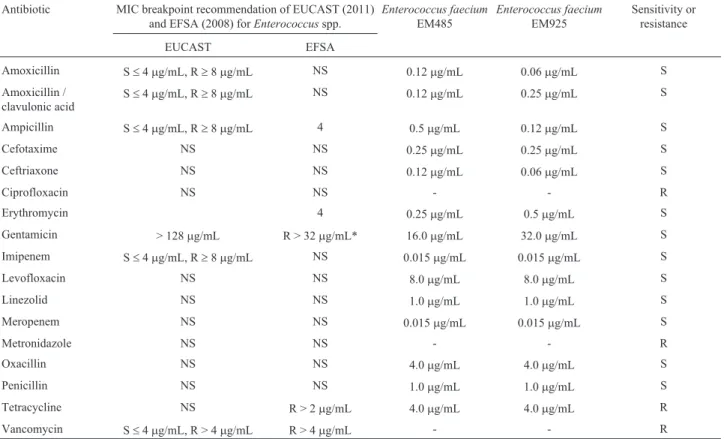
According to the results of the disc diffusion method (data not shown), the two studied E. faecium strains, EM485 and EM925, demonstrated susceptibility to penicil-lin G, ampicilpenicil-lin, chloramphenicol, tetracycpenicil-line,
erythro-mycin, and rifampicin. On the other hand, both strains showed high-level resistance to ciprofloxacin, vancomy-cin, and metronidazole (MIC values > 256.0mg/mL), and also presented a MIC value for tetracycline (4.0mg/mL) higher than the MIC breakpoint defined by EFSA (2008) for this antibiotic (Table 2). Resistance to streptomycin, co-trimoxazole (the MICs for these two antibiotics were not determined in this study) and metronidazole were also detected for both strains with the disc diffusion method. In relation to gentamicin, although the two strains showed re-sistance in the disc diffusion method (inhibition zone < 12 mm), and the MIC values were 16.0 and 32.0mg/mL forE. faeciumEM485 andE. faeciumEM925, respectively, not exceeding the MIC breakpoint established for
Enterococcus spp. (EFSA 2008). Both strains presented MIC values of 4.0mg/mL for oxacillin and 8.0mg/mL for levofloxacin. The resistance ofE. faeciumEM485 andE. faecium EM925 to co-trimoxazole and metronidazole could be an acquired feature considering the resistance to antibiotics commonly reported forEnterococcusspp. in the literature, and it deserves further investigation. Favaroet al.
(2014) reported that fourE. faeciumstrains isolated from feta cheese (from Bulgaria) were susceptible to ampicillin and penicillin and 2 of them were susceptible to van-comycin, which are the most clinically relevant antibiotics
Table 2- Antimicrobial susceptibility.
Antibiotic MIC breakpoint recommendation of EUCAST (2011) and EFSA (2008) forEnterococcusspp.
Enterococcus faecium EM485
Enterococcus faecium EM925
Sensitivity or resistance
EUCAST EFSA
Amoxicillin S£4mg/mL, R³8mg/mL NS 0.12mg/mL 0.06mg/mL S
Amoxicillin / clavulonic acid
S£4mg/mL, R³8mg/mL NS 0.12mg/mL 0.25mg/mL S
Ampicillin S£4mg/mL, R³8mg/mL 4 0.5mg/mL 0.12mg/mL S
Cefotaxime NS NS 0.25mg/mL 0.25mg/mL S
Ceftriaxone NS NS 0.12mg/mL 0.06mg/mL S
Ciprofloxacin NS NS - - R
Erythromycin 4 0.25mg/mL 0.5mg/mL S
Gentamicin > 128mg/mL R > 32mg/mL* 16.0mg/mL 32.0mg/mL S
Imipenem S£4mg/mL, R³8mg/mL NS 0.015mg/mL 0.015mg/mL S
Levofloxacin NS NS 8.0mg/mL 8.0mg/mL S
Linezolid NS NS 1.0mg/mL 1.0mg/mL S
Meropenem NS NS 0.015mg/mL 0.015mg/mL S
Metronidazole NS NS - - R
Oxacillin NS NS 4.0mg/mL 4.0mg/mL S
Penicillin NS NS 1.0mg/mL 1.0mg/mL S
Tetracycline NS R > 2mg/mL 4.0mg/mL 4.0mg/mL R
Vancomycin S£4mg/mL, R > 4mg/mL R > 4mg/mL - - R
for curing infections involving multiple antibiotic-resistant enterococcus strains. In a study of Schirruet al.(2012), the majority of the tested antibiotics inhibited the growth of fourE. faeciumstrains isolated from goat’s milk to some extent (from Sardenia, Italy). In the same study, five of the tested antibiotics (cotrimazin, cotrimixazol, nalidixic acid, oxacillin and sulphonamid) had no inhibitory effect on the studiedE. faeciumstrains. In addition, variable results have been reported for interactions betweenE. faeciumstrains and cefalotin, cefpime, ceftiofur oflaxacin, tazobactam, amoxicillin + clavulanic acid and ampicillin + sulbactam (Schirruet al., 2012).
Antibiotic resistance is a fundamental issue in the safety evaluation ofEnterococcusspp. for use in food prod-ucts. Acquired resistance located in plasmids and trans-posons is the main concern (Giraffa, 2002; Franzet al., 2005). Moreover, intrinsic resistance to some antibiotics is a feature commonly found amongEnterococcusstrains iso-lated from foods, as reported by Franzet al.(2005).
Antibiotic resistance in genusEnterococcusis con-troversial. Enterococci have a long history of application in food production (Schillingeret al., 1996) due to their favor-able metabolic activities (lipolysis, esterolysis, citrate utili-zation, etc.) that contribute to the typical taste and flavor of fermented foods (Centenoet al., 1996; Giraffa and Car-minati, 1997; Manolopoulouet al., 2003). However, some
Enterococcusstrains are pathogens for humans and other animals, and vancomycin-resistantEnterococcusspp. are frequently resistant to many antibiotics commonly used in veterinary and human medicine (Landman and Quale, 1997).
In the application of enterococci in foods, we need to pay special attention because of the possible spread of ge-netic determinants of resistance from these bacteria, which are generally located in conjugative plasmids or trans-posons prone to genetic exchange (Hasmanet al., 2005; Zanellaet al., 2006). Multi-resistance has been more com-monly reported forE. faecalisdue to its notorious ability to acquire and transfer antibiotic-resistance genes (Çitak et al., 2004; McBrideet al., 2007). Moreover, Gomeset al.
(2008) reported that the prevalence of antibiotic resistance was higher forE. faecaliscompared toE. faeciumisolates. The results presented in the literature indicate that foods cannot be ruled out as a potential source for spreading anti-biotic-resistant strains. (Abriouelet al., 2008). Toméet al.
(2008) reported resistance to chloramphenicol by a strain of
E. faeciumisolated from cold-smoked salmon evaluated for its potential application as biopreservative. However, it is important to consider that starter cultures and potential probiotic LAB may be a potential reservoir of antibiotic re-sistance genes and that horizontal gene transfer to the other bacteria present in the human GIT is a possible scenario (Dickset al., 2011; Teuber, 1999; Salyerset al., 2004).
Genes for virulence, biogenic amines and antibiotic resistance
Verification of virulence factors inEnterococcusspp. by bio-molecular and bio-chemical approaches is important due to the risk of genetic transfer because these genes are usually located in conjugative plasmids (Eaton and Gasson, 2001). In tests of the genes adhesion of collagen protein, aggregation substance, cytolysin, endocarditis antigen, enterococcal surface protein, gelatinase, hyaluronidase, histidine decarboxylase, ornithine decarboxylase, tyrosine decarboxylase, vancomycin A and vancomycin B, the only positive results were generated by PCR targeting the tyro-sine decarboxylase and vancomycin B genes inE. faecium
EM485 andE. faecium EM925 (Table 1). In general, the observed frequency of positive results for the virulence fac-tors studied was lower than that reported in other studies on
Enterococcus isolated from foods (Gomes et al., 2008; Barbosaet al., 2010) and lower when compared to studies with clinical isolates (Eaton and Gasson, 2001; Franzet al., 2001; Barbosaet al., 2010).
The investigation of virulence factors inEnterococci
strains with potential application in food preservation is of foremost importance because such strains may contain sev-eral determinants of pathogenicity. Virulence factors may act as colonization factors by promoting the adhesion of bacteria to host cells, or as invasion factors that promote the invasion of epithelial cells, which disrupts the immune sys-tem (de Souza, 2003). Several cell wall surface proteins play roles in enterococcal pathogenicity, including aggre-gation substance, enterococcal surface protein, and colla-gen-binding components (Hendrickxet al., 2009). Some extracellular proteins, such as hyaluronidase, may interact with lymphocyte receptors and be responsible for the in-duction of auto-immune diseases (de Souza, 2003). Cyto-lysin is an exotoxin with bifunctional function as a bacteriocin and by presenting hemolytic effects (Haas et al., 2002). Expression of the aggregation substance protein facilitates close contact between cells, conjugation and pos-sible transfer of virulence plasmids (Hendrickx et al., 2009). The aggregation substance protein may have a role in the translocation of enterococci into epithelial cells (Franz and Holzapfel, 2004).Enterococcussurface protein is a cell wall-anchored protein with a special role in biofilm formation (Hendrickxet al., 2009). ACE (angiotensin con-verting enzyme) proteins facilitate the binding of
vanco-mycin resistance types have been phenotypically and geno-typically identified, and two of them, VanA and VanB, may be located in transferable plasmids (Courvalin, 2006).
Hydrophobicity
Several mechanisms are involved in the adhesion of microorganisms to intestinal epithelial cells. The hydro-phobic nature of the outermost surface of microorganisms is involved in the attachment of bacteria to host tissue. The determination of microbial adhesion to hexadecane as a way to estimate the ability of a strain to adhere to epithelial cells is a valid qualitative phenomenological approach (Kiely and Olson, 2000).
Both strains presented low levels of hydrophobicity (8.18% forE. faeciumEM485 and 11.33% forE. faecium
EM925) determined as adhesion to n-hexadecane. Cell sur-face hydrophobicity is a non-specific interaction between microbial cells and their host. The initial interaction may be weak, is often reversible and it precedes subsequent adhe-sion processes mediated by more specific mechanisms involving cell-surface proteins and lipoteichoic acids (Gra-natoet al., 1999; Rojas et al., 2002; Ross and Jonsson, 2002). Bacterial cells with high hydrophobicity usually present strong interactions with mucosal cells. The same range of hydrophobicity has been reported forE. faecium
isolated from feta cheese (from Bulgaria), presenting levels of hydrophobicity between 7.92% and 10.23% (Favaroet al.2014). In another study (Todorovet al., 2011), hydro-phobicity values between 12.6% and 14.7% were recorded for E. faecium ET05, ET12 and ET88. These values are lower than that recorded for L. rhamnosus GG (53.3%, Todorovet al., 2008). Hydrophobicity may assist in adhe-sion, but it is not a prerequisite for strong adherence. Hydrophobicity varies among genetically closely related species and even among strains of the same species (Schar-Zammaretti and Ubbink, 2003).
Beneficial and technological properties
Growth in milk and viability in milk acidified with lactic acid
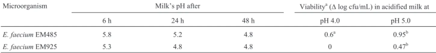
E. faeciumEM485 and EM925 both grew in milk and were able to change milk’s pH after 6, 24 and 48 h of incu-bation. No decrease in cell counts was observed when
E. faeciumEM485 andE. faeciumEM925 were maintained in milk acidified to pH 4.0 or to pH 5.0 at 5 °C for 30 days (Table 3). These technological characteristics are
interest-ing for posterior use in acidic products such as yogurts and acidified milk. The acidifying activity ofE. faecium iso-lated from goat’s milk (from Sardenia, Italy) reported by Schirru et al. (2012) was generally low. A good acid-producing starter culture needs to reduce the pH of milk from its normal value of approximately 6.6 to 5.3 in 6 h us-ing an inoculum of 10%, and in general, enterococci exhibit low milk acidifying ability (Schirruet al., 2012).
Effect of simulated gastric and intestinal conditions on the viability of selected strains
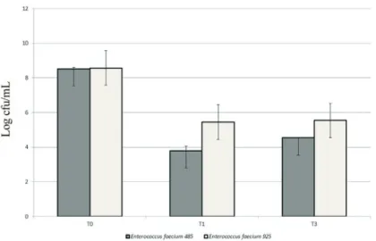
WhenE. faeciumEM485 andE. faeciumEM925 was exposed to simulated gastric and small intestine conditions, good survival rates were recorded for both strains (Figure 1). Such results are not surprising becauseEnterococcus
spp. have a good chance of surviving acidic and bilious conditions (Todorovet al., 2011). A cell count reduction of 3.98 log cfu/mL was recorded forE. faeciumEM485 after exposure to simulated gastric and small intestine condi-tions. Similar results were recorded forE. faeciumEM925, with a reduction in cell number of 3.03 log cfu/mL (Fig-ure 1).
Probiotics must survive in the acidic gastric environ-ment if they are to reach the small intestine and colonize the host, thereby imparting their benefits.Enterococcus spe-cies are considered intrinsically resistant to acid (Tannock, 2004). Although there are differences between species and strains, organisms generally exhibit increased sensitivity at pH values below 3.0 (Ronkaet al., 2003). Hence, acid tolerance is accepted as one of the desirable properties used to select potentially probiotic strains. Gastric transit studies of probiotics have been conducted using both simulated gastric juice and animal and human gastric juices (Charteris
et al., 1998; Gardiner et al., 1999). Both of these ap-proaches have limitations; the former fails to capture the in-fluence of dietary and nonacidic constituents of gastric secretions on probiotic survival, and the latter is restricted by the availability of fresh material (Charteriset al., 1998). In addition, the exploitation of rich media, such as acidified MRS medium, may offer protection to bacteria by provid-ing energy and metabolic precursors. The use of food provid- ingre-dients to enhance probiotic survival through the GIT has been extensively studied (Charteriset al., 1998; Gardineret al., 1999). However, little data are available describing the effects of individual food components and the underlying
Table 3- Growth in milk and viability in milk acidified with lactic acid.
Microorganism Milk’s pH after Viabilitya(Dlog cfu/mL) in acidified milk at
6 h 24 h 48 h pH 4.0 pH 5.0
E. faeciumEM485 5.8 5.2 4.8 0.6a 0.95b
E. faeciumEM925 5.3 4.8 4.8 0 0.47b
aDifference between counts at time 0 and after 30 days of cold storage (5 °C) in milk acidified at different pH values. b
mechanisms of action whereby they enhance the survival of LAB (Charalampopouluset al., 2003).
Production ofb- galactosidase
The ability of microorganisms to ferment lactose in milk is an important technological property of LAB with po-tential applications in the dairy industry. Hydrolysis of lac-tose, which confers taste, texture and nutritional value to milk and milk derivatives is carried out by the enzymes
b-D-galactosidase (EC 3.2.1.23) and/or phospho-b -D-galac-tosidase (EC 3.2.1.85), and it has been described in various organisms such as bacteria, yeasts and molds (Zárate and Chaia, 2012). In addition to their technological importance, both the pure enzymes and the viable microorganisms that contain them have been used to alleviate intestinal disorders such as lactose intolerance. This condition occurs worldwide among the adult population and has been treated successfully by the incorporation of microorganisms, mainly lactobacilli and/or bifidobacteria, into dairy products as a source of
b-galactosidase for the intra-intestinal hydrolysis of lactose and the modulation of colonic microbiota (Zárate and Chaia, 2012). In this sense,b-galactosidase activity is a beneficial characteristic for probiotics or LAB with application in the dairy industry.
The production of b-galactosidase by E. faecium
EM485 andE. faeciumEM925 was confirmed by employing sterile filter paper disks impregnated with o
-nitrophenyl-b-D-galactopyranose (ONPG Disks, Fluka, Buchs, Switzer-land). In previous studies,b-galactosidase production has been reported in different strains of Enterococcus spp. (Todorovet al., 2010; Favaroet al., 2014).
Deconjugation of bile salts
In our study we found thatE. faecium EM485 and
E. faeciumEM925 were similar in their ability to dejugate bile salts. Both strains grow on MRS agar plates
con-taining 0.5% (w/v) sodium salts of taurocholic acid (TC), taurodeoxycholic acid (TDC), glycocholic acid (GC), or glycodeoxycholic acid (GDC). However, deconjugation was recorded only for TDC and GDC.
The major route of cholesterol excretion from humans and other mammals is through feces. Cholesterol is the pre-cursor of primary bile salts that are formed in the liver and are stored as conjugated bile salts in the gall bladder for se-cretion in the gastrointestinal tract (Corzo and Gilliland, 1999). A small fraction of bile salts that is not absorbed is lost as free bile salts in feces. Free bile salts are less soluble than conjugated bile salts, resulting in lower absorption in the intestinal lumen (Center, 1993). At the physiological pH of the intestinal lumen, deconjugated bile salts can be trans-ported through the epithelium (Wonget al., 1994) and into the blood stream of the host, or they can precipitate. Thus, in a steady-state situation, deconjugation of bile acids can re-duce serum cholesterol levels by increasing the formation of new bile acids that are needed to replace those that have es-caped the enterohepatic circulation (Reynieret al., 1981). Experiments with germ-free rats have shown that bile salt deconjugation by B. longum increases bile salt excretion (Chikaiet al., 1987).
Proteolytic activity
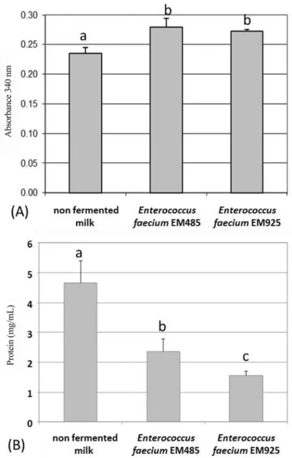
The reduction in protein concentration in extracellular enzymatic extract by E. faecium EM485 and E. faecium
EM925 grown in milk for 24 h in comparison with the con-trol (Figure 2) shows that the two strains display proteolytic activity. However, this enzymatic activity was very low when compared to that reported by Donkoret al.(2007) for
Lb. caseiL26, which presented OD340 nmabove 1.80 after 24 h of fermentation in MRS at 42 °C. Churchet al.(1983) observed OD340 nm above of 0.40 after Streptococcus (St.)
lactisC2 fermentation in milk for 24 h at 22 °C.
tivity and protein concentration values, were 18.73 ± 3.90 U/mg protein and 24.55±2.57 U/mg protein, respectively, without a significance difference between them (p > 0.05,t-test). As shown in Figure 2, the enzymatic activ-ity of both strains is located in the extracellular extract. This finding is consistent with Arizcumet al. (1997) reporting that the enzymatic activity ofEnterococcusspp. was located mainly in the extracellular extract fraction.
The importance of the protease activity is that it is re-lated to the good growth of LAB in milk and to casein hydro-lysis during cheese ripening. A study of Schirruet al.(2012)
reported low proteolytic activity in 2 of 4 testedE. faecium
strains isolated from goat’s milk (from Sardenia, Italy). However, even though lower values have been reported for otherEnterococcusspp. strains (Pepeet al., 2003), the result was similar to those produced by several LAB strains de-scribed in Madrauet al.(2006) as promising autochthonous starter cultures for the production of Pecorino sardo cheese.
Aggregation
corded for E. faecium EM485 was 80 ± 2% and for
E. faeciumEM925 it was 78±3% (Figure 3). Favaroet al.
(2014) reported much lower levels of auto-aggregation for
E. faeciumisolated from feta cheese (from Bulgaria) rang-ing from 8±1 to 10 ±2. In another study conducted by Todorov et al. (2008), strain-specificity in auto-aggre-gation was also observed forL. pentosusST712BZ andL. paracaseiST284BZ. High levels of co-aggregation were observed betweenE. faeciumEM485 and bothEscherichia coliINCQS 00033 (78±2%) andC. perfringensINCQS 00130 (81±2%) and also betweenE. faeciumEM925 and both Escherichia coli INCQS 00033 (74 ± 4%) and C. perfringensINCQS 00130 (84±3%) (Figure 3). Different degrees of co-aggregation have been measured with L. ivanoviisubsp.ivanoviiATCC 19119,L. innocuaATCC 33090, L. monocytogens ATCC 7644 and E. faecium
strains as reported by Favaro et al. (2014). High auto-aggregation would facilitate the exclusion of these patho-genic species from the GIT. Low levels of co-aggregation with pathogens may play an important role in preventing the formation of biofilms, in this way eliminating the patho-gen from the GIT. On the other hand, higher co-aggregation levels with pathogens will facilitate antibacterial action by
E. faeciumEM485 and E. faeciumEM925 and facilitate pathogen exclusion from the human GIT.
Conclusions
To the best of our knowledge, this is the first study re-porting on the properties ofE. faeciumstrains isolated from artisanal Coalho cheeses from Brazil that addresses their technological potential, beneficial properties, and safety with respect to the presence of genes encoding virulence factors, biogenic amines and antibiotic resistance. Con-sidering the results,E. faeciumstrains EM485 and EM925
are safe regarding the presence of genes associated with virulence factors and biogenic amine production, and they present technological properties compatible with use in a mixed starter culture for dairy products. However, the re-sistance to antimicrobials that was detected deserves fur-ther investigation.
Acknowledgments
To EMBRAPA for their financial support of this pro-ject (02.09.01.024.00). To Prof. Maria Teresa Destro and Dr. Eb Chiarini (University of São Paulo, São Paulo, Brazil) for providingListeria monocytogenesstrains used in this study. To Dr. Flávia Carolina Alonso Buriti for pro-viding technical assistance. Dr. Todorov received grants from CNPq (310203/2010-4) and FAPESP (2012/11571-6).
References
Abriouel H, Ben Omar N, Molinos ACet al.(2008) Comparative analysis of genetic diversity and incidence of virulence fac-tors and antibiotic resistance among enterococcal popula-tions from raw fruit and vegetable foods, water and soil, and clinical samples. Int J Food Microbiol 123:38-49.
Archimbaud C, Shankar N, Forestier Cet al.(2002) In vitro adhe-sive properties and virulence factors of Enterococcus
faecalisstrains. Res Microbiol 153:75-80.
Arizcum C, Barcina Y, Torre P (1997) Identification and charac-terization of proteolytic activity ofEnterococcusspp. Iso-lated from milk and Roncal and Idiazábal chesee. Int J Food Microbiol 38:17-24.
Avram-Hananel L, Stock J, Parlesak Aet al. (2010)E. durans
strain M4-5 isolated from human colonic flora attenuates in-testinal inflammation. Dis Colon Rectum 53:1676-1686. Barbosa J, Gibbs PA, Teixeira P (2010) Virulence factors among
enterococci isolated from traditional fermented meat
ucts produced in the North of Portugal Food Control 21:651-656.
Botes M, Van Reenen CA, Dicks LMT (2008) Evaluation of
Enterococcus mundtiiST4SA andLactobacillus plantarum
423 as probiotics by using a gastro-intestinal model with in-fant milk formulations as substrate. Int J Food Microbiol 128:362-370.
Bradford MM (1976) A rapid and sensitive method for the quan-titation of microgram quantities of protein utilizing the prin-ciple of protein-dye binding. Ann Biochem 72:248-254. Bybee SN, Scorza AV, Lappin MR (2011) Effect of the probiotic
Enterococcus faeciumSF68 on presence of diarrhea in cats
and dogs housed in an animal shelter. J Vet Intern Med 25:856-860.
Centeno JA, Menéndez S, Rodriguez-Otero JL (1996) Main mi-crobial flora present as natural starters in Cebreiro raw cow’s-milk cheese (Northwest Spain). Int J Food Microbiol 33:307-313.
Center SA (1993) Serum bile acid in companion animal medicine. In: Micheal SL (ed) Gastroenterology: 1990s. Saunders, Philadelphia, pp. 625-657.
Charalampopoulos D, Pandiella SS, Webb C (2003) Evaluation of the effect of malt, wheat and barley extracts on the viability of potentially probiotic lactic acid bacteria under acidic con-ditions. Int J Food Microbiol 82:133-141.
Charteris WP, Kelly PM, Morelli Let al.(1998) Antibiotic sus-ceptibility of potentially probioticLactobacillusspecies. J Food Protect 61:1636-1643.
Chikai T, Nakao H, Uchida K (1987) Deconjugation of bile-acids by human intestinal bactéria implanted in germ-free rats. Lipids 22:669-671.
Church FC, Swaisgood HE, Porter DHet al.(1983) Spectrophoto-metric assay usingo-phthaldealdehyde for determination of proteolysis in milk and isolated milk proteins. J Dairy Sci 66:1219-1227.
Çitak S, Yucel N, Orhan S (2004) Antibiotic resistance and inci-dence ofEnterococcusspecies in Turkish white cheese. Int J Dairy Technol 57:27-35.
Corzo G, Gilliland SE (1999) Bile salt hydrolase activity of three strains of Lactobacillus acidophilus. J Dairy Sci 82:472-480.
Courvalin P (2006) Vancomycin resistance in gram-positive cocci. Clin Inf Dis 42:S25-S34.
de Sousa CP (2003) Pathogenicity mechanisms of procaryotic cells: evolutionary view. Braz J Infect Dis 7:23-31. Dicks LMT, Todorov SD, Franco BDGM (2011) Current status of
antibiotic resistance in lactic acid bacteria. In: Bonilla AR, Muniz KP (eds) Antibiotic Resistance: Causes and Risk Factors, Mechanisms and Alternatives. Pharmacology - Re-search, Safety Testing and Regulation. Nova Publisher, New York, pp 379-425.
Donkor ON, Henriksson A, Vasiljevic Tet al.(2007) Proteolytic activity of dairy lactic acid bacteria and probiotics as deter-minant of growth and in vitro angiotension-converting en-zyme inhibitory activity in fermented milk. Lait 86:21-38. dos Santos KMO, Vieira ADS, Rocha CRCet al.(2014) Brazilian
artisanal cheeses as a source of beneficial Enterococcus
faeciumstrains: Characterization of the bacteriocinogenic
potential. Ann Microbiol 64:1463-1471.
Eaton TJ, Gasson MJ (2001) Molecular screening of
Enterococcusvirulence determinants and potential for
ge-netic exchange between food and medical isolates. Appl En-viron Microbiol 67:1628-1635.
EFSA (2008) Update of the criteria used in the assessment of bac-terial resistance to antibiotics of human or veterinary impor-tance. Prepared by the Panel on Additives and Products or Substances used in Animal Feed. EFSA J 732:1-15. Favaro L, Basaglia M, Casella Set al.(2014) Bacteriocinogenic
potential and safety evaluation of non starterEnterococcus
faecium strains isolated from home made white brine
cheese. Food Microbiol 38:228-239.
Foulquié-Moreno MR, Sarantinopoulos P, Tsakalidou Eet al.
(2006) The role and application of enterococci in food and health. Int J Food Microbiol 106:1-24.
Franz CM, Muscholl-Silberhorn AB, Yousif NMKet al.(2001) Incidence of virulence factors and antibiotic resistance among Enterococci isolated from food. Appl Environ Microbiol 67:4385-4389.
Franz CMAP, Holzapfel WH (2004) The genusEnterococcus: biotechnological and safety issues. In: Salminen A, von Wright A, Ouwehand A (eds) Lactic Acid Bacteria: Micro-biological and Functional Aspects. Marcel Dekker, Inc, New York, pp. 199-248.
Franz CMAP, Huch M, Abriouel Het al.(2011) Enterococci as probiotics and their implications in food safety. Int J Food Microbiol 151:125-140.
Franz CMAP, Hummel A, Holzapfel WH (2005) Problems re-lated to the safety assessment of lactic acid bacteria starter cultures in probiotics Mitteil Lebensmittel Hyg 96:39-65. Franz CMAP, Stiles ME, Schleifer KHet al.(2003) Enterococci
in foods - a conundrum for food safety. Int J Food Microbiol 88:105-122.
Frazzon APG, Gama BA, Hermes Vet al.(2010) Prevalence of antimicrobial resistance and molecular characterization of tetracycline resistance mediated bytetM andtetL genes in
Enterococcus spp. isolated from food in Southern Brazil.
World J Microbiol Biotechnol 26:365-370.
Gardiner GE, Ross RP, Wallace JMet al.(1999) Influence of a probiotic adjunct culture of Enterococcus faeciumon the quality of cheddar cheese. J Agric Food Chem 47:4907-4916.
Giraffa G (2002) Enterococci from foods. FEMS Microbiol Rev 26:163-171.
Giraffa G (2003). Functionality of enterococci in dairy products. Int J Food Microbiol 88:215-222.
Giraffa G, Carminati D (1997) Control ofListeria monocytogenes
in the rind of Taleggio, a surface-smear cheese, by a bac-teriocin from Enterococcus faecium 7C5. Sci Aliment 17:383-391.
Girish KS, Kemparaju K (2007) The magic glue hyaluronan and its eraser hyaluronidase: A biological overview. Life Sci 80:1921-1943.
Gomes BC, Esteves CT, Palazzo ICVet al.(2008). Prevalence and characterization of Enterococcus spp. isolated from Brazilian foods. Food Microbiol 25:668-675.
Haas W, Shepard BD, Gilmore MS (2002) Two-component regu-lator of Enterococcus faecaliscytolysin responds to quo-rum-sensing autoinduction. Nature 415:84-87.
Hasman H, Villadsen AG, Aarestrup FM (2005) Diversity and stability of plasmids from glycopeptide-resistant
Enterococcus faecium (GRE) isolated from pigs in
Den-mark. Microb Drug Resist 11:178-184.
Hendrickx APA, Willems RJL, Bonten MJMet al.(2009) LPxTG surface proteins of enterococci. Trends Microbiol 17:423-430.
Kayaoglu G, Orstavik D (2004) Virulence factors of
Enterococcus faecalis: Relationship to endodontic disease.
Crit Rev Oral Biol Med 15:308-320.
Kiely LJ, Olson NF (2000) The physicochemical surface charac-teristics ofLactobacillus casei. Food Microbiol 17:277-291. Laemmili UK (1970) Cleavage of structural proteins during the assembly of the head of bacteriophage T4. Nature 227:680-685.
Landman D, Quale JM (1997) Management of infections due to resistant enterococci: a review of therapeutic options. J Anti-microb Chemoth 40:161-170.
Madrau MA, Mangia NP, Murgia MAet al.(2006) Employment of Autochthonous Microflora in Pecorino Sardo Cheese Manufacturing and Evolution of Physicochemical Parame-ters during Ripening. Int Dairy J 16:876-885.
Manolopoulou E, Sarantinopoulos P, Zoidou Eet al.(2003) Evo-lution of microbial populations during traditional Feta cheese manufacture and ripening. Int J Food Microbiol 82:153-161.
Martin-Platero AM, Valdivia E, Maqueda Met al.(2009) Charac-terization and safety evaluation of enterococci isolated from Spanish goats’ milk cheeses. Int J Food Microbiol 132:24-32.
McBride SM, Fischetti VA, LeBlanc DJet al.(2007) Genetic Di-versity amongEnterococcus faecalis. PLoS ONE 2:e582. Montalban-Lopez M, Sanchez-Hidalgo M, Valdivia E et al.
(2011) Are bacteriocins underexploited? NOVEL applica-tions for OLD antimicrobials. Curr Pharmacol Biotechnol 12:1205-1220.
Moraes PM, Perin LM, Tassinari Ortolani MBet al.(2010) Proto-cols for the isolation and detection of lactic acid bacteria with bacteriocinogenic potential. LWT - Food Sci Technol 43:1320-1324.
Ogier J-C, Serror P (2008) Safety assessment of dairy microor-ganisms: The Enterococcus genus. Int J Food Microbiol 126:291-301.
Ortolani MBT, Yamazi AK, Moraes PMet al.(2010). Microbio-logical auality and safety of raw milk and soft cheese and de-tection of autochthonous lactic acid bacteria with antagonis-tic activity against Listeria monocytogenes, Salmonella
spp., and Staphylococcus aureus. Foodborne Path Dis 7:175-180.
Pepe O, Villani F, Oliviero Det al.(2003) Effect of proteolytic starter cultures as leavening agents of pizza dough. Int J Food Microbiol 84:319-326.
Pinto MGV, Franz CMAP, Schillinger U et al. (2006)
Lactobacillusspp. with in vitroprobiotic properties from
human faeces and traditional fermented products. Int J Food Microbiol 109:205-214.
Raz I, Gollop N, Polak-Charcon Set al.(2007) Isolation and char-acterisation of new putative probiotic bacteria from human colonic flora. Br J Nutr 97:725-734.
Reynier MO, Montel JC, Gerolami Aet al.(1981) Comparative effects of cholic, chenodeoxycholic & ursodeoxycholic ac-ids on micellar solubilization and intestinal absorption of cholesterol. J Lipid Res 22:467-473.
Rivas P, Alonso J, Moya Jet al.(2005) The impact of hospi-tal-acquired infections on the microbial etiology and prog-nosis of late-onset prosthetic valve endocarditis Chest 128:764-771.
Rojas M, Ascencio F, Conway PL (2002) Purification and charac-terization of a surface protein fromLactobacillus fermentum
104R that binds to porcine small intestinal mucus and gastric mucin. Appl Environ Microbiol 68:2330-2336.
Ronka E, Malinen E, Saarela Met al.(2003) Probiotic and milk technological properties ofLactobacillus brevis. Int J Food Microbiol 83:63-74.
Ross S, Jonsson H (2002) A high-molecular mass cell-surface protein fromLactobacillus reuteri1063 adheres to mucus components. Microbiol 148:433-442.
Salyers AA, Gupta A, Wang Y (2004) Human intestinal bacteria as reservoirs for antibiotic resistance genes. Trends Micro-biol 12:412-416.
Schar-Zammaretti P, Ubbink J (2003) The cell wall of lactic acid bacteria: surface constituents and macromolecular confor-mations. Bioph J 85:4076-4092.
Schillinger U, Geisen R, Holzapfel WH (1996) Potential of antag-onistic microorganisms and bacteriocins for the biological preservation of foods. Trends Food Sci Technol 7:158-164. Schirru S, Todorov SD, Favaro Let al.(2012) Sardinian goat’s milk as source of bacteriocinogenic potential protective cul-tures. Food Control 25:309-320.
Tannock GW (2004) Can the GUT microflora of infants be modi-fied by giving probiotics to mothers? J Ped Gastroenterol Nutr 38:244-246.
Teuber M (1999) Spread of antibiotic resistance with food-borne pathogens. Cell Molec Life Sci 56:755-763.
Todorov SD, Botes M, Guigas Cet al.(2008) Boza, a natural source of probiotic lactic acid bacteria. J Appl Microbiol 104:465-477.
Todorov SD, Dicks LMT (2008) Evaluation of lactic acid bacteria from kefir, molasses and olive brine as possible probiotics based on physiological properties. Annals Microbiol 58:661-670.
Todorov SD, Furtado DN, Saad SMIet al.(2011) Potential bene-ficial properties of bacteriocin-producing lactic acid bacteria isolated from smoked salmon J Appl Microbiol 110:971-986.
Todorov SD, Wachsman M, Tomé Eet al.(2010) Characterisa-tion of an antiviral pediocin-like bacteriocin produced by
Enterococcus faecium. Food Microbiol 27:869-879.
Tomé E, Gibbs PA, Teixeira PC (2008) Growth control ofListeria
innocua 2030c on vacuum-packaged cold-smoked salmon
by lactic acid bacteria Int J Food Microbiol 121:285-294. Tsakalidou E, Anastasiou R, Vandenberghe Iet al.(1999)
Cell-wall-bound proteinase ofLactobacillus delbrueckiisubsp.
lactis ACA-DC 178: characterization and specificity for
b-casein. Appl Environ Microbiol 65:2035-2040.
Vankerckhoven V, Van Autgaerden T, Vael C, Lammens Cet al.
asa1,gelE,cylA,esp, andhylgenes in enterococci and sur-vey for virulence determinants among European hospital isolates of Enterococcus faecium. J Clinic Microbiol 42:4473-4479.
Vinderola G, Capellini B, Villarreal Fet al.(2008) Usefulness of a set of simple in vitro tests for the screening and identifica-tion of probiotic candidate strains for dairy use. LTW - Food Sci Technol 41:1678-1688.
Vinderola GC, Costa GA, Regenhardt Set al.(2002) Influence of compounds associated with fermented dairy products on the growth of lactic acid starter and probiotic bacteria. Int Dairy J 12:579-589.
Wong WH, Oelkers P, Craddock ALet al. (1994) Expression cloning and characterization of the hamster ileal
sodium-dependent bile acid transporter. J Biol Chem 269:1340-1347.
Zanella RC, Lima MJC, Tegani LSet al.(2006) Emergence of VanB phenotype-vanA genotype in vancomycin-resistant enterococci in Brazilian hospital. Braz J Microbiol 37:117-118.
Zárate G, Chaia AP (2012) Influence of lactose and lactate on growth and b-galactosidase activity of potential probiotic
Propionibacterium acidipropionici. Anaerobe 18:25-30.
Associate Editor: Eduardo Cesar Tondo