HEMATOPOIETIC STEM CELL TRANSPLANTATION
A Doctoral THESIS
SUBMITTED TO THE
UNIVERSIDADE NOVA DE LISBOA
BY
CRISTINA JOÃO
PhD in Medicine (Hematology)
" Th e r e a r e t w o obj e ct s in m e dica l e du ca t ion :
t o h e a l t h e sick a n d a dva n ce t h e scie n ce ."
- Dr. Charles H. Mayo
Pr e fa ce a n d Ack n ow le dgm e n t s………..………. 13
Abst r a ct ………... 18
Re su m o………. 23
Ré su m é ... 28
Ch a pt e r I Ge n e r a l I n t r odu ct ion ………. 34
1.1. The role of B cells and immunoglobulin in T cell development 34
1.2. Immune Reconstitution after autologous hematopoietic stem cells transplantation and after treatment with rituximab. The cases of Non-Hodgkin Lymphomas and Multiple Myeloma 42 1.3. Measurement of T cell receptor diversity 55 Ch a pt e r I I Aim s of t h e st u dy……….. 58
Ch a pt e r I I I
Direct measurement of lymphocyte receptor diversity………
Nucleic Acids Resear ch 2003, Vol. 31, No22 e129
61
3.3. Material and methods 66
3.4. Results 71
3.5. Discussion 83
Ch a pt e r I V
B cell-dependent TCR diversification………
Journal of I m m unology 2004,172: 4709- 4716
86
4.1. Abstract 88
4.2. Introduction 89
4.3. Material and methods 9323
4.4. Results 99
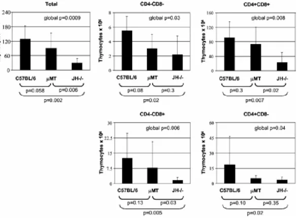
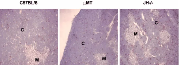
4.4.1. Thymocyte development is perturbed in mice that lack B cells and immunoglobulins
99 4.4.2. Increased apoptosis in the thymic cortex of
mice that lack B cells and immunoglobulins
102 4.4.3. Numbers of recent thymic emigrants are
maintained and thymocyte proliferation are increased in mice that lack B cells and immunoglobulins
103
4.4.4. Contraction of TCR repertoire in mice that lack B cells and/or immunoglobulins
107 4.4.5. B cells precursors in C57BL/6, QM, JH-/- and
μMT thymi
112 4.4.6. T cell diversity in JH-/- mice reconstituted by
adoptive transfer of B cells or administration of immunoglobulin
115
Immunoglobulin promotes the diversity and the function of T cells………
European Journal of I m m unology 2006, 36: 1718- 1728
125
5.1. Abstract 127
5.2. Introduction 128
5.3. Materials and Methods 130
5.4. Results 136
5.4.1. B cell deficient mice and mice with quasi-monoclonal B cells have a severely contracted peripheral T cell receptor repertoire
136
5.4.2. Oligoclonal expansion on JH-/- T cells 140 5.4.3. Increased in vivo proliferation of T cells on B
cell deficient mice
143 5.4.4. Delayed rejection of H-Y incompatible grafts by
JH-/- and QM mice
5.4.5. Exclusion of other immunological factors that, besides contracted TCR diversity, may
decrease T cell function
148 151
5.4.6. Recovery of TCR diversity and T cell function 154
5.5. Discussion 158
Ch a pt e r V I
Early lymphocyte recovery after autologous stem cell transplantation predicts superior survival in mantle-cell lymphoma………
Bone Mar r ow Transplant at ion 2006, 37: 865- 871
162
6.1. Abstract 164
6.2. Introduction 165
6.4.1. Patient characteristics 171 6.4.2. Prognostic factors for progression and survival 173 6.4.3. Discrimination of ALC-15 in MCL patients as
predictor of PFS
180
6.5. Discussion 181
6.6. Acknowledgements 186
Ch a pt e r V I I
Re cove r y of polyclon a l im m u n oglobu lin se r u m le ve ls t o n or m a l le ve ls a ft e r a u t ologou s st e m ce ll t r a n spla n t a t ion pr e dict s dise a se fr e e su r viva l in pa t ie n t s w it h m u lt iple m ye lom a ………..
Manuscript in Preparat ion
187
7.1. Abstract 189
7.2. Introduction 191
7.3. Patients and Methods 193
7.4. Results 196
7.4.1. Patient characteristics 196 7.4.2. Progression and survival 198 7.4.3.Multivariate analysis of prognostic factors 200
7.5. Discussion 203
Ch a pt e r V I I I
H ost im m u n e com pe t e n ce pr e dict s a su pe r ior pr ogr e ssion - fr e e su r viva l in n on - H odgk in ’s lym ph om a pa t ie n t s t r e a t e d w it h Rit u x im a b a n d I n t e r le u k in - 1 2 ………..
Manuscript Subm it t ed
206
8.1. Abstract 208
8.4. Results 217 8.4.1. Patient characteristics 217 8.4.2. Progression and survival 219 8.4.3.Sub-analysis of follicular lymphomas 225 8.4.4. The role of ALC on CD4 counts 226
8.5. Discussion 228
Ch a pt e r I X
D iscu ssion , Ge n e r a l Con clu sion s a n d Fu t u r e D ir e ct ion s…..…….. 232
Ch a pt e r I
Figu r e 1 – “ T cells developm ent in t he t hym us.” ………...
Figu r e 2 – “ Mechanism s of act ion of rit uxim ab and ways t o
increase it s clinical efficacy.” ………...
39
46
Ch a pt e r I I I
Figu r e 1 – “ Est ablishing t he relat ionship bet w een num ber of
gene chip hybridizat ion sit es and sam ple diversit y.” ………
Figu r e 2 – “ Repr oducibilit y of m et hod for analysis of r ecept or
diversit y.” ………...
Figu r e 3 – “ Analysis of B cell diver sit y using t he gene chip
m et hod.” ………
Figu r e 4 – “ Analysis of hum an T cell diversit y using gene
chips.” ………
72
74
78
81
Ch a pt e r I V
Figu r e 1 – “ Thym ocyt e num bers in C57BL/ 6, µMT, and JH- / -
m ice.” ………...
Figu r e 2 – “ Apopt osis in t he t hym us.” ……….
Figu r e 3 – “ Recent t hym ic em igrant s.” ……….
Figu r e 4 – “ DNA cont ent of t hym ocyt es fr om C57BL/ 6, µMT, and
JH- / - m ice.” ………..
Figu r e 5 – “ TCR Vβ diversit y.” ………
100 103 105
of t hym ic B cells and B cell precursors.” ……….. 114
Ch a pt e r V
Figu r e 1 – “TCR Vβ diver sit y quant ified in splenocyt es.” ………….
Figu r e 2 – “ Num ber of T cells ( A) and presence of follicular
dendrit ic cells net w or k in lym ph nodes ( B) in C57BL/ 6, QM and
JH- / - m ice.”……….
Figu r e 3 – “ Mem ory phenot ype and in vivo proliferat ion st udies
in t he spleen of B cell com pr om ised m ice com pared t o wild t ype.”
Figu r e 4 – “ Kaplan Meier sur vival curves for H- Y incom pat ible
skin graft s in C57BL/ 6, QM and JH- / - m ice.” ………..
Figu r e 5 – “ A) I n vit r o pr oliferat ion abilit y of CD4+ T cells
against polyclonal st im uli in C57BL/ 6, QM and JH- / - m ice; B) I n
vit ro proliferat ion response of DO 11.10 T cells t o OVA ( 323- 339)
present ed by wild t ype or QM B cells; C) Fract ion of CD4+/ CD25+
splenocyt es m easured by flow cyt om et ry analysis.” ………...….
Figu r e 6 – “ Survival of H- Y incom pat ible skin gr aft s in QM m ice
w here TCR Vβ diversit y was reconst it ut ed com pared t o
non-reconst it ut ed QM m ice ( A) and in wild t ype m ice ( C57BL/ 6)
w here TCR Vβ diversit y w as decreased by t hym ect om y ( B)
com pared t o w ild t ype.” ……….
137
138
147
150
153
156
Ch a pt e r V I
num ber of lym phocyt es at day 15.“ ……….
Figu r e 2 – “ Progression free survival for 42 pat ient s w it h m ant le
cell lym phom a aft er ASCT as a funct ion of recover y of absolut e
num ber of lym phocyt es at day 15.“ ……….………...
174
175
Ch a pt e r V I I
Figu r e 1 – “ Progression free survival analysis of I gG MM
pat ient s aft er ASCT.” ………
Figu r e 2 – “ Progression free survival analysis in t he sub group of I gG MM pat ient s who were in response at day 100 aft er
ASCT.” ………
199
200
Ch a pt e r V I I I
Figu r e 1 – “ Progr ession free survival for 41 pat ient s w it h non
Hodgkin lym phom a from t he t im e of rit uxim ab/ I L- 12 t herapy as
a funct ion of r ecover y of absolut e num ber of lym phocyt es“ ……..…
Figu r e 2 – “ Progr ession free survival for 41 pat ient s w it h non
Hodgkin lym phom a from t he t im e of rit uxim ab/ I L- 12 t herapy as
a funct ion of recovery of CD4 lym phocyt e” ………
222
Ch a pt e r I
Ta ble 1 – “ Cyt okines and gr ow t h fact ors exploit ed t o increase
pat ient s im m unit y” ………
Ta ble 2 – “ Reconst it ut ion of B and T cell num bers and funct ion
aft er ASCT” ………..
47
50
Ch a pt e r I V
Ta ble 1 – “ Mean concent rat ion of serum I g ± SD in C57BL/ 6,
QM, µMT, JH- / - m ice and in JH- / - m ice reconst it ut ed w it h B cells
or aft er adm inist rat ion of I gG” ……….
Ta ble 2 – “ Reconst it ut ion of TCR diver sit y in JH- / - m ice aft er
adopt ive t ransfer of bone m arrow B cells or adm inist rat ion of
I gG.” ………
101
117
Ch a pt e r V
Ta ble 1 – “ Num ber of Jβ fam ilies represent ed in t he cloned and
sequenced TCR Vβ 8.1 – Cβ region in C57BL/ 6 and JH- / - m ice.“ …
Ta ble 2 – “ Com parison of t he sequences bet w een t he end of Vβ
8.1 region and t he beginning of t he t w o m ore frequent Jβ
sequences ( J
β
2.6
and Jβ 2.1) used for bot h C57BL/ 6 and JH- / -st rains.”………
141
Ta ble 1 – “ Baseline charact erist ics of pat ient s according t o t he
recovery of absolut e lym phocyt e count at day 15 aft er ASCT
( ALC- 15) .” ……….
Ta ble 2 – “ Univariat e analysis for pat ient s w it h m ant le cell
lym phom a – overall survival and progression free survival rat es
were indicat ed.” ………
Ta ble 3 – “ Mult ivariat e analysis for pat ient s w it h m ant le cell
lym phom a – overall survival and progression free survival rat es
were indicat ed. Mult ivariat e m odel cont ains all t he prognost ic
fact ors list ed.” ……….
174
177
179
Ch a pt e r V I I
Ta ble 1 – “ Pat ient s charact erist ics“ ……….………..
Ta ble 2 – “ Univariat e analysis for PFS on pat ient s w it h I gG MM
subj ect t o ASCT.“ ……….
Ta ble 3 – “ Mult ivariat e analysis for PFS on pat ient s w it h I gG MM
subj ect t o ASCT.” ………
197
201
202
Ch a pt e r V I I I
Ta ble 1 – “ Pat ient s charact erist ics“ ……….………..
Ta ble 2 – “ Univariat e analysis for PFS in pat ient s w it h NHL
subj ect t o t reat m ent w it h r it uxim ab/ I L- 12.” ………
Ta ble 3 – “ Mult ivar iat e analysis for PFS in pat ient s w it h NHL
t reat ed w it h rit uxim ab/ I L- 12.” ……….
218
221
This dissertation presents the results of my work on immune reconstitution, developed at the Mayo Clinic, Rochester, Minnesota, USA from 2002 until 2006. This work was first developed in collaboration with Dr. Jeffrey Platt and Professor Marília Cascalho and their research group, at the Transplantation Biology Division, and continued at the Hematology Division at the Mayo Clinic in Rochester, MN, USA, under the supervision of Dr. Luis Porrata and Professor Svetomir Markovic.
Along this period I relied on the close and fundamental mentorship of Prof. Maria Gomes da Silva, from Faculdade de Ciências Médicas da Universidade Nova de Lisboa. Prof. Maria Gomes da Silva has been my mentor during the Hematology Residency and Fellowship at the Instituto Português de Oncologia de Lisboa since 1999. She recognized my interest on scientific research and supported me on the decision to interrupt the Hematology Fellowship and dedicate myself fulltime to scientific research for a period of time. Prof. Maria Gomes da Silva has supported all my work and her friendship and guidance during these years have been invaluable for me.
their work. After my first visit to their laboratory, in September 2001, I applied for and was awarded with a Portuguese PhD fellowship from
Fundação para a Ciência e Tecnologia and integrated Dr. Platt’s team at the Mayo Clinic in June 2002.
I found the Mayo Clinic a place where biomedical research was performed with high quality standards of humanism, scientific reasoning and academic life. Mayo Clinic at Rochester, Minnesota, comprises an integrated pluridisciplinar clinic and two hospitals, staffed by more than 2,500 physicians and scientists who conduct wide-ranging research to improve patient care, while training the next generation of medical scholars. I further integrated the group of those scholars when I obtained there a Master degree in Clinical Research. It was at Mayo Clinic that I developed the studies that conducted to this dissertation.
I owe the opportunity of being trained at a very prestigious academic institution to all who allowed that training and supported this project. I deeply thank Professor António Parreira who always supported and instigated me to combine clinical work and scientific research. He is a model of a scholar leader, working to improve the scientific level of the Portuguese Medicine and Science.
lymphocytes andimmunoglobulin exert upon T lymphocyte development and function.
After two years of basic research in the area of immune reconstitution, which originated 3 publications in high impact factor scientific journals, and after starting the Mayo Clinic Master Program in Clinical Research, I began to work with Dr. Luis Porrata and Professor Svetomir Markovic at the Hematology Division of Mayo Clinic. At that time, I felt it was essential that part of my PhD studies were devoted to the clinical questions raised by my previous work.
Dr. Porrata and Professor Markovic were investigating the importance of the recovery of a normal number of lymphocytes after autologous stem cell transplantation. They previously showed that an early recovery of lymphocyte numbers after autologous stem cell transplantation in several hematological and solid neoplasms was independently associated with a prolonged survival. We hypothesized that their findings could be explained by my previous observations on T cell reconstitution under the influence of B lymphocytes. Our collaborative work soon originated two manuscripts included in this dissertation.
me to Professor Markovic and Dr. Porrata, who helped and lead me to pursue clinical scientific research after the conclusion of the first part of this work. I thank their availability and expert discussion of scientific questions and their continuous intellectual support and alliance.
I am also sincerely grateful to Prof. António Coutinho, director of the Instituto Gulbenkian de Ciência (IGC). After returning from the United States, Professor Coutinho invited me to start a new research project as an independent junior investigator at IGC, granting me a new “research house”. He has always incentivated and discussed my work and critically looked at my data.
To my colleagues and friends at the Mayo Clinic, Brenda Ogle, Gregory Brunn, Cecilia Rietz, Claudia Ferreira, Catarina Cortesão, Pedro Geraldes, Hilal Maradit-Kremers, Dianne Khurana, Vini Khurana and Xiaosheng Wu, I deeply thank all the support, friendship and intellectual discussions.
I thank with all my heart my family and close friends, who always accompanied and supported this work.
As determined in the “Decreto Lei nº 388/70, artigo 8º, parágrafo 2”, the results included in this thesis are already published or are being prepared to submit for publication, in the following manuscripts:
1.
Ogle B., Cascalho M., João C., Taylor W., West J. L., Platt L. J. “Direct measurement of lymphocyte receptor diversity”. N u cle ic Acids Re se a r ch, 31:e139, 2003.My contributions to this paper were to design, perform, analyse and describe
the experiments related to the immunization of the mice. I wrote the part of
the manuscript related with the immunization; I commented the rest of the
manuscript and actively participated in writing the discussion.
2.
João C., Ogle B., Gay-Rabinstein C., Platt J. L., Cascalho M., “Bcell-dependent TCR diversification”. Jou r n a l of I m m u n ology, 172:4709-4716, 2004.
My contributions to this paper were to design, perform, analyse and describe
the majority of the experiments. I wrote the manuscript and helped to
submit it.
3.
João C., Ogle B., Geyer S., “Immunoglobulin promotes the diversityand the function of T cells”. Eu r ope a n Jou r n a l of I m m u n ology , 36:1718-1728, 2006.
My contributions to this paper were to design, perform and analyse the vast
majority of the experiments. I wrote the manuscript and submitted the
4.
João C., Porrata L.F., Inwards D. J., Ansell S., Micallef I., Johnston P., Gastineau D.A., Markovic S. N., “Early lymphocyte recovery after autologous stem cell transplantation predicts superior survival in Mantle Cell Lymphoma”. Bon e M a r r ow Tr a n spla n t a t ion, 37:865-871, 2006.My contributions to this paper were to conceive the statistical strategy, to
analyse the data set and write the manuscript.
5.
João C., Geyer S., Markovic S. N., Gertz M., Lacy M., Dispenzieri A.,Kumar S., Hayman S., Gastineau D., Porrata L. P., “Recovery of polyclonal immunoglobulin serum levels to normal levels after autologous stem cell transplantation predicts disease free survival in patients with multiple myeloma”. In preparation, 2007.
My contributions to this paper were to design the analysis and the data set,
collect the data, conceive the statistical analysis, analyse the data and write
the manuscript.
6.
João C., Porrata L.F., Witzig T., Kurtin P., Micallef I., Markovic S.N.,Erlichman C., Novak A., Ansell S., “Absolute lymphocyte count and CD4 count predict a superior progression-free survival in non-Hodgkin’s lymphoma patients treated with Rituximab and Interleukin-12”. Submitted, 2007.
My contributions to this paper were to elaborate and perform the analysis and
The investigation of the web of relationships between the different elements of the immune system has proven instrumental to better understand this complex biological system. This is particularly true in the case of the interactions between B and T lymphocytes, both during cellular development and at the stage of cellular effectors functions. The understanding of the B–T cells interdependency and the possibility to manipulate this relationship may be directly applicable to situations where immunity is deficient, as is the case of cancer or immune suppression after radio and chemotherapy.
obtained demonstrated that the diversity of the T cell compartment is increased by the presence of polyclonal immunoglobulin. Polyclonal immunoglobulin, and particularly the Fab fragments of the molecule, represent the most diverse self-molecules in the body and its peptides are presented by antigen presenting cells to precursor T cells in the thymus during its development. This probably contributes significantly to the generation of receptor diversity.
reconstitution in the context of AIDS, cancer, autoimmunity and post myeloablative treatments.
We also studied the impact of a robust immunity for the response to treatment with the antibody anti CD20, rituximab, in patients with non-Hodgkin’s lymphoma (NHL) (Chapter VIII). Patients with higher absolute counts of CD4+ T lymphocytes respond better (in terms of longer progression free survival) to rituximab compared to patients with lower number of CD4+ T lymphocytes. These observations highlight again the fact that a competent immune system is required for the clinical benefit of rituximab therapy in NHL patients.
In conclusion, the work presented in this dissertation demonstrates, for the first time, that diverse B cells and polyclonal immunoglobulin promote T cell diversification in the thymus and improve T lymphocyte function. Also, it shows that in the setting of immune reconstitution, as after autologous stem cell transplantation for mantle cell lymphoma and in the setting of immune therapy for NHL, the absolute lymphocyte counts are an independent factor predicting progression free and overall survival.
O estudo da rede de inter-relações entre os diversos elementos do sistema immune tem-se mostrado um instrumento essencial para uma melhor compreensão deste complexo sistema biológico. Tal é particularmente verdade no caso das interacções entre os linfócitos B e T, quer durante o desenvolvimento celular, quer ao nível das funções celulares efectoras. A compreensão da interdependência entre linfócitos B e T e a possibilidade de manipular esta relação pode ser directamente aplicável a situações em que a imunidade está deficiente, como é o caso das doenças neoplásicas ou da imunossupressão após radio ou quimioterapia.
presença no timo de péptidos mais diversos, como a imunoglobulna policlonal, induzisse a génese de precursores T mais diversos.
Demonstrámos que a diversidade do compartimento T é aumentado pela presença de imunoglobulina policlonal. A imunoglobulina policlonal, e particularmente os fragmentos Fab desta molécula, representam as moléculas autólogas mais diversas presentes nos organismos vertebrados. Estes péptidos são apresentados por células apresentadoras de antigénio às células precursoras T no timo, durante o desenvolvimento celular T. Tal, provavelmente, contribui para a génese da diversidade dos receptores.
Além disso, a reconstituição da diversidade dos linfócitos T em ratinhos com uma diversidade de reportório T diminuída, induzida pela administração de fragmentos Fab de imunoglobulina, conduz a um aumento da diversidade dos RCT e a uma diminuição significativa da sobrevivência dos enxertos cutâneos (Capítulo V). Estes resultados sugerem que o aumento do reportório de células T contribui para uma melhoria das funções celulares T e poderão ter implicações importantes na terapêutica e reconstitutição imunológica em contexto de SIDA, neoplasias, autoimunidade e após tratamentos mieloablativos.
Baseado nos resultados anteriores, decidimos testar a hipótese clínica de que doentes com neoplasias hematológicas sujeitos a transplantação de precursores hematopoiéticos e com recuperação imunológica precoce após transplante teriam uma sobrevivência mais longa do que doentes que não recuperassem tão bem a sua imunidade.
após transplante de presursores hematopoiéticos, sobre a sobrevivência de doentes com neoplasias hematológicas.
Do mesmo modo, estudámos o efeito que a recuperação de níveis séricos normais de imunoglobulina policlonal tem na sobrevivência de doentes com outras neoplasias hematológicas de linfócitos B, como o mieloma múltiplo, após transplante autólogo de precursos hematopoiéticos (Capítulo VII). A sobrevivência livre de doença dos 110 doentes com mieloma múltiplo analizados está associada com a sua capacidade de recuperar níveis séricos normais do compartmento policlonal de imunoglobulina. Estes resultados pioneiros indicam a importância da imunoglobulina policlonal para a génese de competência imunológica.
Também estudámos o impacto de um sistema imunitário eficiente sobre a resposta ao tratamento com o anticorpo anti CD20, rituximab, em doentes com linfoma não Hodgkin (LNH) (Capítulo VIII). Os resultados mostram que doentes com valores mais elevados de linfócitos T CD4+ respondem melhor (em termos de maior sobrevida livre de doença) ao rituximab, do que doentes com valores mais baixos. Estas observações ilustram a necessidade de um sistema imunitário competente para o benefício clínico da terapêutica com rituximab em doentes com LNH.
L'étude du réseau d'interrelations entre les divers éléments du système immune s'est montré un instrument essentiel pour une meilleure compréhension de ce système biologique complexe. C'est particulièrement vrai dans le cas des interactions entre les lymphocytes B et T, soit pendant le développement cellulaire soit à l'étape des fonctions cellulaires d'effecteurs. La compréhension de l'interdépendance entre des lymphocytes B et T et la possibilité de manipuler cette relation peuvent être directement applicables à des situations où l'immunité est déficiente, comme c'est le cas du cancer ou d’immunosuppression après la radiothérapie et la chimiothérapie
récepteurs T (Chapitres IV et V). L'hypothèse évalué était si la présence dans le thymus de peptides plus diverses, comme l'immunoglobuline polyclonale, induirait la génération de précurseurs T plus divers.
Les résultats obtenus ont démontré que la diversité du compartiment de cellules T est augmentée par la présence d'immunoglobuline polyclonale. L'immunoglobuline polyclonale, et particulièrement les fragments Fab de cette molécule, représente les auto-molécules les plus divers dans les organismes vertébrés. Ces peptides sont présentés par des cellules présentateurs d'antigène à cellules précurseur T dans le thymus, pendant le développement cellulaire T. Ceci probablement contribue significativement, à la génération de la diversité des récepteurs.
Nous avons aussi démontré que la présence d'un répertoire plus divers de lymphocytes T est associée avec une fonction immunologique T plus efficace
in vivo puisque des petits rat avec des récepteurs de cellules T (RCT) plus diverses rejettent des transplants cutanés discordants pour antigènes m inor
de histocompatibilité plus rapidement que des rats avec un répertoire T moindre (Chapitre V).
diminution de la fonction lymphocytaire T. D'autre part, la reconstitution de la diversité des lymphocytes T dans des rats avec une diversité diminuée du répertoire T, réussie par l’administration de fragments Fab d'immunoglobuline, conduit à une augmentation de la diversité de RCT et à une diminution significative de la survie des greffes cutanées (Chapitre V). Ces résultats suggèrent que l'augmentation de la diversité du répertoire de cellules T contribue à une amélioration des fonctions cellulaires T. Ces résultats pourront avoir des implications importantes dans la thérapeutique et la reconstitution immunologique dans le contexte de SIDA, néoplasies, auto-immunité et après traitements myéloablatives.
que malades que n'ont pas récupéré de comptages lymphocytaire aussi élevés. Ces résultats démontrent l'effet positif de la reconstitution immunologique robuste, après transplant de précurseurs hématopoïétiques pour la survie des malades avec des néoplasies hématologiques.
Dans un cadre de recherche clinique, cette dissertation inclut aussi l'étude de l'effet que la récupération de niveaux sériques normaux d'immunoglobuline polyclonale sur la survie de malades avec autres néoplasies hématologiques de lymphocyte B, comme le myélome multiple, après transplant autologue de précurseurs hématopoïétiques (Chapitre VII). La survie libre de maladie chez 110 malades avec myélome multiple a été associée à la capacité de récupération des niveaux sériques normaux du compartiment polyclonale d'immunoglobuline. Ces résultats indiquent, à nouveau, l'importance de l'immunoglobuline polyclonale pour la génération de la compétence immunologique.
En conclusion, le travail présenté dans cette dissertation démontre que les cellules B et l'immunoglobuline polyclonale promeuvent la diversité de cellules T dans le thymus et améliorent la fonction lymphocytaire T périphérique.
General Introduction
The role of B cells and immunoglobulin in T cell development
The immune system is one of the most complex biological systems in vertebrate animals and it can be systematically divided in innate and adaptive. The innate immunity includes the components of the immune system that do not recombine its receptors to produce a diverse repertoire. The basic protective strategy of an innate immune system allows the organism to constitutively produce generic, germ line-encoded receptors. These receptors recognize conserved patterns on different classes of pathogens and trigger an inflammatory response that limits pathogen invasion [1]. The innate compartment includes cellular elements (natural killer cells, macrophages and inflammatory cells) and all the physical barriers protecting the body.
clonal amplification, cellular differentiation, and production of antibodies with the same antigen binding specificity [3].
Several subtypes of T cells have been considered: helper T cells (T cells expressing surface CD4 molecules), cytotoxic T cells (T cells expressing surface CD8 glycoprotein), memory T cells, regulatory T, NKT cells and γδ T cells. All these cell types have different and specifically recognized functions. Helper T cells could be seen as the major driving force and the main regulators of the immune defense. Their primary task is to activate B cells and cytotoxic T cells [11]. However, the helper T cells themselves must be activated. This occurs by means of action of antigen presenting cells, namely dendritic cells, macrophages or B cells. Once activated, helper T cells proliferate and produce cytokines that activate B and T cells as well as other immune cellular elements.
The cytotoxic CD8+ T cells are specialized in recognizing and killing cells of the body infected by viruses and bacteria, and also cancer cells.
Memory T cells are a subset of antigen-specific T cells that persist for long periods after an infection has resolved and rapidly expand if they are exposed again to the same antigens. Memory cells may be either CD4+ or CD8+, and can be functionally divided in central and effector memory T cells [12, 13].
as CD4+CD25+FoxP3+ T cells) arise in the thymus, whereas the adaptive T regs may also originate during a normal immune response [15, 16].
Natural Killer T cells (NKT cells) bridge the adaptive with the innate immune system. They do not use major histocompatibility complex (MHC) molecules to recognize peptide antigens but employ CD1 to recognize glycolipid antigen [17].
γδ T cells represent a small subset of T cells (around 5%) that express a surface TCR made up of one γ- and one δ-chain and are mainly found in the gut. These T cells are not MHC restricted and seem to be able to recognize whole proteins rather than peptides [18].
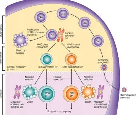
A previous study, using mice engineered to express monoclonal T cells with or without monoclonal B cells, found that mice producing both T cells and B cells (called monoclonal B-T mice or MBT mice) have 2-fold more thymocytes and 35-fold more peripheral CD4+ T cells than mice that make only T cells (called monoclonal T cell mice or MT mice) [19]. One possible explanation for these observations is that B cells might in some way promote the development of T cells. This work launched further investigations on T-B relationships during cell development.
to promote T cell development (reviewed by Hergen Spits in [20]). The process of T cell development includes not only the generation of thymocytes but also their maturation to promote the ability to recognize self-MHC without autoimmunity. TCR genes recombine to yield approximately 1015 different TCRs [21]. From these TCRs, only around 108 to 1011 are selected in humans [22, 23] and around 106 in mice [24]. This process is called
positive selection and allows the survival of the thymocytes in which TCRs recognize self-peptides presented by self-MHC molecules with an intermediate affinity. In contrast, thymocytes with TCRs that recognize self-peptides with a high affinity are negatively selected and die by apoptosis, protecting the organism from autoimmunity. Also, thymocytes which TCRs not recognizing self peptides-MHC complexes or recognizing them with a very low affinity die by lack of stimulus via the TCR (neglect). The fact that each T cell precursor, through its TCR, can only recognize self-peptides presented by self-MHC molecules is named “restriction” [25, 26]. These two processes (selection and restriction) warrant that only T cells that recognize the self and are not highly self-reactive are “selected”, and survive to egress to the periphery.
General Introduction
do not express either CD4 or CD8 and are called double negative (DN). In this phase, thymocytes rearrange the TCR β chain and undergo proliferation and expansion. After rearranging the β chain, through a stochastic process of VDJ gene segment recombination, thymocytes begin to express membrane CD4 and CD8 molecules, becoming double positives (DP) (see Figure 1).
Figure 1 – T cells development in the thymus.
The T cells precursors enter the thymus by the cortico-medullar junction and, in contact with thymic epithelial cells, undergo processes of selection and restriction in order to survive. Before leaving the thymus as mature/naïve T cells, the T cells’ precursors rearrange the DNA encoding the TCR chains and acquire different membrane markers.
Arstila el al. calculated that the
β
chain rearrangements can originate at least 106 differentβ
chains and theα
chain rearrangement can originate arepertoire of ~0.5 × 10⁶ different chains [22]. These different
α
andβ
chainspair together to form a heterodimer; it is estimated that one
β
chain pairs, on average, with 25 differentα
chains, generating a final T cell receptor repertoire which diversity was predicted to be around 107 in humans [22].Since only one percent of thymocytes that begin development reach maturity, the critical step in modelling the T cell repertoire must be positive selection. Positive selection must select T cells capable of recognizing self-MHC with intermediate affinity and these selected T cells also must be capable of recognizing “foreign” peptides associated with that MHC. Thus, as a consequence of T cell development, T cells should be able to interact with peptides from the range of bacteria, viruses and toxins that threaten health. It is generally believed that the combined diversity of the T cell repertoire and positive selection process will enable a response towards any pathogenic “challenge”.
hypothesis is that TCR cross-react with many peptides. However, too much cross reactivity between TCR molecules would promote autoimmunity. Thus, although it happens, cross reactivity per se should be limited.
Immune Reconstitution after autologous hematopoietic stem
cells transplantation and after treatment with rituximab.
The cases of Non-Hodgkin Lymphomas and Multiple Myeloma
There are several neoplastic diseases where high-dose treatments have proved to be more effective than conventional dose therapies in what concerns response rate, progression free survival and overall survival. This is the case of mantle cell lymphoma and multiple myeloma that were studied in Chapter VI, VII, and VIII. When extremely high doses of chemotherapy and/or radiotherapy are administered, fatal hematopoietic toxicity needs to be prevented by the infusion of hematopoietic stem cells, either autologous or allogeneic. Allogeneic hematopoietic stem cells have the advantage of the immunologic effect of graft versus tumor, through the recognition of different HLA class I and II antigens and co-stimulatory molecules on tumor cells that lead to the generation of alloreactive T cells [28] .
faster. There is no opportunity for graft-versus-host disease, since the donor and recipient are the same.
Allogeneic HSC donors must have major histocompatibility antigens that, at least partially, match the recipient’s. Even when a good match is found, the recipient will require high levels of immunosuppression to prevent graft-versus-host disease. Allogeneic transplant donors may be related (sibling) or unrelated volunteers. Allogeneic stem cell transplantation aims, not only to the rescue of hematopoiesis, but also to improve the immunological effect graft versus tumor, as a means of promoting engraftment, accelerating immune recovery and strengthening the anti-neoplastic effects.
High dose chemotherapy followed by autologous hematopoietic stem cell transplantation (ASCT) has been associated with prolonged disease-free survival in numerous malignancies including relapsed non-Hodgkin’s lymphoma [29], acute myelogenous leukemia [30], multiple myeloma [31] and a number of solid tumors [32, 33]. Approximately 105,000 autologous hematopoietic stem cell transplants were performed until 2004 for malignant diseases based on several American registries published by the Center of International Blood and Marrow Transplant Research [34]. Of those, 27,419 were performed for NHL and 20,423 for plasma cell disorders [34].
that comprise 90% of those entities, includes indolent (mostly follicular, lymphocytic, marginal zone and lymphoplasmocytic) and aggressive (mostly diffuse large cell, mantle cell, Burkitt, and lymphoblastic) histological forms [35]. Indolent diseases are generally incurable but follow a prolonged course, with frequent need for therapy, while aggressive forms can be cured in a significant proportion but have a global shorter survival. Currently, treatment for most forms of B-cell NHL at diagnosis and/or at relapse includes chemotherapy associated with the anti B-cell monoclonal antibody Rituximab, a chimeric murine/human monoclonal antibody directed to the CD20 antigen that has been shown to induce complement- and antibody-dependent cell mediated lysis, as well as apoptosis of tumor cells and sensitization to the cytotoxic effects of anti-neoplastic drugs [36]. Another treatment option for Non Hodgkin lymphomas is high dose chemotherapy followed by hematopoietic stem cells transplant (HSCT), autologous or allogeneic. This is usually an option in the case of aggressive non-Hodgkin lymphomas, as it is the case of diffuse large B cell and mantle cell lymphomas.
the treatment of choice for the fit and young patients [31, 41, 42]. The use of allogeneic stem cell transplants to exploit the effect of graft versus myeloma effect has been recently shown effective, as survival of patients with newly diagnosed myeloma is superior among recipients of a hematopoietic stem-cell autograft followed by a stem-cell allograft from an HLA-identical sibling compared to recipients of tandem stem-cell autografts [42].
Even so, the clinical efficacy of ASCT is limited by delayed immune recovery, resulting in infectious complications and probably contributing to high tumor relapse rates (40 to 70%) [43-45].
Besides autologous stem cell transplantation many other treatment options exist to treat hematological malignancies. These options include chemotherapy, radiotherapy, radioimmunotherapy and biological agents such as antibodies, intracellular molecules inhibitors and immunomodulators. As previously mentioned, for the most common subtypes of non-Hodgkin lymphomas, treatment at diagnosis and/or at relapse includes poly-chemotherapy regimens associated with the anti B-cell monoclonal antibody, Rituximab [46, 47].
As a consequence, circulating B cells remain undetectable for at least 6 months after treatment and recover to normal levels between 6 and 9 months later [49, 50]. The homeostatic and clinical consequences of these changes are not yet clear.
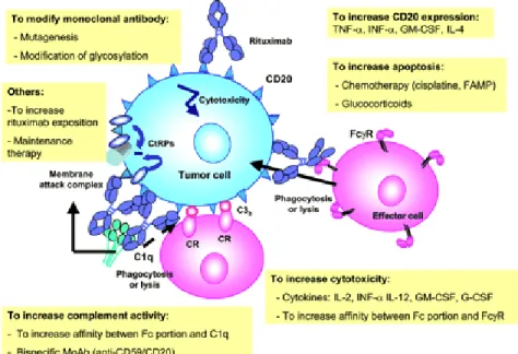
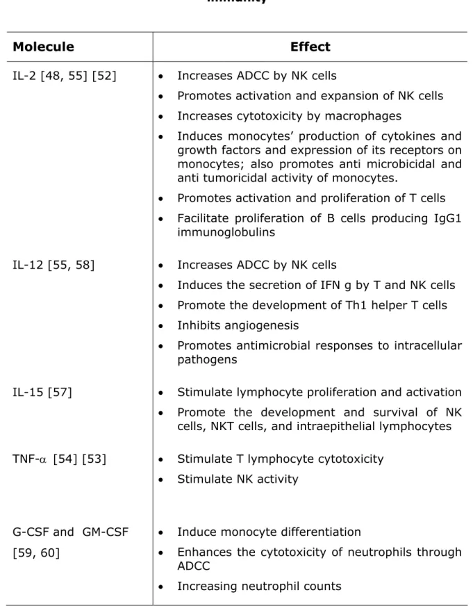
Several ways to simultaneously increase the activity of rituximab and to stimulate immunity have been tried exploring the role of cytokines and growth factors like IL-2, IL-4, IL-12, IL-15, TNF-α, G-CSF, GM-CSF [48, 51-55] [56, 57]. These molecules act on T and B cells, macrophages and NK cells activating its proliferation and activity.
The effects of some of these immunomodulators are outlined in Figure 2 and listed in Table 1.
Figure 2 - Mechanisms of action of rituximab and ways to
increase its clinical efficacy. FAMP indicates fludarabine
Table 1 – Cytokines and growth factors exploited to increase patients immunity
Molecule Effect
IL-2 [48, 55] [52] • Increases ADCC by NK cells• Promotes activation and expansion of NK cells
• Increases cytotoxicity by macrophages
• Induces monocytes’ production of cytokines and growth factors and expression of its receptors on monocytes; also promotes anti microbicidal and anti tumoricidal activity of monocytes.
• Promotes activation and proliferation of T cells
• Facilitate proliferation of B cells producing IgG1 immunoglobulins
IL-12 [55, 58] • Increases ADCC by NK cells
• Induces the secretion of IFN g by T and NK cells
• Promote the development of Th1 helper T cells
• Inhibits angiogenesis
• Promotes antimicrobial responses to intracellular pathogens
IL-15 [57] • Stimulate lymphocyte proliferation and activation
• Promote the development and survival of NK cells, NKT cells, and intraepithelial lymphocytes TNF-α [54] [53] • Stimulate T lymphocyte cytotoxicity
• Stimulate NK activity
G-CSF and GM-CSF [59, 60]
• Induce monocyte differentiation
• Enhances the cytotoxicity of neutrophils through ADCC
As previously explained, the ability of the immune system to cope with diverse and fast evolving pathogens is achieved by the development of a vast and diverse repertoire of antigen receptors in lymphocytes that can potentially recognize any possible foreign antigens. Since B cell NHL are neoplasms of immune system, this function is usually altered. Furthermore, the majority of treatments options per se alter the dynamics of the immune system and cause a profound impact, in general decreasing the immunity. In fact, NHL patients frequently demonstrate clinical and biological signs of immune suppression before treatment. Chemotherapy alone strongly contributes to lymphocytopenia, significantly depleting circulating T cells, which recovery depends on multiple factors, including the intensity of the regimens used.
This finding, in part, explains the patients’ susceptibility to infections for a prolonged period of time post-transplant.
The reconstitution of the immune system after myelotoxic therapies has been mostly studied in the setting of hematopoietic transplantation. It may be analyzed from two distinct points of view: (1) the numerical recovery of cellular elements; and (2) the functional recovery of cellular interactions. Following hematopoietic stem cells transplantation there is a relatively rapid reappearance of hematopoietic and lymphoid cells. However, functional recovery of immune cells occurs at a much slower rate (Table 2). Complete reconstitution of the normal humoral and cellular (T and B cell) immunity may be delayed beyond one year following transplantation or, in certain settings, it may never fully recover, specially in what concerns the diversity of the T and B cell repertoire [66-71].
Table 2 - Reconstitution of B and T cell numbers and function after ASCT (adapted from [74])
B-cell Reconstitution Post-ASCT T-cell Reconstitution Post-ASCT
Circulating B cells
Low from 3 to 18 months
Circulating T cells
Serum CD4 Low for years
IgM Low for up to 6
months
CD8 Low for 3 to 18 months
IgG Low from 12 to
18 months
T-cell proliferation Low for 3
months to 5 years
IgA Low for up to 36
months
Cytokine production
Low for 6 months to 5 years
I n vivo antibody response:
Response to IL-2 Low for 7
months to 5 years
T dependent antigen
Low for months to years
Cytotoxic T cells Low for 2
months to 5 years
T independent antigen
Low for years
I n vit r o B cell responses to polyclonal stimulator
Low for months to years
activated killer cells and cytotoxicity [75-78].
The study of immune reconstitution after treatment with rituximab is limited to a few very recent published studies. Those studies report that, after treatment with the monoclonal antibody and during the reconstitution phase (3 to 15 months), the majority of the peripheral blood B cells have both an immature, naïve phenotype (CD38hiCD27-CD24hiIgD-/+) and function, being less responsive to proliferative stimuli and more prone to apoptosis when in culture [49, 50, 79, 80]. Studies of the impact on T cell immunity after treatment with rituximab are very scarce in the literature. It was recently published that a significant increase in activated CD4+ and CD8+ T cells, as well as CD25brightFOXP3+ regulatory cells, happens after treatment with rituximab [81].
Previous studies of our group at the Mayo Clinic have shown that early recovery of lymphocytes post-ASCT is critical to the clinical outcome (disease free and overall survival) in several hematological and non-hematological malignancies [82-85]. In agreement with others [86, 87], the analysis of the recovery of lymphocyte numbers at day 15 post-transplant provided evidence that timely host immune system recovery may impact on human tumor cell biology. Therefore, the therapeutic goal in ASCT should be to maximize hematopoietic engraftment and to improve immunological recovery. Effective methods that allow an accelerated immune reconstitution
The possibility of manipulating the immune system with IvIg
infusion
The therapeutic applications of human immunoglobulins have been investigated for decades. The largest clinical impact of intravenous polyclonal immunoglobulin therapy (IVIG) probably occurrs in the setting of immunodeficiency [88-90], autoimmunity and inflammation [91, 92] and infectious disorders [93, 94] [95]. Its rationale was based on studies of the mechanisms of action of the molecule, that justified these applications [96, 97].
The immunomodulatory effects of immunoglobulins are also related to several well established actions, including the modulation of the expression and function of Fc receptors, the interference with complement activation and cytokine networks, the provision of anti-idiotypic antibodies and the modification of the activation, differentiation and effector functions of T and B lymphocytes cells and dendritic cells [91, 98].
peripheral lymphoid organs [103], and negative effects on regulatory T cells [104].
Regarding the effect of B cells on lymphoid organogenesis, it is recognized that expression of CXCL13, a B cell chemokine, can generate organized lymphoid tissue including B cells and T cells [105]. B cell expression of LTα1β2 contributes to the development and migration of follicular dendritic cells [106, 107]. B cell expression of LTα1β2 is also necessary for the development of T cell zones in the spleen [108]. In the same way, differentiation of the “follicular-associated epithelium” in gut associated lymphoid tissue requires the presence of B cells [109].
It has been recently suggested that B cells have a role on the regulation of regulatory T cells. Some reports refer a positive effect of B cells via B7 on stimulating regulatory T cells [110], while others point to a negative effect of the B cell population, which may block regulatory T cells effect [104]. This is a novel field of research deserving further investigation.
Measurement of T cell receptor diversity
T and B cells generate diverse antigen-specific receptors through gene recombination; both cell repertoires are shaped during their development by recognizing self-antigens. Several estimations of the T and B cells repertoire diversity have been accomplished using various available methods. However, the different methodologies have their specific limitations, varying with each technical tool applied.
The availability of an accurate instrument to measure the diversity of the T cell repertoire is essential to examine the complexity of the immune system, either to understand its ability to recognize a diverse range of antigen determinants or to assess its limitation in the setting of lymphopenia and other situations of repertoire contractions.
Also, the availability of an accurate and direct measurement of T cell receptor repertoire is crucial for the comparison of different T cell receptor population’s repertoires and the testing of different approaches to improve T cell development, in particular the generation of TCR diversity.
diversity is based on Cot analysis (a method based on the principle that the time required for a DNA sample to re-anneal is related to the sequence complexity of the sample) and PCR, may lead to serious decreases in specificity due to possible erroneous amplifications of impure PCR products. Also, amplification cycles might decrease the diversity of the final PCR product, owing to the loss of sequences that are amplified less efficiently; the consequence may be an erroneous assessment of the true cellular diversity. Another method used to assess the T cell repertoire diversity is the frequency analysis of a given TCR clone based on techniques of limiting dilution [124]. This method is quite laborious and based on the not yet proven hypothesis that the frequency of a certain clonotype is representative of the frequency of all clones.
Lastly, a strategy frequently used in clinical research to access diversity is the analysis of lymphocyte populations by flow cytometry [125, 126]. This method is even less sensitive to detect alterations than the PCR-based methods, since the antibodies employed detect only the “constant” antigenic determinants shared by many lymphocyte receptor clones. Diversity evaluated by flow cytometry is, at best, inferred from the result.
In conclusion, several are the methods available to assess repertoire diversity but a direct, accurate, quantitative and easily used method is essential to examine and better understand the complexity of the immune system, both in physiologic and pathological situations, as the work
Aims of the study
The aims of this work and the hypothesis underneath these objectives were:
1) To develop a quantitative method to directly assess the T cell repertoire diversity.
We hypothesize that it is possible to direct assess a cellular repertoire diversity by hybridization of all lymphocyte receptor-specific RNAs in a given sample to oligonucleotides on a gene chip as the number of sites undergoing hybridization out of the >400,000 available sites on a gene chip corresponds to the level of diversity.
2) To determine the role of polyclonal and monoclonal B cells and immunoglobulins upon T cell development, mostly the generation of the T cell repertoire diversity, using a mouse model where B cells have different levels of BCR diversity.
3) To examine the importance of the T cell repertoire diversity to peripheral T cells function through in vit ro and in vivo testing.
We hypothesize that the function of T cells may be improved if the diversity of the T cell repertoire is increased, since a larger range of distinct antigens would be detected by a more diverse T cell compartment as compared to a less diverse T cell repertoire.
4) To study the impact of lymphocyte and polyclonal immunoglobulins recovery in the survival of patients with certain hematological neoplasms, namely mantle cell lymphoma and multiple myeloma patients subjected to autologous stem cell transplantation.
We hypothesize that the kinetics of the recovery of lymphocyte number during immune reconstitution may predict the robustness of the immune system translating in longer survival of the patients under these conditions; autologous hematopoietic stem cell transplantation provides a useful model to evaluate a regenerating immune system.
5) To analyse the impact of the lymphocytes counts and sub-populations before treatment with rituximab in the progression free survival of patients with non-Hodgkin lymphoma receiving this antibody.
CHAPTER III
Brenda M. Ogle1,2, Marilia Cascalho,1,3,4 Cristina M. Joao1, William Taylor5, Lori J. West6 and Jeffrey L. Platt1,3,4,7
1Transplantation Biology Program, 2Departments of Physiology, 3Immunology
and 4Pediatrics, the 5Cancer Center and 7Surgery Mayo Clinic, Rochester, Minnesota
6The Hospital for Sick Children Research Institute and the University of
Toronto, Toronto, ON, Canada
This work was supported by grants from the National Institutes of Health (HL46810 and HL52297).
3.1. Abstract
The ability to mount an immune defense against infectious microorganisms and their products and against tumors is believed to be a direct function of lymphocyte diversity. Because the diversity of lymphocyte receptor genes is >1000-fold more diverse than the entire genome and varies between genetically identical individuals, measuring lymphocyte diversity has been a daunting challenge. We developed a novel technique for measuring lymphocyte diversity directly using gene chips. We reasoned and here demonstrate that the frequency of hybridization of nucleic acids coding for lymphocyte receptors to the oligonucleotides on a gene chip varies in direct proportion to diversity. We applied the technique to detect changes in lymphocyte diversity in mice with known B cell alterations and in persons with known T cell repertoire defects. This approach is the first to provide direct analysis of lymphocyte receptor diversity and should facilitate fundamental study of the adaptive immune system and clinical efforts to assess immunological diseases. In addition this approach could be more broadly applied for example, to measure diversity of viral quasispecies.
Keywords: lymphocyte antigen receptor gene rearrangement, T
3.2. Introduction
While the total number of lymphocytes in the blood can be directly measured, the diversity of the lymphocyte compartment, on which immunocompetence is based, cannot. In the absence of direct measures of lymphocyte diversity, various indirect means for estimating diversity have been used. For example, antibodies against variable (V)-region families have been used to characterize lymphocyte populations by flow cytometric analysis [125, 127]. Since this approach detects “constant” antigenic determinants shared by many lymphocyte receptor clones, diversity is at best inferred from the result. As another example, nucleic acids encoding lymphocyte receptors can be amplified by PCR using constant region (C) and V-family specific primers [122]. Like FACS analysis, this approach does not differentiate between individual clones of the same family and may fail to detect balanced narrowing (or expansion) of the repertoire.
methodologies effectively detect clonal expansion within a V family; however, because several thousand V-J family combinations for lymphocyte receptors exist, all V-J combinations cannot be analyzed routinely. Since only a small fraction of V-J combinations are analyzed, the choice of which is random, the actual diversity of the TCR repertoire may not be quantified.
Still another means to assess lymphocyte diversity is based on the tenants of limiting dilution analysis and detects the frequency of a given TCR clone [124]. This method is quite laborious and is based on the assumption that the frequency of the selected clonotype is representative of the frequency of all clones.
3.3. Materials and Methods
3 .3 .1 . I sola t ion of RN A. All mouse strains were raised and maintained with protocols approved by the institutional animal care and use committee of the Mayo Clinic, Rochester, MN. All human samples were obtained in accordance with the institutional review board of the Mayo Clinic, Rochester, MN. Spleens harvested from mice were placed in RPMI and pushed through a 70 μm cell strainer. Lymphocytes were isolated from the resulting suspension of splenocytes or from peripheral blood using Ficoll-paqueTM (Amersham
Biosciences, Piscatanaway, New Jersey) gradient. Total RNA was isolated from the lymphocytes using the Qiagen RNeasy kitTM (Qiagen, Inc., Valencia,
California) per the manufacturer’s instructions. Isolated RNA was resuspended at a concentration of 2 μg/μl.
3 .3 .2 . Ge n e r a t ion of lym ph ocyt e r e ce pt or - spe cific cRN A. First strand cDNA was constructed first as follows. In an RNAse-free microcentrifuge tube, 10 μl of total RNA (20 μg) was mixed with 1 μl (100 pmol/μl) of either:
T7
+C
β
T7+ CJH4
(5’-GGCCAGTGAATTGTAATACGACTCACTATAGGGAGGCGGGAGGAGACGG TGACTGAGGTTCCTTG-3’) for B cell receptor analysis.
This mixture was incubated at 70°C for 10 minutes followed by a quick spin and chill on ice. To this reaction, 4 μl of 5X first strand cDNA buffer (Invitrogen, Inc., Carlsbad, California), 2 μl of 0.1 M DTT, and 1 μl of 10 mM dNTP mix were added and incubated at 37°C for 2 minutes. Next, 2 μl of SuperScript II Reverse TranscriptaseTM (Invitrogen, Inc.) was added and the
total mixture was further incubated at 37°C for 1 hour. Following incubation, the first strand product was placed on ice. For second strand synthesis, the following reagents were added to the first strand product: 91 μl of DEPC-treated water, 30 μl of 5X Second Strand Reaction Buffer (Invitrogen, Inc.), 3
instructions. The purified product was quantified using spectrophotometric analysis applying the convention that 1 OD at 260 nm equals 40 μg/ml of RNA. cRNA was resuspended at a concentration of 1 μg/μl. cRNA was then fragmented to 50-200 bp sizes by combining with 5 μl of 5X fragmentation buffer (Invitrogen, Inc.) in 15 μl of water. The mixture was incubated at 94°C for 35 minutes and put on ice following incubation.
3 .3 .3 . Applica t ion of cRN A t o t h e ge n e ch ip. Equal amounts of cRNA from different samples were hybridized to U95B gene chips (Affymetrix, Inc., Santa Clara, California). While the ideal gene chip might be constructed using random oligonucleotides, we reasoned that chips containing known but unselected expressed sequence tags from human genes would share less homology with mouse lymphocyte receptor RNA and could be used instead. And in fact, duplicate experiments performed on U95C chips yielded comparable results, suggesting that a random oligonucleotide chip may not add benefit.
summed. First, the standard curve was generated (from hybridization of samples with known numbers of different oligos). Next, test samples were assessed and based on the number of hits, the diversity was extrapolated from the standard curve.
3 .3 .5 . ELI SA for de t e ct ion of a n t i- KLH a n t ibodie s. Mice were immunized by intraperitoneal injection with 25 μg of KLH (Keyhole Limpet Hemocyanin, Sigma, St. Louis, MO) in incomplete Freund’s adjuvant. A boost of 10 μg of KLH was administered 20 days later. After an additional two weeks the mice were sacrificed and serum and splenocytes were isolated. Purified KLH (3 μg/ml in phosphate buffered saline (PBS); 50 μl/well) was added to wells of ninety-six well flat bottom microtiter plates (Nunc-Immuno 96 Micro well – MaxisorpTM, Nalge Nunc International, Rochester, NY). ELISA was developed as described [129]. Plates were read using a microplate reader (Power Wave XTM, Bio Tek Instruments, Winooski, VT) and analyzed
using KC4 – Kineticalc software. Samples were analyzed in triplicate in three independent experiments.
at a value of p < 0.05. Immune responsiveness was compared between control (C57) and various mutant mice (QM, JH-/-) using unpaired Student’s t
3.4. Results
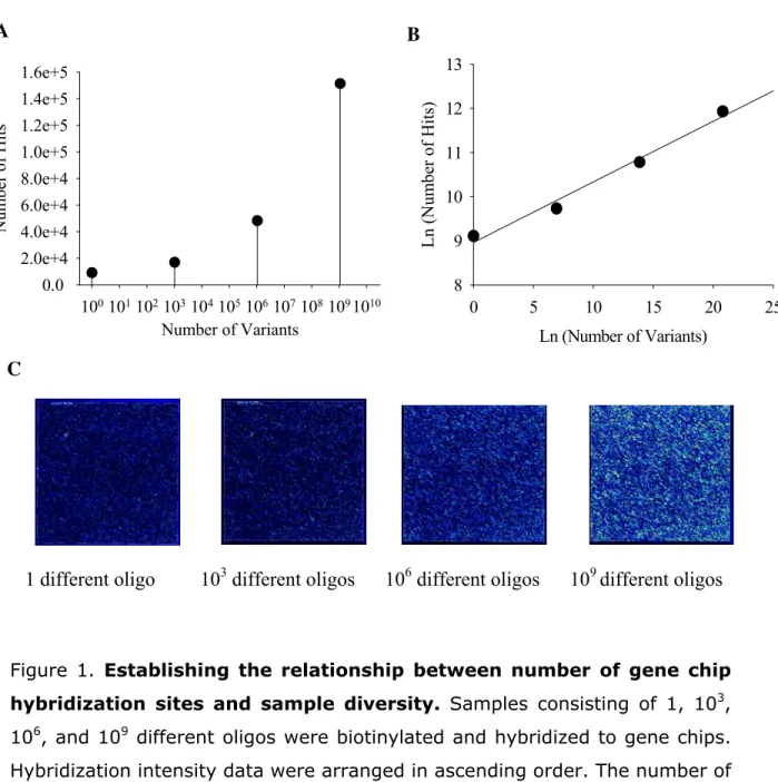
1 different oligo 10
3different oligos 10
6different oligos 10
9different oligos
Figure 1. Establishing the relationship between number of gene chip
hybridization sites and sample diversity. Samples consisting of 1, 103,
106, and 109 different oligos were biotinylated and hybridized to gene chips. Hybridization intensity data were arranged in ascending order. The number of probe locations with intensity above background (i.e., number of hits) was summed and compared to the number of different oligos initially applied to the gene chip (i.e., number of variants) (a) Relationship between number of hits and number of variants. The number of hits increases with the number of variants, indicating that the human gene chip can be used to detect random oligos. (b) Linear relationship between number of hits and number of
Number of Variants
1001011021031041051061071081091010
Number of Hits 0.0 2.0e+4 4.0e+4 6.0e+4 8.0e+4 1.0e+5 1.2e+5 1.4e+5 1.6e+5
Ln (Number of Variants)
0 5 10 15 20 25
L n (Num b er of Hits ) 8 9 10 11 12 13
A
B
variants. The natural log of both axes yielded a linear relationship between hits and variants. (c) Visual hybridization of random oligos to gene chips. Scans of the gene chips afford rapid inspection of the "hit" profile.
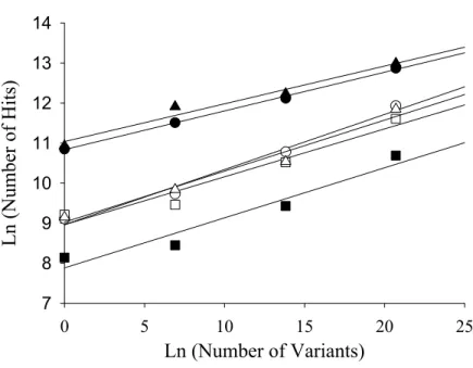
Figure 2. Reproducibility of method for analysis of receptor diversity.
Samples obtained as described in Figure 1 were studied in six separate experiments (black circle,-●-)(white circle, -○-)(black square, -■-)(white square, -□-)(black triangle, -▲-)(white triangle, -∆-) to test reproducibility.
Ln (Number of Variants)
0 5 10 15 20 25
L
n
(N
umb
er of H
it
s)
The slopes of the standard curves were the same statistically; however, the y intercept varied from experiment to experiment.
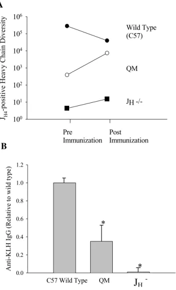
To test whether variations in lymphocyte diversity could be measured directly, we the method to the study of B cells in mice. We used murine B cells for this purpose because diversity of these cells can be measured, at least in principle, through analysis of Ig gene expression and because we have available mutant mice with defined variations in B cell antigen receptor repertoire.
We compared diversity of B cell antigen receptors in wild type (C57Bl/6) mice with the diversity of B cell receptors in quasi-monoclonal (QM) and JH-/- mice.
The QM mice were generated by gene-targeted replacement of the endogenous JH elements with a VDJ rearranged region from a
of the J kappa gene segments and, therefore, cannot assemble Ig heavy or
kappa light chains [131]. These animals are B cell deficient although they do have surface Ig-negative precursor B cells (B220+/CD43+ pro-B cells) that assemble lambda light chain genes at a low level.
To test changes in lymphocyte diversity, we compared gene chip hybridization of B cell heavy chain-specific cRNA from splenocytes of wild type, QM and JH-/- mice. The heavy chain specificity of the cRNA was gained
via generation of first strand cDNA from isolated RNA using a JH4-specific
custom primer. A primer to the JH4 region of the heavy chain was used to
detect diversity in one region of the heavy chain. While the results do not represent diversity of the entire heavy chain population they might provide a baseline from which a change could be detected. Following second strand DNA synthesis, the double-stranded product was biotinylated via in vit ro
transcription (IVT). The IVT product (cRNA) was purified and 10 μg of each sample and 10 μg of each standard were hybridized to individual gene chips. The hybridization intensities obtained from the JH-/- mice (which lack B cell
receptors) were used to set the background threshold, above which hybridization sites (hits) were counted. Sample diversity was extrapolated from the standard curve.
As the results shown indicate, wild type mice expressed more than 105 (2.8 x
105) different B cell J
expected, QM JH4-positive heavy chain diversity was much less than wild type
diversity, though well above background levels.
We then tested the ability of this system to detect changes in diversity following antigenic challenge with keyhole limpet hemocyanin (KLH).
Following immunization with proteins such as KLH, heavy chain diversity is thought to decrease due to oligoclonal expansion of high affinity clones. In contrast, immunization of QM mice with KLH (an antigen that does not bind with the QM antibody) causes diverse non-QM B cells to expand at the expense of the predominant QM B cells, thereby increasing heavy chain diversity [132]. Consistent with these theoretical concepts, JH4-positive heavy
chain diversity in wild type mice decreased by greater than one order of magnitude, from 2.8 x 10⁵ to 4.0 x 10⁴, and diversity in QM mice increased (7.5 x 10³) following immunization with KLH
Figure 3. Analysis of B cell diversity using the gene chip method. Splenocytes were harvested from 3-4 week old JH-/-, QM and WT mice and
mononuclear cells were isolated on Ficoll-paque gradients. Total RNA was isolated from the lymphocytes and first strand cDNA was generated using a primer designed to bind the constant region of the mouse heavy chain JH4
region plus the T7 polymerase promoter. The custom primer promoted amplification of JH4-heavy chain-specific RNA only. Equal amounts of the in
A n ti -K LH IgG (R el at iv
e to w
il d ty pe) 0.0 0.2 0.4 0.6 0.8 1.0 1.2
C57 Wild Type QM Jh
-/-*
*
Pre PostImmunization Immunization JH4
-posi
tive Heavy Chain Diversi
![Table 2 - Reconstitution of B and T cell numbers and function after ASCT (adapted from [74])](https://thumb-eu.123doks.com/thumbv2/123dok_br/15745503.637061/50.918.125.800.234.821/table-reconstitution-b-cell-numbers-function-asct-adapted.webp)