FGFR1 is an independent prognostic factor and can
be regulated by miR-497 in gastric cancer progression
Gang Xie, Qi Ke, Yu Zu Ji, An-qun Wang, Meng Jing and li-li Zou
Department of Pathology, Mianyang Central Hospital, Mianyang, Sichuan Province, China
Abstract
Fibroblast growth factor receptor 1 (FGFR1) has been reported in gastric cancer to be a prognostic factor. However, miR-497-targeted FGFR1 has not been explored in the carcinogenesis of gastric cancer. The present study intended to revalidate the prognostic significance of FGFR1 in patients with gastric cancer, and the mechanism of miR-497-regulated FGFR1 was investigated in gastric cancer cell proliferation and apoptosis. The messenger RNA (mRNA) and protein levels were assayed by RT-qPCR and western blotting, respectively. The targeted genes were predicted by a bioinformatics algorithm and confirmed by a dual luciferase reporter assay. Cell proliferation was analyzed by CCK-8 assay. Annexin V-FITC/PI staining was used to evaluate the apoptosis in AGS and SGC-7901 cells. FGFR1 was frequently up-regulated in gastric cancer tissues and associated with poor overall survival in patients with gastric cancer. Interestingly, FGFR1 loss-of-function resulted in a significant growth inhibition and apoptosis in AGS and SGC-7901 cells. In addition, we found that miR-497 was inhibited in gastric cancer tissues and cell lines, while overexpression of miR-497 could suppress proliferation and induce apoptosis in AGS and SGC-7901 cells. Importantly, bioinformatics analysis and experimental data suggested that FGFR1 was a direct target of miR-497, which could inhibit FGFR1 expression when transfected with miR-497 mimics. Furthermore, we found that overexpression of FGFR1 reversed the growth inhibition and apoptosis of miR-497 mimics in AGS and SGC-7901 cells. Thesefindings suggested that overexpression of miR-497 inhibited proliferation and induced apoptosis in gastric cancer through the suppression of FGFR1.
Key words: FGFR1; miR-497; Apoptosis; Post-transcriptional; Gastric cancer
Introduction
The fibroblast growth factor receptor (FGFR) family is composed of four members, FGFR1/2/3/4, which are activated by interacting with fibroblast growth factors (FGFs), resulting in the activation of downstream signal-ing pathways includsignal-ing mitogen-activated protein kinase signal transducer and activator of transcription and PI3K/ Akt/mTOR signaling (1,2). FGFR1 belongs to receptor tyrosine kinases and possesses various biological func-tions, such as proliferation, survival, migration, and dif-ferentiation (3). FGFR1 is expressed on a number of different cell types including tumor cells (4,5). Intrigu-ingly, overexpression of FGFR1 has been observed in multiple human malignancies (6–8). A meta-analysis
and clinical survey suggest that FGFR1 is emerging as a diagnostic and prognostic marker for non-small-cell lung cancer (NSCLC) (9,10). However, its functional con-tributions have not been completely clarified in gastric cancer.
MicroRNAs (miRNAs) are short, noncoding, single-stranded RNAs (18-25 nucleotides) that have been verified to suppress protein translation by binding to the 30-untranslated regions of target gene (11). miRNAs
function as a novel class of post-transcriptional reg-ulators participating in a variety of biological processes, including angiogenesis, tumor proliferation, and metas-tasis (12). For example, miR-338-3p inhibits migration and induces apoptosis in gastric cancer cells by blocking protein-tyrosine phosphatase 1B expression (13). miR-148b-3p inhibits gastric cancer metastasis by inhibiting the Dock6/ Rac1/Cdc42 axis (14). However, the underlying molecular mechanism of miR-497 in gastric cancer progression by targeting FGFR1 is rarely investigated.
miR-497 belongs to the miR-15 family and negatively regulates angiogenesis in tumors (15). Numerous studies have suggested that miR-497 can serve as a tumor suppressor in a variety of tumors, including renal cell carcinoma (16), NSCLC (17), breast cancer (18), and hepatocellular carcinoma (19). In gastric cancer, lncRNA-GACAT3 and XIST promote cell growth and invasion by negatively regulating miR-497 expression (20,21). Over-expression of miR-497 inhibits gastric cancer cells growth and metastasisin vitroandin vivo(22).
In the present study, we aimed to evaluate the prognostic significance of FGFR1 in patients with gastric
Correspondence: An-qun Wang:<anqun_wang11@sina.com> Received June 1, 2018 | Accepted September 21, 2018
cancer, and the mechanism of miR-497-regulated FGFR1 in gastric cancer cell proliferation and apoptosis.
Material and Methods
Seventy-four pairs of gastric cancer tissues and adjacent non-tumor tissues were collected from patients who had undergone surgery at the Mianyang Central Hospital (China) between January 2009 and June 2014. All of the patients were recruited according to the histo-pathological evaluation without radiotherapy or chemother-apy before surgical operation. The tissues were immediately stored in liquid nitrogen after surgery. Informed consent forms were obtained from the patients. This study was approved by the Ethics Committee of the Mianyang Central Hospital (China).
Cell culture
Normal human GES-1 cell line andfive gastric cancer cell lines (MGC-803, AGS, HGC-27, SGC-7901, and BGC-823) were purchased from the Cell Bank of Type Culture Collection of Chinese Academy of Sciences (China). Cells were cultured in Dulbecco’s modified Eagle’s medium (DMEM; Invitrogen, USA) with 5% fetal bovine serum (Thermo Scientific HyClone, China), in a humidified incu-bator (Thermo, USA) with 5% CO2, 95% air.
Cell transfection and plasmid constructs
miR-NC and miR-497 were synthesized by RiboBio (China). Cells were seeded in 6-well plates and transfected with miR-NC and miR-497 using Lipofectamine 2000 (Invitrogen; Thermo Fisher Scientifc, Inc., USA) for 48 h at 37°C according to the manufacturer’s protocol.
Short hairpin RNA (shRNA) was constructed to spe-cifically target FGFR1 by shRNA design tools (http:// rnaidesigner.thermofisher.com/rnaiexpress/). Using BLAST (http://blast.ncbi.nlm.nih.gov/Blast.cgi), we verified that the designed shRNA targeted only the FGFR1. Sh-FGFR1 and sh-NC were synthesized by GenePharma (China).
A mammalian expression plasmid (pReceiver-M02-ERBB3) designed to specially express the full-length open reading frame of human FGFR1 without miR-497 respon-sive 30-UTR was purchased from GeneCopoeia (USA).
An empty plasmid served as a negative control. Over-expressed FGFR1 plasmid (vector-FGFR1) and control (vector-NC) were transfected into cells using Lipofecta-mine 2000 (Invitrogen; Thermo Fisher Scientifc, Inc.) for 48 h at 37°C according to the manufacturer’s protocol.
CCK-8 assay
The cell viability of AGS and SGC-7901 cells (1104) was detected using a CCK-8 assay kit (Japan). The absorbance was measured at 450 nm using a Spectra Max M5 plate reader (Molecular Devices, USA). The CCK-8 proliferation assay was performed as previously described (23).
Flow cytometry for apoptosis
AGS and SGC-7901 cells were incubated with different conditions for 48 h. Annexin V-FITC/PI kit (Becton, Dickinson and Company, USA) was used to stain cells for 15 min, and then cell apoptosis assay was performed by flow cytometry assay (FACScan, BD Biosciences, USA) and analyzed by CELL Quest 3.0 software (BD Biosciences).
Luciferase reporter gene assay
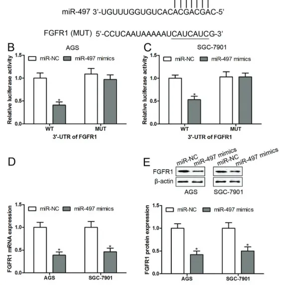
The putative binding sites between miR-497 and FGFR1 were obtained using the online prediction soft-wares Targetscan (www.targetscan.org) and miRanda (www.microrna.org). The wild-type (WT) and mutant-type (MUT) 30-UTR of FGFR1 (0.5 mg) were inserted into the
multiple cloning sites of the luciferase expressing pMIR-REPORT vector (Ambion; Thermo Fisher Scientific, Inc.) and co-transfected with miR-NC or miR-497 (100 nM), and then luciferase activity was evaluated with luciferase reporter assay kit (Beyotime Institute of Biotechnology, China) according to the manufacturer’s protocol.
Immunohistochemical staining
The paraffin-embedded tumor tissues were cut into 3-mm sections and mounted on glass slides for staining with immunoperoxidase, and the procedures of immuno-histochemical staining of FGFR1 (Cat. No. ab59194; dilution: 1:50; Abcam, UK) were performed as previously described (24). The slides were visualized under a microscope (Leica DM 2500; Leica, Germany). Image Pro-Plus 6 software (Media Cybernetics, Inc., USA) was used for the analysis of the integrated optical density in the tumor tissues and corresponding adjacent normal tissues of immunohistochemical positive staining.
Reverse transcription-quantitative polymerase chain reaction (RT-qPCR)
miRNA RT-qPCR: The total RNA was isolated using RNAiso (Takara, China). miRNA was subsequently reverse-transcribed to cDNA using the miRNA-specific stem-loop reverse-transcription primer (Ribobio, China). The relative expression levels of miRNA were calculated using the 2-DDCt method (25) and normalized to the internal control U6, using miRNA-specific primers (RiboBio, China). The reaction conditions were performed according to the instructions from Ribobio Co., Ltd with SYBR Green qPCR Mix (BioRad, USA).
the 2-DDCtmethod (25) and normalized to glyceraldehyde 3-phosphate dehydrogenase (GAPDH). The primers were used as follows: FGFR1: forward primer 5’-CACATCGA GGTGAACGGGAGTAAG-30and reverse primer 50-CGCA
TCCTCAAAGGAGACATTCC-30; GAPDH: forward primer
50-GCACCGTCAAGCTGAGAAC-30and reverse primer 50
-TGGTGAAGACGCCAGTGGA-30.
Western blotting
Proteins were extracted with radio immunoprecipita-tion assay (RIPA) buffer (Cat. No. P0013B; Beyotime Institute of Biotechnology, China). Fifty micrograms of protein for each sample was separated on a 10% SDS-PAGE gel and transferred to nitrocellulose membranes (BioRad Laboratories, Inc., USA). After blocking with 5% non-fat dry milk at room temperature for 2 h, the membranes were incubated with the primary antibody of FGFR1 (Cat. No. ab59194; dilution: 1: 2,000; Abcam, UK) at room temperature for 2 h. Following three washes with Tris-HCL-buffered saline with Tween-20 (TBST), the mem-branes were incubated with the appropriate horseradish peroxidase-conjugated secondary antibody (cat.no: sc-516102; dilution: 1:10,000; Santa Cruz Biotechnology, USA) at room temperature for 2 h and visualized by chemilumi-nescence (Thermo Fisher Scientific, Inc.).b-actin (Cat. No. sc-130065; 1: 2,000; Santa Cruz Biotechnology) was used as the control antibody. Signals were analyzed with Quantity Onessoftware version 4.5 (Bio Rad Laboratories, Inc.).
Statistical analysis
Data are reported as means±SE. Statistical analysis
was performed using IBM SPSS Statistics Version 19.0 (USA) and GraphPad Prism Version 7.0 (USA). Student’s
t-test was used to analyze two-group differences. Inter-group differences were analyzed by one-way analysis of variance, followed by a post-hoc Tukey’s test for multiple comparisons. Overall survival was calculated using the Kaplan-Meier method with the log-rank test applied for comparison. Spearman’s rank analysis was used to identify the correlation between the expression levels of miR-497 and FGFR1 gastric cancer. P values less than 0.05 were considered to indicate a statistically significant difference.
Results
FGFR1 was up-regulated in gastric cancer and associated with poor prognosis
FGFR1 signaling plays a crucial role in maintaining cancer properties, such as cell survival, angiogenesis, and metastasis, in a variety of cancers (8,26). Several studies have reported that FGFR1 is overexpressed in gastric cancer tissues and may be considered as a prognostic factor (7,27,28). Consistent with these results, our findings demonstrated that the expression of FGFR1 was increased in 82.4% (63/74) of patients with gastric cancer (Figure 1A). In addition, the mRNA levels of FGFR1
were significantly up-regulated in the gastric cancer tissues compared to the corresponding adjacent normal tissues (Figure 1B). The immunohistochemical staining analysis also showed a significant increase in the levels of FGFR1 in tumor tissues compared with normal tissues (Figure 1C and D). In addition, the association between FGFR1 mRNA expression and overall survival prognosis was evaluated by the Kaplan-Meier survival curve in patients with gastric cancer. Patients were segregated into a high expression group and a low expression group according to the fold-change of FGFR1 in tumor tissue to normal tissue more than 2-fold. The findings showed that patients with high mRNA expression of FGFR1 resulted in a significantly poorer prognosis compared with patients with a low level of FGFR1 (Figure 1E). We also found that high expression of FGFR1 was correlated with TNM stages, lymph nodes metastasis, and distant metastasis or recurrence, but not correlated with gender, age, and tumor size (Table 1).
FGFR1 knockout inhibited proliferation and induced apoptosis in gastric cancer cells
FGFR1 is implicated in the carcinogenesis of gastric cancer, and FGFR1 inhibitor, L16H50, and L6123 can suppress cell proliferation and migration in gastric cancer cells (6,29). In our study, shRNA was designed to inhibit FGFR1 expression by post-transcriptional gene silencing to explore the functions of FGFR1 in gastric cancer cell proliferation and apoptosis. We found that FGFR1 mRNA and protein levels were significantly up-regulated in all of the gastric cancer cell lines compared with normal GES-1 cells (Figure 2A and B). Intriguingly, the mRNA and protein levels of FGFR1 were markedly higher in AGS and SGC-7901 cells than in MGC-803, HGC-27, and BGC-823 (Figure 2A and B). Hence, AGS and SGC-7901 cells were used on our further experiments. Sub-sequently, sh-NC or sh-FGFR1 was transfected into AGS and SGC-7901 cells. The results demonstrated that both mRNA and protein expression of FGFR1 were signifi -cantly declined with sh-FGFR1 transfection compared with sh-NC administration (Figure 2C); the inhibition rate of sh-FGFR1 to FGFR1 expression was 80%. After trans-fected with sh-NC or sh-FGFR1, the cell proliferation and apoptosis were measured by CCK-8 and annexin V-FITC/ PI staining assay, respectively. The results demonstrated that transfection of sh-FGFR1 suppressed proliferation (Figure 2D) and induced apoptosis (Figure 2E) in AGS and SGC-7901 cells.
Overexpression of miR-497 inhibited proliferation and induced apoptosis in gastric cancer cells
SGC-7901 cells to measure the effect of miR-497 on cell proliferation and apoptosis. First, we found that transfec-tion with miR-497 mimics led to an increase of miR-497 expression in AGS and SGC-7901 cells (Figure 3C). Further investigations showed that overexpressed miR-497 significantly inhibited cell proliferation (Figure 3D and E) and induced apoptosis (Figure 3F, G and H) in AGS and SGC-7901 cells compared with the control group.
FGFR1 was a direct target of miR-497
Based on Targetscan and miRanda, we found the 30-UTR of FGFR1 contained a putative target sequence to
bind with miR-497 (Figure 4A). To confirm these, either the WT or MUT sequence of ACE was transfected into the luciferase-reporter plasmids, and then the plasmids were co-transfected with miR-497 mimics into AGS and SGC-7901 cells. We found that miR-497 mimics were able to
decrease the luciferase enzyme activity in AGS and SGC-7901 cells transfected with WT 30-UTR-reporter, while
miR-497 mimics had no obvious effect on the luciferase enzyme activity in AGS and SGC-7901 cells transfected with MUT 30-UTR-reporter (Figure 4B and C).
Consis-tently, overexpression of miR-497 significantly inhibited the mRNA (Figure 4D) and protein (Figure 4E) expression of FGFR1 in AGS and SGC-7901 cells compared with transfected with miR-NC. These results indicated that miR-497 directly suppressed FGFR1 expression by targeting its 30-UTR.
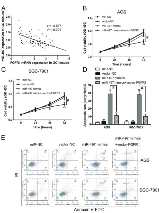
Overexpressed FGFR1 reversed miR-497-induced growth inhibition and apoptosis
Based on the above results, we concluded that FGFR1 might act as an antagonist to miR-497 in gastric cancer cell proliferation and apoptosis. We also found that the expression levels of FGFR1 and miR-497 were inversely correlated in the 74 gastric cancer tissues (r=–0.577,
Po0.001; Figure 5A). To further verify these conclusions,
we performed a co-expression assay with miR-497 mimics and FGFR1 co-transfected into AGS and SGC-7901 cells. The results indicated that overexpression of FGFR1 reversed the growth inhibition (Figure 5B and C) and apoptosis (Figure 5D and E) of miR-497 mimics in AGS and SGC-7901 cells. These findings suggest that over-expression of miR-497 inhibited proliferation and induced apoptosis in gastric cancer through the suppression of FGFR1.
Discussion
In our present study, FGFR1 was up-regulated in gastric cancer tissues and cell lines and associated with poor survival prognosis, metastasis, and TNM stage, suggesting that FGFR1 played a crucial role in the carcinogenesis of gastric cancer. As an oncogene, FGFR1 knockout mediated cell growth inhibition and apoptosis in gastric cancer cells. Importantly, we found a post-transcriptional regulatory mechanism of miR-497 as a tumor suppressor, which had been reported in many cancer types (17,20,30). miR-497 targeted to inhibition of FGFR1 expression blocked gastric cancer cells proliferation and induced apoptosis, indicating that miR-497 and FGFR1 may serve as a potential therapeutic target for the treatment of gastric cancer.
Emerging evidence has suggested that FGFR1 plays crucial roles in the pathological processes of gastric cancer, including cell proliferation, metastasis, recurrence, and poor prognosis (7,27,28). The prognostic significance of FGFR1 in gastric cancer wasfirstly reported by Murase et al. (27) suggesting that high expression of FGFR1 is associated with poor survival in patients with gastric cancer. Subsequently, further study shows that high expression of FGFR1 is associated with tumor progres-sion and survival in diffuse-type gastric cancer, but not in intestinal-type gastric cancer (7). Consistent with those results, we observed that high expression of FGFR1 was also correlated with lymph node metastasis,
Table 1.Correlation between clinicopathological factors andfibroblast growth factor receptor 1 (FGFR1) expression levels in gastric cancer patients.
Variable n FGFR1-Low
(n = 33)
FGFR1-High (n = 41)
P value
Gender 0.974
Male 45 20 25
Female 29 13 16
Age (years) 0.980
o60 36 16 20
^60 38 17 21
Tumor size (cm) 0.284
p3 42 21 21
43 32 12 20
TNM stages 0.013
I-II 33 20 13
III-IV 41 13 28
Distant metastasis or recurrence o0.001
Negative 38 25 13
Positive 36 8 28
Lymph nodes metastasis 0.001
Negative 35 23 12
Positive 39 10 29
recurrence, higher TNM stage, and poor overall survival. However, the comparative analysis of the expression of FGFR1 in gastric cancer tissues and adjacent non-tumor tissues or normal cells and gastric cancer cell lines had not been presented in these studies (7,27). In our study, we found that FGFR1 was significantly
increased in gastric cancer tissues compared to adjacent non-tumor tissues. In addition, the expression of FGFR1 was up-regulated in gastric cancer cell lines compared to normal GES-1 cells. Based on these results, we hypothe-sized that FGFR1 may be an oncogene in the progression of gastric cancer. To verify this conclusion, sh-RNA was
Figure 2.mRNA (A) and protein (B) expression offibroblast growth factor receptor 1 (FGFR1) in normal human GES-1 cells andfive gastric cancer cell lines measured by RT-qPCR and western blotting, respectively. After transfection with shRNA to inhibit FGFR1 expression, the mRNA and protein expression of FGFR1 were measured by RT-PCR and western blotting, respectively (C). Cell viability was measured by CCK-8 assay (D), and cell apoptosis byflow cytometry in AGS and SGC-7901 cells (E). Data are reported as means
used to inhibit the expression of FGFR1in vitro, and found that cell proliferation was inhibited and apoptosis was increased in AGS and SGC-7901 cells after knockout of FGFR1 by si-RNA. Similar results were reported in lung cancer (31), hepatocellular carcinoma (32), and prostate cancer (33). Therefore, wefirmly believe that FGFR1 as an
oncogene can serve as an independent prognostic factor and a very promising therapeutic target for patients with gastric cancer.
Another important finding was that miR-497, known as a tumor suppressor (18), directly targeted the 30-UTR
of FGFR1 to inhibit the process of translation, which is a
classical post-transcriptional mechanism involved in cell growth inhibition and apoptosis in AGS and SGC-7901 cells. A previous study has demonstrated that miR-497 has a lower expression in gastric cancer tissues than non-tumorous tissues (34). In multidrug-resistant human gastric cancer cell line SGC7901, the expression of miR-497 is down-regulated (35). Similarly, Li et al. (22) also showed that miR-497 is decreased in both gastric cancer tissues and cell lines. Our study showed the same outcomes: the expression of miR-497 was inhibited in gastric cancer tissues and cell lines.In vitroandin vivo
observations indicate that restoration of miR-497 can
block the growth of gastric cancer cells (22). We also found that overexpressed miR-497 inhibited gastric cancer cells proliferation and induced apoptosisin vitro. Based on thesefindings, miR-497 may function as a tumor suppres-sor miRNA in gastric cancer.
Our study also found that FGFR1 was a direct target of miR-497, and we were the first to explore the functional roles of miR-497 and FGFR1 in gastric cancer cells proliferation and apoptosis. Overexpression of FGFR1 reversed miR-497 mimics inducing growth inhibition and apoptosis, as well as the expression levels of FGFR1 and miR-497 were inversely correlated in the tumor tissues.
Thus, we proposed the possibility that FGFR1 and miR-497 had exactly the opposite role in the progression and development of gastric cancer.
Taken together, our results showed that miR-497 inhibited FGFR1 expression and consequently induced growth inhibition and apoptosis in gastric cancer cells.
Experimental validations delineated a novel regulatory mechanism that miR-497 could serve as tumor sup-pressor in the progression of gastric cancer by blocking FGFR1 expression. More importantly, miR-497 and FGFR1 might contribute to the development of prognostic indices and therapeutic targets for patients with gastric cancer.
Figure 5.Correlation between miR-497 andfibroblast growth factor receptor 1 (FGFR1) by Spearman’s rank correlation analysis (A). After transfection with miR-497 mimics or miR-497 mimics combined with vector-FGF1, cell viability was measured by CCK-8 assay (B andC), and cell apoptosis was detected byflow cytometry in AGS and SGC-7901 cells (DandE). Data are reported as means±SE. *Po0.05, **Po0.01;#P
References
1. Yuan H, Li ZM, Shao J, Ji WX, Xia W, Lu S. FGF2/FGFR1 regulates autophagy in FGFR1-amplified non-small cell lung cancer cells. J Exp Clin Cancer Res 2017; 36: 72, doi: 10.1186/s13046-017-0534-0.
2. Dieci MV, Arnedos M, Andre F, Soria JC. Fibroblast growth factor receptor inhibitors as a cancer treatment: from a biologic rationale to medical perspectives.Cancer Discov 2013; 3: 264–279, doi: 10.1158/2159-8290.CD-12-0362.
3. Turner N, Grose R. Fibroblast growth factor signalling: from development to cancer.Nat Rev Cancer2010; 10: 116–129, doi: 10.1038/nrc2780.
4. Kang M, Garg V, Hadjantonakis AK. Lineage establishment and progression within the inner cell mass of the mouse blastocyst requires FGFR1 and FGFR2.Dev Cell2017; 41: 496–510. e5, doi: 10.1016/j.devcel.2017.05.003.
5. Guo Q, Sun Y, Yu S, Bai H, Zhao J, Zhuo M, et al. Programmed cell death-ligand 1 (PD-L1) expression and fibroblast growth factor receptor 1 (FGFR1) amplification in stage III/IV lung squamous cell carcinoma (SQC).Thorac Cancer2017; 8: 73–79, doi: 10.1111/1759-7714.12399. 6. Wu J, Du X, Li W, Zhou Y, Bai E, Kang Y, et al. A novel
non-ATP competitive FGFR1 inhibitor with therapeutic potential on gastric cancer through inhibition of cell proliferation, survival and migration.Apoptosis 2017; 22: 852–864, doi: 10.1007/s10495-017-1361-7.
7. Inokuchi M, Murase H, Otsuki S, Kawano T, Kojima K. Different clinical significance of FGFR1-4 expression between diffuse-type and intestinal-type gastric cancer. World J Surg Oncol2017; 15: 2, doi: 10.1186/s12957-016-1081-4.
8. Lu T, Li Z, Yang Y, Ji W, Yu Y, Niu X, et al. The Hippo/YAP1 pathway interacts with FGFR1 signaling to maintain stem-ness in lung cancer. Cancer Lett 2018; 423: 36–46, doi: 10.1016/j.canlet.2018.02.015.
9. Xie FJ, Lu HY, Zheng QQ, Qin J, Gao Y, Zhang YP, et al. The clinical pathological characteristics and prognosis of FGFR1 gene amplification in non-small-cell lung cancer: a meta-analysis. Onco Targets Ther 2016; 9: 171–181, doi: 10.2147/OTT.S91848.
10. Seo AN, Jin Y, Lee HJ, Sun PL, Kim H, Jheon S, et al. FGFR1 amplification is associated with poor prognosis and smoking in non-small-cell lung cancer.Virchows Arch2014; 465: 547–558, doi: 10.1007/s00428-014-1634-2.
11. Guay C, Regazzi R. Circulating microRNAs as novel bio-markers for diabetes mellitus.Nat Rev Endocrinol2013; 9: 513–521, doi: 10.1038/nrendo.2013.86.
12. Ma F, Zhang L, Ma L, Zhang Y, Zhang J, Guo B. MiR-361-5p inhibits glycolytic metabolism, proliferation and invasion of breast cancer by targeting FGFR1 and MMP-1.J Exp Clin Cancer Res2017; 36: 158, doi: 10.1186/s13046-017-0630-1.
13. Sun F, Yu M, Yu J, Liu Z, Zhou X, Liu Y, et al. miR-338-3p functions as a tumor suppressor in gastric cancer by targeting PTP1B.Cell Death Dis2018; 9: 522, doi: 10.1038/s41419-018-0611-0.
14. Li X, Jiang M, Chen D, Xu B, Wang R, Chu Y, et al. miR-148b-3p inhibits gastric cancer metastasis by inhibiting the
Dock6/Rac1/Cdc42 axis.J Exp Clin Cancer Res2018; 37: 71, doi: 10.1186/s13046-018-0729-z.
15. Yang M, Li CJ, Sun X, Guo Q, Xiao Y, Su T, et al. MiR-497 approximately 195 cluster regulates angiogenesis during coupling with osteogenesis by maintaining endothelial Notch and HIF-1alpha activity.Nat Commun2017; 8: 16003, doi: 10.1038/ncomms16003.
16. Pengcheng S, Ziqi W, Luyao Y, Xiangwei Z, Liang L, Yuwei L, et al. MicroRNA-497 suppresses renal cell carcinoma by targeting VEGFR-2 in ACHN cells.Biosci Rep2017; 37: pii: BSR20170270, doi: 10.1042/BSR20170270.
17. Huang C, Ma R, Yue J, Li N, Li Z, Qi D. MiR-497 suppresses YAP1 and inhibits tumor growth in non-small cell lung cancer. Cell Physiol Biochem 2015; 37: 342–352, doi: 10.1159/ 000430358.
18. Li D, Zhao Y, Liu C, Chen X, Qi Y, Jiang Y, et al. Analysis of MiR-195 and MiR-497 expression, regulation and role in breast cancer.Clin Cancer Res2011; 17: 1722–1730, doi: 10.1158/1078-0432.CCR-10-1800.
19. Zhang L, Yu Z, Xian Y, Lin X. microRNA-497 inhibits cell proliferation and induces apoptosis by targeting YAP1 in human hepatocellular carcinoma.FEBS Open Bio2016; 6: 155–164, doi: 10.1002/2211-5463.12032.
20. Feng L, Zhu Y, Zhang Y, Rao M. LncRNA GACAT3 promotes gastric cancer progression by negatively regulating miR-497 expression.Biomed Pharmacother2018; 97: 136–142, doi: 10.1016/j.biopha.2017.10.074.
21. Ma L, Zhou Y, Luo X, Gao H, Deng X, Jiang Y. Long non-coding RNA XIST promotes cell growth and invasion through regulating miR-497/MACC1 axis in gastric cancer. Oncotarget2017; 8: 4125–4135, doi: 10.18632/oncotarget. 13670.
22. Li W, Jin X, Deng X, Zhang G, Zhang B, Ma L. The putative tumor suppressor microRNA-497 modulates gastric cancer cell proliferation and invasion by repressing eIF4E.Biochem Biophys Res Commun2014; 449: 235–240, doi: 10.1016/j. bbrc.2014.05.011.
23. Sun CC, Zhang L, Li G, Li SJ, Chen ZL, Fu YF, et al. The lncRNA PDIA3P interacts with miR-185-5p to modulate oral squamous cell carcinoma progression by targeting cyclin D2.Mol Ther Nucleic Acids2017; 9: 100–110, doi: 10.1016/ j.omtn.2017.08.015.
24. Gong J, Wang ZX, Liu ZY. miRNA1271 inhibits cell prolifera-tion in neuroglioma by targetingfibronectin 1.Mol Med Rep 2017; 16: 143–150, doi: 10.3892/mmr.2017.6610. 25. Livak KJ, Schmittgen TD. Analysis of relative gene
expres-sion data using real-time quantitative PCR and the 2(-Delta Delta C(T)) Method. Methods 2001; 25: 402–408, doi: 10.1006/meth.2001.1262.
26. Hu P, Chen H, McGowan EM, Ren N, Xu M, Lin Y. Assessment of FGFR1 over-expression and over-activity in lung cancer cells: a toolkit for anti-FGFR1 drug screening. Hum Gene Ther Methods2018; 29: 30–43, doi: 10.1089/ hgtb.2017.104.
28. Wen D, Li S, Ji F, Cao H, Jiang W, Zhu J, et al. miR-133b acts as a tumor suppressor and negatively regulates FGFR1 in gastric cancer. Tumour Biol 2013; 34: 793–803, doi: 10.1007/s13277-012-0609-7.
29. Xu C, Li W, Qiu P, Xia Y, Du X, Wang F, et al. The therapeutic potential of a novel non-ATP-competitivefibroblast growth factor receptor 1 inhibitor on gastric cancer.Anticancer Drugs 2015; 26: 379–387, doi: 10.1097/CAD.0000000000000195. 30. Yang G, Xiong G, Cao Z, Zheng S, You L, Zhang T, et al.
miR-497 expression, function and clinical application in cancer.Oncotarget2016; 7: 55900–55911, doi: 10.18632/ oncotarget.10152.
31. Yang J, Zhao H, Xin Y, Fan L. MicroRNA-198 inhibits proliferation and induces apoptosis of lung cancer cells via targeting FGFR1.J Cell Biochem2014; 115: 987–995, doi: 10.1002/jcb.24742.
32. Wang L, Bo X, Zheng Q, Xiao X, Wu L, Li B. miR-296 inhibits proliferation and induces apoptosis by targeting FGFR1 in human hepatocellular carcinoma. FEBS Lett 2016; 590: 4252–4262, doi: 10.1002/1873-3468.12442.
33. Feng S, Shao L, Yu W, Gavine P, Ittmann M. Targeting fibroblast growth factor receptor signaling inhibits prostate cancer progression.Clin Cancer Res2012; 18: 3880–3888, doi: 10.1158/1078-0432.CCR-11-3214.
34. Guo J, Miao Y, Xiao B, Huan R, Jiang Z, Meng D, et al. Differential expression of microRNA species in human gastric cancer versus non-tumorous tissues.J Gastroenterol Hepatol 2009; 24: 652–657, doi: 10.1111/j.1440-1746.2008.05666.x. 35. Zhu W, Zhu D, Lu S, Wang T, Wang J, Jiang B, et al.