Universidade Nova de Lisboa
Instituto de Higiene e Medicina Tropical
The role of detoxification in the mosquito
Anopheles
gambiae
response to
Plasmodium
infection
Rute Castelo Félix
Licenciada em Biologia pela Universidade de Évora
Dissertação apresentada para cumprimento dos requisitos necessários à obtenção do grau de Doutor no Ramo de Ciências Biomédicas, Especialidade em Parasitologia, realizada sob orientação científica do Prof. Dr. Henrique Silveira
Orientador: Prof. Dr. Henrique Silveira Unidade de Parasitologia Médica Instituto de Higiene e Medicina Tropical
Co-Orientador: Profª. Drª. Vera Ribeiro
Centro de Biomedicina Molecular e Estrutural Universidade do Algarve
Comissão Tutorial: Dr. João Pinto
Unidade de Parasitologia Médica Instituto de Higiene e Medicina Tropical
O trabalho foi financiado pela Fundação para a Ciência e Tecnologia, através da bolsa de doutoramento SFRH/BD/28024/2006 e dos projectos de investigação POCTI/SAU-IMI/59489/2004 e PTDC/SAUMII/102596/2008.
iii
v
A
cknowledgements
The present thesis would not have been possible without the important collaboration of several institutions and people to whom I would like to thank:
To the Instituto de Higiene e Medicina Tropical and to Centro de Malária e outras Doenças Tropicais for providing me with the necessary conditions to carry out my work. To the UEI Parasitologia Médica, where I developed my work, especially to Prof. Dr. Virgílio Estólio do Rosário for all the support as director of the unit during that time.
To the Centro de Biomedicina Molecular e Estrutural, Universidade do Algarve for accepting me and giving me the necessary conditions to carry on my work.
To the Liverpool School of Tropical Medicine, for receiving me as a temporary student allowing me to perform the microarray analysis, which was fundamental to the beginning of this work.
To Fundação para a Ciência e Tecnologia (FCT) for the financial support provided by a PhD fellowship grant (SFRH/BD/28024/2006) and research funds from projects POCTI/SAU-IMI/59489/2004 and PTDC/SAUMII/102596/2008.
To Prof. Dr. Henrique Silveira, my supervisor, that encouraged and challenged me throughout the development of this thesis. His guidance, insights and discussions gave me the opportunity to grow as a researcher.
To Prof. Dr. Vera Marques, from CBME, University of Algarve, for accepting me as a student and for all the good advices during the development of this work.
To Dr. João Pinto, for accepting to be a member of my tutorial commission and for accompanying this work.
vi
maintaining the A. gambiae insectary and to Dinora Ferreira, for her friendship and contagiously daily joy.
A special thanks to Patrícia Machado and to Cristina Mendes for all their support, for the late talks in the lab, for listening and always being there whenever I needed, for brighten life in the lab, making my life so much easier.
To my colleagues Jorge Correia and Ana Ribeiro, for all the support in the lab, for the healthy work related discussions and for always having time for a coffee.
To all my friends for the support and friendship during these years, lending a sympathetic ear instead of running away, cheering me up in the most difficult moments.
To my parents and sister for all the support, strength, motivation, practical advice, love and confidence entrusted in me.
vii
Resumo
O papel da destoxificação na resposta do mosquito Anopheles gambiae
à infecção por Plasmodium
Rute C. Félix
PALAVRAS-CHAVE: Malária, mosquito vector, Anopheles gambiae, parasita, Plasmodium berghei, infecção, enzimas de detoxificação, citocromos P450, tubulinas
A malária, uma das doenças mais devastadoras que ocorrem em África é causada por um parasita do género Plasmodium e é transmitida aos humanos por mosquitos vectores
do género Anopheles durante a refeição de sangue. Apesar da resposta do mosquito à
infecção por Plasmodium ter vindo a ser intensamente estudada nos últimos anos, as interacções entre o mosquito vector e o parasita são muito complexas e, estão longe de serem completamente compreendidas. Este estudo tem como objectivo principal contribuir para o conhecimento da resposta do mosquito à infecção por Plasmodium, focando-se no papel das enzimas de detoxificação. Para atingir este objectivo realizou-se uma análirealizou-se transcriptómica com microarrays, com o intuito de identificar alterações
de transcrição de enzimas de detoxificação no mosquito Anopheles gambiae em
resposta à infecção por Plasmodium. Esta análise permitiu identificar alterações na
expressão de 254 genes de destoxificação no estômago e corpo gordo de A. gambiae
viii
regiões promotoras dos citocromos P450: CYP6M2 e o CYP6Z1. Este estudo obteve
ix
Abstract
The role of detoxification in the mosquito Anopheles gambiae response
to Plasmodium infection
Rute C. Félix
KEYWORDS: Malaria, mosquito vector, parasite, Anopheles gambiae, Plasmodium berghei, infection, detoxification enzymes, P450 cytochromes, tubulins
Malaria, one of the most devastating diseases in Africa, is caused by protozoan parasites of the genus Plasmodium and is transmitted to humans by mosquito vectors of the genus Anopheles during their blood meal. Although the mosquito responses to Plasmodium infection have been intensely studied in the last years, the interactions between the mosquito vector and the malaria parasite are extremely complex and are far from being totally understood. This study aims to contribute for the knowledge of the complex
mosquito response to Plasmodium, focusing on the role of detoxification enzymes. To
achieve this, a microarray-based transcriptional profiling was performed to identify
transcriptional changes in detoxification enzymes in the mosquito Anopheles gambiae
in response to Plasmodium infection. This analysis allowed a comprehensive knowledge of the transcription profile of 254 detoxification genes in the midgut and fat body of A. gambiae during the ookinete invasion of the midgut epithelium and during the sporozoites release from the oocysts. The results showed that the ookinete invasion of the midgut epithelium caused a higher number of genes to be differentially expressed in both tissues, being the mosquito midgut the most affected tissue in both phases of the parasite invasion. From all the relevant differentially expressed detoxification genes, tubulins and P450 cytochromes stood out and were chosen as targets for further study. Tubulins were selected because their function in the response to Plasmodium invasion is not well defined yet. P450 cytochromes were selected because they were described to
be differentially expressed in response to Plasmodium as well as to other infections. A
reverse genetic analysis by RNA silencing and injection of tubulin inhibitors was used to identify and characterize the role of tubulins during the development of parasite infection and their possible association with P450 cytochromes. The silencing and co-silencing of tubulins caused an increase in the infection rate and intensity. Nevertheless, although this increase was consistent it was not significant. On the other hand the injection of paclitaxel, a tubulin inhibitor, significantly increased the infection rate and intensity, further suggesting the involvement of tubulins in the response to Plasmodium
infection. This work also showed that the co-silencing of tubulinA and tubulinB and the
x
xi
Abbreviations
A. Anopheles
AP-1 Activator protein 1
B2M Beta-2-microglobulin
bp Base pairs
C/EBP CCAAT-enhancer-binding proteins
cDNA complementary deoxyribonucleic acid
CPR Cytochrome P450 reductase
CYP Cytochrome P450
DDT Dichlorodiphenyltrichloroethane
DEPC Diethylpyrocarbonate
DMSO Dimethyl sulfoxide
DNA Deoxyribonucleic acid
dsRNA Double-stranded ribonucleic acid
F Fisher’s Exact test
FAD Flavin adenine dinucleotide
FMN Flavin mononucleotide
GATA GATA transcription factors
GST Glutathione S-transferase
KD Knock down
MW Mann-Whitney test
NADPH Nicotinamide adenine dinucleotide phosphate
NF-KB Nuclear factor kB
NO Nitric oxide
xii
PBS Phosphate- buffered saline
PCR Polymerase chain reaction
RNA Ribonucleic acid
ROS Reactive oxygen species
RNOS Reactive nitrogen oxide species
RPS7 Ribosomal protein S7
RT-qPCR Quantitative Real time – Polymerase chain reaction
SEM Standard error of the mean
SOD Superoxide dismutase
tuba tubulinA
tubB tubulinB
WHO World Health Organization
Nucleotide Bases
A Adenine
C Cytosine
G Guanine
T Thymine
M A or C
N Any nucleotide (A, C, G or T)
R Purine (A or G)
Y Pyrimidine (C or T)
K G or T
W A or T
xiii
Table of Contents
Acknowledgements ... v
Resumo ... vii
Abstract ... ix
Abbreviations ... xi
Table of Contents ... xiii
List of Figures ... xvii
List of Tables ... xix
Chapter 1 – Introduction ... 1
Malaria ... 3
Parasite ... 3
Malaria, an ongoing problem ... 5
Anopheles gambiae mosquito ... 6
Detoxification enzymes ... 6
Cytoskeleton genes ... 7
P450 promoter regions ... 7
Aims of this thesis ... 9
Specific objectives ... 9
References ... 10
Chapter 2 - The role of Anophelesgambiae P450 cytochrome in insecticide resistance and infection ... 13
Introduction ... 15
Insecticide resistance ... 15
Target site resistance ... 16
Metabolic resistance ... 17
Insect P450 cytochromes... 19
Nomenclature ... 19
Structure ... 19
Microssomal / mitochondrial ... 20
xiv
Anopheles gambiae P450 cytochromes and malaria infection ... 25
Conclusion... 29
References ... 31
Chapter 3 - Plasmodium infection alters Anopheles gambiae detoxification gene expression ... 39
Abstract ... 41
Backgroung ... 41
Results and Discussion ... 42
Microarray ... 42
Genes differentially expressed in infected versus uninfected mosquitoes at day 1 post blood meal ... 42
Genes differentially expressed in infected versus uninfected mosquitoes 11 days post blood meal ... 43
Genes that show a different response between Plasmodium midgut epithelium invasion and release of sporozoites into the hemolymph ... 43
Conclusions ... 46
Methods ... 47
Mosquitoes ... 47
P. berghei infection of mosquitoes ... 47
Tissue collection ... 48
Microarray analysis ... 48
Quantitative RT-PCR ... 48
References ... 49
Additional file 1 ... 51
Additional file 2 ... 52
Additional file 3 ... 56
Additional file 4 ... 57
Additional file 5 ... 58
Chapter 4 - The Interplay Between Tubulins and P450 Cytochromes During Plasmodium berghei Invasion of Anopheles gambiae Midgut ... 59
Abstract ... 61
xv
Material and Methods ... 62
Ethics Statement ... 62
Mosquitoes ... 62
dsRNA synthesis ... 62
Silencing genes ... 62
Tubulin inhibitors injection of mosquitoes ... 62
P. berghei infection of mosquitoes ... 62
Tissue collection ... 62
Quantitation of gene expression ... 62
Statistical analysis ... 63
Results ... 63
Effect of Silencing CPR in P. berghei infection ... 63
Effect of tubulins silencing in P. berghei infection ... 63
Effect of tubulins inhibitors injection in P. berghei infection ... 64
Effect of CPR silencing in P450 cytochromes expression ... 64
Effect of tubulins silencing in P450 cytochromes expression ... 64
Effect of tubulins inhibitors injection in P450 cytochromes expression ... 65
Discussion ... 65
References ... 68
Supporting table 1 ... 69
Chapter 5 - Promoter analysis of three P450 cytochromes in Anopheles gambiae . 71 Abstract ... 73
Introduction ... 74
Materials and methods ... 75
DNA extraction ... 75
Construction of reporter plasmids ... 75
Cell culture ... 76
Transfection ... 76
Cell challenges ... 76
Dual-luciferase reporter assays ... 77
Statistical analysis ... 77
Results ... 78
Identification of transcription factor binding sites ... 78
Expression of CYP6M2, CYP6Z1 and CYP6Z2 promoter regions ... 80
xvi
Discussion ... 86
Literature cited ... 90
Supplemental Table 1 ... 95
Supplemental Material ... 96
A) Putative transcription factors binding sites for CYP6M2 promoter region ... 96
B) Putative transcription factors binding sites for CYP6Z1 promoter region ... 97
C) Putative transcription factors binding sites for CYP6Z2 promoter region ... 98
xvii
List of Figures
Chapter 1 – Introduction
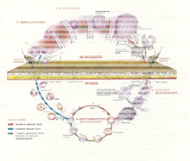
Figure 1 – Life cycle of the Plasmodium.
Chapter 2 - The role of Anopheles gambiae P450 cytochrome in insecticide resistance and infection
Figure 1 - Catalytic mechanism of P450 enzymes.
Chapter 3 - Plasmodium infection alters Anopheles gambiae detoxification gene expression
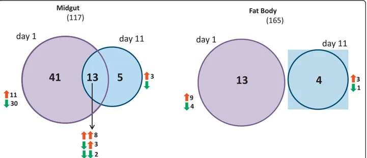
Figure 1 - Differential expression of detoxification genes in the midgut and fat body at day 1 and day 11 post feeding with a P. berghei infected or an uninfected blood meal.
Figure 2 - Heat diagrams showing genes that responded differently between the event of Plasmodium invasion into the midgut epithelium (day 1 post feeding) and the release of sporozoites into the hemolymph (day 11 post feeding).
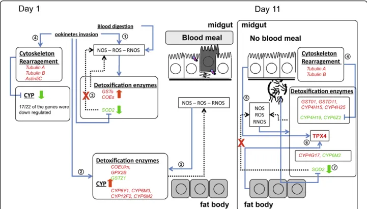
Figure 3 - Hypothetical scenario of Anopheles gambiae detoxification response to Plasmodium berghei infection with focus on the detoxification enzymes.
Chapter 4 – The Interplay between tubulins and P450 cytochromes during Plasmodium berghei invasion of Anopheles gambiae midgut
Figure 1 - Effect of silencing CPR (A), tubA, tubB, or co-silencing tubA and tubB (B) on P. berghei infection at 8 days after an infected blood meal.
Figure2 - Effect of tubulin inhibitors on P. berghei infection at 8 days after an infected blood meal.
xviii
Figure 5 - Effect of tubulin inhibitors in P450 cytochromes mRNA expression levels in midguts.
Chapter 5 - Promoter analysis of three P450 cytochromes in Anopheles gambiae
Figure 1 - Scheme of putative transcription factor binding sites in the promoter region of CYP6Z1, CYP6M2 and CYP6Z2.
Figure 2 - Normalised luciferase activities of A. gambiae CYP6M2 promoter region following transfection of luciferase reporter constructs into Sua 5.1* cells.
Figure 3 - Normalised luciferase activities of A. gambiae CYP6Z1 promoter region following transfection of luciferase reporter constructs into Sua 5.1* cells.
xix
List of Tables
Chapter 3 - Plasmodium infection alters Anopheles gambiae detoxification gene expression
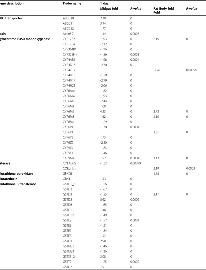
Table 1 - Genes differentially expressed (p<0.001) between infected and uninfected mosquitoes on day 1 after infection.
Table 2 - Genes differentially expressed (p<0.001) between infected and uninfected mosquitoes on day 11 after infection.
Chapter 4 – The Interplay between tubulins and P450 cytochromes during Plasmodium berghei invasion of Anopheles gambiae midgut
Introduction
3
Malaria
Malaria is a severe mosquito-borne disease, which persists today as one of the most widespread and devastating parasitic infections affecting the human population. There were an estimated 225 million cases of malaria in 2009 that accounted for approximately 781000 deaths, most of them of children under five years old living in sub-Saharan Africa (WHO, 2010).
Malaria is caused by protozoan parasites from the genus Plasmodium, which are
transmitted to humans when female mosquitoes of the genus Anopheles feed on human
blood.
Parasite
Plasmodium spp. are obligate parasites. They have a complex life cycle involving two hosts: a vertebrate host and a mosquito vector. In the mosquito vector parasites develop their sexual life cycle (sporogonic cycle) and in the vertebrate host they complete their asexual life cycle, which can be divided in hepatic and erythrocytic, the latter being responsible for the malaria symptoms. Plasmodium life cycle (adapted from (Knell, 1991)) is shown in detail in Figure 1.
The sporogonic cycle starts when Plasmodium enters the midgut with the blood meal.
4
mature into schizonts and undergo
merozoites, initiating new cycles of erythrocyte invasion, maturation and rupture which causes the symptoms of the malaria illness. Some merozoites differentiate into gametocytes that are taken up by the mosquit
(Knell, 1991).
Figure 1 – Life cycle of Plasmodium
mature into schizonts and undergo a series of divisions, forming new invasive merozoites, initiating new cycles of erythrocyte invasion, maturation and rupture which causes the symptoms of the malaria illness. Some merozoites differentiate into gametocytes that are taken up by the mosquito in a blood meal initiating a new cycle
Plasmodium (adapted from (Knell, 1991)).
Introduction
5
Malaria, an ongoing problem
In all tropical and subtropical parts of the world, malaria maintains a high prevalence. The hot and humid weather, the poor health and sanitary system existent in these parts of the globe and the interruption/ceasing of control programs contribute to the success of the development and reproduction of the mosquito vector. This, together with i) the absence of an efficient protective vaccine, ii) the rapid spread of parasite resistance to anti-malarial drugs and iii) the increasing resistance to insecticides of the mosquito vector, perpetuates the occurrence and, in some countries, is accountable for the increase of malaria cases, highlighting the fragility of malaria control and the need to maintain/increase control programs and the implementation of novel and more effective approaches.
6
Anopheles gambiae mosquito
Anopheles gambiae is the major African vector for transmission of malaria. It became a
suitable model for the study of innate immunity as A. gambiae mounts efficient local
and systemic immune responses against Plasmodium infection and its genome has been sequenced (Holt et al., 2002) and since then regularly annotated.
The combination A. gambiae and the rodent parasite Plasmodium berghei is one of the most used model systems for laboratory assays of parasite infections. The existence of poor or incompatible vector-parasite combinations suggests that specific molecular and cellular interactions are essential for a vector-parasite system to become established and subsequently co-evolve (Alavi et al., 2003). Of all the interactions between the mosquito and the parasite, the ones that occur during the three main bottlenecks (where the parasite numbers are largely reduced and its development can be disrupted) are the most important (Blandin and Levashina, 2004). The extreme parasite losses are attributed to efficient mosquito immune responses, which can completely block parasite development.
Detoxification enzymes
Anopheles gambiae becomes infected with the malaria parasite by taking a blood meal. The blood meal alone triggers the transcriptional regulation of several mosquito genes required to blood digestion causing metabolic changes which induce a state of oxidative
stress (Vlachou et al., 2005, Kumar et al., 2003). A Plasmodium infected blood meal
Introduction
7 endogenous compounds like steroids (Feyereisen, 1999, Scott, 1999). An actualized description about insect P450 cytochromes and their putative role in A. gambiae is reviewed in chapter 2.
The exact role of detoxification enzymes in the mosquito response to Plasmodium
infection is not well known. The impact of Plasmodium infection on the expression of
detoxification genes in the midgut and fat body of A. gambiae mosquitoes is described
in chapter 3. Two time points, decisive for parasite invasion: day 1 post-infection (p.i.) (when the ookinete is traversing the midgut epithelium) and day 11 p.i. (when the sporozoites are release to the hemolymph) were analysed.
Cytoskeleton genes
During Plasmodium infection differential regulation of cytoskeleton genes has been observed (Vlachou et al., 2005, Marinotti et al., 2005, Abrantes et al., 2008). The cytoskeleton rearrangement was described as a protective mechanism during ookinete
invasion of the midgut in A. gambiae (Vlachou et al., 2005). Tubulin disarray could be
responsible for the regulation of several P450 cytochromes, causing their suppression or induction during the mosquito response to parasite invasion. Similar to the association described in mammals (Dvorak et al., 2005). An attempt to clarify the role of tubulins on parasite invasion and their interplay with P450 cytochromes is described in chapter 4.
P450 promoter regions
8
Introduction
9
Aims of this thesis
Malaria is not a problem from the past. Even today the number of new malaria cases is rising in some of the poorest tropical and subtropical countries. It is increasingly important to take efficient measures and to find new strategies to control malaria transmission. For this it is essential to understand the biology of the parasite and the mosquito and specially the interactions between them. Thus, the complete knowledge of the mosquito immune response to the invading parasite is essential. The A. gambiae innate immunity has been extensively studied over the last years, and yet there are still several unknown mechanisms of protection.
The present work aimed to study the role of detoxification genes of A. gambiae in
response to P. berghei. As there are many detoxification enzymes in the genome of A.
gambiae and they have a great variety of functions, the study focused on the role of P450 cytochromes.
Specific objectives
1 – Identification of the A. gambiae mosquitoes detoxification enzymes, which are differentially expressed during P. berghei invasion of midgut epithelium and sporozoite egress from the oocyst.
2 – Characterization of the role of A. gambiae tubulins in the response to Plasmodium
infection and its interaction with the regulation of P450 cytochromes.
10
References
Abrantes, P., Dimopoulos, G., Grosso, A. R., Do, R. V. & Silveira, H. 2008. Chloroquine mediated modulation of Anopheles gambiae gene expression. PLoS.One., 3, e2587.
Alavi, Y., Arai, M., Mendoza, J., Tufet-Bayona, M., Sinha, R., Fowler, K., Billker, O., Franke-Fayard, B., Janse, C. J., Waters, A. & Sinden, R. E. 2003. The dynamics of interactions between Plasmodium and the mosquito: a study of the infectivity of Plasmodium berghei and Plasmodium gallinaceum, and their transmission by Anopheles stephensi, Anopheles gambiae and Aedes aegypti. Int.J.Parasitol., 33, 933-943.
Blandin, S. & Levashina, E. A. 2004. Mosquito immune responses against malaria parasites. Curr.Opin.Immunol., 16, 16-20.
David, J. P., Strode, C., Vontas, J., Nikou, D., Vaughan, A., Pignatelli, P. M., Louis, C., Hemingway, J. & Ranson, H. 2005. The Anopheles gambiae detoxification chip: a highly specific microarray to study metabolic-based insecticide resistance in malaria vectors. Proc.Natl.Acad.Sci.U.S.A, 102, 4080-4084.
Djouaka, R. F., Bakare, A. A., Coulibaly, O. N., Akogbeto, M. C., Ranson, H., Hemingway, J. & Strode, C. 2008. Expression of the cytochrome P450s, CYP6P3 and CYP6M2 are significantly elevated in multiple pyrethroid resistant populations of Anopheles gambiae s.s. from Southern Benin and Nigeria. BMC.Genomics, 9, 538.
Dong, Y., Manfredini, F. & Dimopoulos, G. 2009. Implication of the mosquito midgut microbiota in the defense against malaria parasites. PLoS.Pathog., 5, e1000423.
Dvorak, Z., Ulrichova, J. & Modriansky, M. 2005. Role of microtubules network in
CYP genes expression. Curr.Drug Metab, 6, 545-552.
Feyereisen, R. 1999. Insect P450 enzymes. Annu.Rev.Entomol., 44, 507-533.
Introduction
11 Holt, R. A., Subramanian, G. M., Halpern, A., Sutton, G. G., Charlab, R., Nusskern, D. R., Wincker, P., Clark, A. G., Ribeiro, J. M., Wides, R., Salzberg, S. L., Loftus, B., Yandell, M., Majoros, W. H., Rusch, D. B., Lai, Z., Kraft, C. L., Abril, J. F., Anthouard, V., Arensburger, P., Atkinson, P. W., Baden, H., De, B. V., Baldwin, D., Benes, V., Biedler, J., Blass, C., Bolanos, R., Boscus, D., Barnstead, M., Cai, S., Center, A., Chaturverdi, K., Christophides, G. K., Chrystal, M. A., Clamp, M., Cravchik, A., Curwen, V., Dana, A., Delcher, A., Dew, I., Evans, C. A., Flanigan, M., Grundschober-Freimoser, A., Friedli, L., Gu, Z., Guan, P., Guigo, R., Hillenmeyer, M. E., Hladun, S. L., Hogan, J. R., Hong, Y. S., Hoover, J., Jaillon, O., Ke, Z., Kodira, C., Kokoza, E., Koutsos, A., Letunic, I., Levitsky, A., Liang, Y., Lin, J. J., Lobo, N. F., Lopez, J. R., Malek, J. A., Mcintosh, T. C., Meister, S., Miller, J., Mobarry, C., Mongin, E., Murphy, S. D., O'brochta, D. A., Pfannkoch, C., Qi, R., Regier, M. A., Remington, K., Shao, H., Sharakhova, M. V., Sitter, C. D., Shetty, J., Smith, T. J., Strong, R., Sun, J., Thomasova, D., Ton, L. Q., Topalis, P., Tu, Z., Unger, M. F., Walenz, B., Wang, A., Wang, J., Wang, M., Wang, X., Woodford, K. J., Wortman, J. R., Wu, M., Yao, A., Zdobnov, E. M., Zhang, H., Zhao, Q., et al. 2002. The genome
sequence of the malaria mosquito Anopheles gambiae. Science, 298, 129-149.
Knell, A. J. 1991. Malaria: A publication of the tropical programme of the Wellcome
Trust, Oxford, Oxford University Press
Kumar, S., Christophides, G. K., Cantera, R., Charles, B., Han, Y. S., Meister, S., Dimopoulos, G., Kafatos, F. C. & Barillas-Mury, C. 2003. The role of reactive oxygen species on Plasmodium melanotic encapsulation in Anopheles gambiae. Proc.Natl.Acad.Sci.U.S.A, 100, 14139-14144.
Marinotti, O., Nguyen, Q. K., Calvo, E., James, A. A. & Ribeiro, J. M. 2005. Microarray analysis of genes showing variable expression following a blood meal in Anopheles gambiae. Insect Mol.Biol., 14, 365-373.
Molina-Cruz, A., Dejong, R. J., Charles, B., Gupta, L., Kumar, S., Jaramillo-Gutierrez, G. & Barillas-Mury, C. 2008. Reactive oxygen species modulate Anopheles
gambiae immunity against bacteria and Plasmodium. J.Biol.Chem., 283,
12
Pinto, S. B., Lombardo, F., Koutsos, A. C., Waterhouse, R. M., Mckay, K., An, C., Ramakrishnan, C., Kafatos, F. C. & Michel, K. 2009. Discovery of Plasmodium modulators by genome-wide analysis of circulating hemocytes in Anopheles
gambiae. Proc.Natl.Acad.Sci.U.S.A, 106, 21270-21275.
Ranson, H., Claudianos, C., Ortelli, F., Abgrall, C., Hemingway, J., Sharakhova, M. V., Unger, M. F., Collins, F. H. & Feyereisen, R. 2002. Evolution of supergene families associated with insecticide resistance. Science, 298, 179-181.
Scott, J. G. 1999. Cytochromes P450 and insecticide resistance. Insect
Biochem.Mol.Biol., 29, 757-777.
Vlachou, D., Schlegelmilch, T., Christophides, G. K. & Kafatos, F. C. 2005. Functional genomic analysis of midgut epithelial responses in Anopheles during
Plasmodium invasion. Curr.Biol., 15, 1185-1195.
Chapter 2 - The role of
Anopheles
gambiae
P450 cytochrome in insecticide
resistance and infection
This chapter was accepted for publication as a book chapter:
The role of Anopheles gambiae P450 cytochromes in insecticide resistance and infection
15
Introduction
Anopheles gambiae is the major vector of malaria transmission in sub-Saharan Africa where the disease is responsible for the highest morbidity and mortality worldwide. Malaria, nowadays, is still a major burden causing the death of nearly one million people each year, mostly children under the age of five, and affecting those living in the poorest countries (World Health Organization [WHO], 2010).
Currently, the major obstacles to malaria eradication are the absence of a protective vaccine, the spread of parasite resistance to anti-malarial drugs and the mosquito resistance to insecticides. Controlling mosquito vectors is fundamental to reduce mosquito-borne diseases. In fact, it has been one of the most used and effective method to prevent malaria, namely trough insecticides spraying and impregnated bed nets. These methods are highly dependent on a single class of insecticides, the pyrethroids, which are the most frequently used compounds for indoor residual spraying, and the
only insecticide class used for insecticide treated nets (WHO, 2010). The extensive use
of a single class of insecticides further increases the risk of mosquitoes developing resistance, which could rapidly lead to a major public health problem mainly in sub-Saharan countries where insecticidal vector control is being used widely (WHO, 2010). Strategies to control malaria are still not enough to totally eliminate malaria transmission, having yet to overcome several difficulties as the development of parasite drug resistance and mosquito-vector insecticide resistance (Yassine & Osta, 2010). Unfortunately the emergence of mosquito populations capable of withstanding insecticide exposure is threatening the efficiency of these control measures.
Insecticide resistance
16
genetic and biological basis is critical. Insecticide resistance can also lead to outbreaks of human diseases when vectors cannot be controlled. Hence, the elucidation of resistance mechanisms is extremely important for the development of tools to monitor resistance in populations, thereby contributing to mosquito control programs. Although the mechanisms by which insecticides become less effective are similar across all vector taxa, each resistance problem is potentially unique and may involve a complex pattern of resistance foci (Brogdon & McAllister, 1998).
The main forms of resistance mechanisms can be divided in two groups: target site resistance, which occurs when the insecticide no longer binds to its target, and metabolic resistance, which occurs when enhanced levels of modified activities of detoxification enzymes prevent the insecticide from reaching its site of action. Alone or in combination these mechanisms confer resistance, sometimes at high levels, to all classes of insecticides.
Target site resistance
Target site resistance is based on alterations of amino acids in the site of action where the insecticide is supposed to bind, causing the insecticide to be less effective or ineffective at all. Knock down resistance (Kdr) occurs due to a single or multiple substitutions in the sodium channel (Martinez-Torres et al., 1998, Ranson et al., 2000a) and alteration in acetylcholinesterase results in decreased sensitivity to insecticides (Mutero et al., 1994). Insecticide resistance has been reported from many insects including A. gambiae that showed the presence of insensitive acetylcholinesterase in two different populations that were resistant to carbosulfan, a carbamate insecticide (N’Guessan et al., 2003). Mutations at a single codon in the Rdl (resistance to dieldrin) gene have been documented in all dieldrin-resistant insects, and results in conferring both insensitivity to the insecticide and also a decrease rate of desensitisation
(ffrench-Constant et al., 1998). However, in A. gambiae this type of resistance mechanisms was
The role of Anopheles gambiae P450 cytochromes in insecticide resistance and infection
17
Metabolic resistance
Metabolic resistance usually involves over-expression of enzymes capable of detoxifying insecticides or alterations in the amino acids within these enzymes causing alterations in the levels or activities of detoxification proteins. There are three major enzyme families involved in this type of resistance, GST, carboxylesterases and P450 cytochromes. Carboxylesterases are mainly involved in organophosphate and carbamate and to a lesser extent in pyrethroid resistance, while P450 cytochromes are mainly involved in the metabolism of pyrethoids and to a lesser extent, detoxification of organophosphates and carbamates (Hemingway & Ranson, 2000). Glutathione S-transferases are involved in the detoxification of a wide range of xenobiotics, including the organochloride insecticide DDT (Enayati et al., 2005). In A. gambiae metabolic resistance to insecticides can be conferred by elevation in the activity of these three classes of detoxifying enzymes.
The over-expression of carboxylesterases as an evolutionary response to organophosphorus and carbamate insecticide selection pressure has been reported in
several insects, includingmosquitoes (Newcomb et al., 1997; Vulule et al., 1999; Zhu et
al., 1999). Organophosphorus and carbamate inhibit B esterases by rapid esterification of the serine residue in the active site, usually followed by a slow hydrolysis of the new ester bond. Therefore, these insecticides can be considered as inhibitors of esterases, because they are poor substrates which have a high affinity for these enzymes (Hemingway & Karunaratne, 1998). Carboxylesterases in large amounts causes resistance as the insecticides are rapidly sequestered, even before reaching the target-site acetylcholinesterase (Hemingway & Karunaratne, 1998). There are many reports of over expression of carboxylesterases in insecticide resistant mosquitoes including A. gambiae, where enhanced production of carboxylesterases was observed in permethrin-resistant mosquitoes (Vulule et al., 1999).
Glutathione S-transferases are a major class of detoxification enzymes that possess a
wide range of substrates specificities (Enayati et al., 2005). Elevated GST activity has
18
in transcriptional rate, rather than qualitative changes in individual enzymes (Hemingway et al., 2004). The primary function of GSTs is the detoxification of both endogenous and xenobiotic compounds either directly or by catalysing the secondary metabolism of a vast array of compounds oxidised by P450 cytochromes (Wilce & Parker, 1994). GST enzymes metabolise insecticides by facilitating their reductive dehydrochlorination or by conjugation reactions with reduced glutathione to produce water soluble metabolites that are more readily excreted (Wilce & Parker, 1994). They also contribute to the removal of toxic oxygen free radical species produced through the action of pesticides (Enayati et al., 2005). In A. gambiae elevated GST levels were shown to be associated with DDT resistance (Ranson et al., 2001). Furthermore genetic
mapping of the major loci conferring DDT resistance in A. gambiae implicate both cis-
and trans-acting factors in the overexpression of GSTs (Ranson et al., 2000b). GSTs in A. gambiae were also over expressed in a DDT-resistant strain, but only one GSTE2-2 was able to metabolise DDT (Ortelli et al., 2003).
The role of Anopheles gambiae P450 cytochromes in insecticide resistance and infection
19
Insect P450 cytochromes
P450 cytochromes are hemoproteins which act as terminal oxidases in monooxygenase systems. P450 cytochromes, whose name originated on its characteristic absorbance peak at 450 nm that appears when these enzymes are reduced and saturated with carbon-monoxide, constitute one of the oldest and largest super families of enzymes being found in almost all living organisms. In the literature, P450 enzymes are known by several names: cytochromes P450 monooxigenases, mixed functions oxidases, microsomal oxidases and heme thiolate proteins.
Insect P450s play a critical role in the metabolism of a wide variety of endogenous and exogenous compounds such as steroids, fatty acids and a wide range of xenobiotics and have also been implicated in vital processes like growth, development, feeding, reproduction, insecticide resistance and tolerance to plants toxins (Feyereisen, 1999; Scott et al., 1998; Scott, 1999). P450 cytochromes are also intimately involved in the synthesis and degradation of insect hormones and pheromones, including 20-hydroxyecdysone and juvenile hormone (Feyereisen, 1999).
Nomenclature
To distinguish one of these cytochromes among all the P450s, a standardized nomenclature system was implemented (Nelson et al., 1996; Nebert et al., 1991). Each P450 is named with CYP, followed by an Arabical number for the gene family, a letter for the sub-family and another Arabical number for the gene. Cytochromes P450s with share more then 40% of the amino acids are usually grouped into the same family and members with >55% of the amino acids identical are normally grouped in the same sub-family. However, there are exceptions to these rules (Nelson et al., 1996). As it is based on amino acid similarities, no information regarding the function of each P450 should be assumed from its name.
Structure
20
with mitochondrial membranes. This class of enzymes requires both a FAD-containing reductase and an iron sulphur redoxin, and catalyzes several steps in the biosynthesis of steroid. Class II enzymes are the most common in eukaryotes and are found in the endoplasmic reticulum. These enzymes only require an FAD/FMN-containing P450 reductase for transfer of electrons. Their functions are extremely diverse and, in eukaryotes, include aspects of the biosynthesis and catabolism of signalling molecules and steroid hormones (Feyereisen, 1999). Class III enzymes are self-sufficient and
require no electron donor. They are involved in the synthesis of signalling molecules.
Finally, class IV enzymes receive electrons directly from NAD(P)H. Class I and II P450s from all organisms participate in the detoxification or sometimes the activation of xenobiotics and class III and IV enzymes are considered remains of the ancestral forms of P450s involved in detoxification of damaging activated oxygen species (Werck-Reichhart & Feyereisen, 2000).
Most P450s are approximately 500 amino acids long. The core of these proteins is
formed by a four-helix bundle, two sets of β sheets, two helices and a coil called the
“meander”. A characteristic consensus sequence known as the P450 “signature” FXXGXXXCXG, located on the C-terminus of the heme binding region, contains a conserved cysteine that serves as a fifth ligand to the heme iron. There are two other conserved motifs specific of the P450 proteins. One is the DGXXT domain, which corresponds to the proton transfer groove on the distal site of the heme. Another is the EXXR domain, which is probably needed to stabilize the core structure located on the proximal side of heme (Werck-Reichhart & Feyereisen, 2000).
Microssomal / mitochondrial
In insects both mitochondrial and microssomal P450 systems have been described. The majority of P450 in insects are microssomal, located in the endoplasmatic reticulum, and require the flavoprotein NADPH cytochrome P450 reductase as the main electron
donor; however cytochrome b5 is sometimes needed, depending of the substrate and of
The role of
Characterization / Function
Cytochromes P450 enzymes catalyse thousands of different reactions, which are based on the activation of molecular oxygen, with insertion of one of its atoms into the substrate, and reduction of the other to form water (Guengerich, 1991). P450s use electrons from NAD(P)H to catalyse the activation of molecular oxygen, leading to the regiospecific and stereospecific oxidative attack of structurally diverse chemicals (Werck-Reichhart & Feyereisen, 2000).
The interaction that occurs between P450 cytochrome
P450 reductase is better expressed as a cyclic reaction (Guenguerich, 1991 depicted in Figure 1.
Figure 1. Catalytic mechanism of P450 enzymes
The cycle is initiated by the binding of the substrate to the ferric form of the enzyme to form an enzyme-substrate complex, followed by a reduction of the ferric complex by an electron transferred from NADPH via NADPH
binding of molecular oxygen to the reduced complex forms an enzyme
substrate complex followed by the transference of a second electron from NADPH via NADPH-cytochrome P450 reductase or from cytochrome
The role of Anopheles gambiae P450 cytochromes in insecticide resistance and infecti
Cytochromes P450 enzymes catalyse thousands of different reactions, which are based the activation of molecular oxygen, with insertion of one of its atoms into the substrate, and reduction of the other to form water (Guengerich, 1991). P450s use ctrons from NAD(P)H to catalyse the activation of molecular oxygen, leading to the regiospecific and stereospecific oxidative attack of structurally diverse chemicals
Reichhart & Feyereisen, 2000).
The interaction that occurs between P450 cytochromes and the NADPH cytochrome P450 reductase is better expressed as a cyclic reaction (Guenguerich, 1991
Catalytic mechanism of P450 enzymes, where S is the substrate.
The cycle is initiated by the binding of the substrate to the ferric form of the enzyme to substrate complex, followed by a reduction of the ferric complex by an electron transferred from NADPH via NADPH-cytochrome P450 reductase. Next, the binding of molecular oxygen to the reduced complex forms an enzyme
substrate complex followed by the transference of a second electron from NADPH via
cytochrome P450 reductase or from cytochrome b5. A second proton is added,
P450 cytochromes in insecticide resistance and infection
21 Cytochromes P450 enzymes catalyse thousands of different reactions, which are based
the activation of molecular oxygen, with insertion of one of its atoms into the substrate, and reduction of the other to form water (Guengerich, 1991). P450s use ctrons from NAD(P)H to catalyse the activation of molecular oxygen, leading to the regiospecific and stereospecific oxidative attack of structurally diverse chemicals
s and the NADPH cytochrome P450 reductase is better expressed as a cyclic reaction (Guenguerich, 1991) as it is
The cycle is initiated by the binding of the substrate to the ferric form of the enzyme to substrate complex, followed by a reduction of the ferric complex by an cytochrome P450 reductase. Next, the binding of molecular oxygen to the reduced complex forms an enzyme-oxygen-substrate complex followed by the transference of a second electron from NADPH via
22
which results in the breaking of the oxygen-oxygen bond, releasing one atom of oxygen as water. The oxygen atom remaining is transferred to the substrate, originating an oxidized product, which is released, and a ferric form of the enzyme is once more generated. Then the cycle is re-initiated (Guenguerich, 1991).
Diversity and specificity
The huge diversity of P450 cytochromes is probably due to an extensive process of gene duplication and cases of gene amplification, conversion, genome duplication, gene loss and lateral transfer (Werck-Reichhart & Feyereisen, 2000). Due to their extremely diverse functions, they can be found with different patterns of expression in all types of tissues and in almost all types of organisms. Although being expressed in a wide range of tissues, insect P450s have their highest activity associated with midgut, fat body and malpighian tubules (Feyereisen, 1999; Scott, 1999).
Additionally, P450s metabolise a large number of substrates, probably due to the existence of numerous P450 isoforms and to the broad specificity of some isoforms (Scott & Wen, 2001). Nevertheless the substrate specificity and type of reaction catalysed by each P450 cytochrome is still not well understood.
Their diversity enables individual P450 cytochromes to display different expression patterns related to life stages, tissues, inducers/inhibitors and substrates. Specifically, there are P450s that are expressed in all life stages (CYP12 genes) while others are only expressed in adults (CYP6Z1) or in larval stages (CYP6Z3) (Nikou et al., 2003). Although being found expressed in almost all types of tissues, there are P450s which are tissue specific, while others are everywhere (Feyereisen, 1999; Scott et al., 1998; Scott & Wen, 2001). Expression of P450 cytochromes may also be sex specific, as some P450s showed higher levels of expression in male compared with female tissues (Muller et al., 2007; Nikou et al., 2003).
The role of Anopheles gambiae P450 cytochromes in insecticide resistance and infection
23 also differs depending on the P450s. P450s show a vast variation in response to inducers and inhibitors, each P450 can be induced/inhibited by one or several compounds. Some P450s can also remain unaltered while others are induced or repressed (Scott et al., 1998).
Anopheles gambiae P450 cytochromes and insecticide resistance
The A. gambiae genome has 111 annotated P450 cytochromes (Ranson et al., 2002). The great interest in these cytochromes derives from their role in the oxidative metabolism of insecticides, but only in few cases a definitive link between an increased expression of a specific P450 cytochrome and increased insecticide metabolism has been established.
Increasing reports of specific A. gambiae P450 cytochromes being involved in insecticide resistance have been published in the past. The involvement of P450s in pyrethroid resistance started to be demonstrated in A. gambiae from Kenyan villages, in synergistic studies using specific P450 cytochrome inhibitors and also given the detection of increased heme levels in resistant mosquitoes (Vulule et al., 1999).
In 2003, Nikou et al., verified that a P450 cytochrome (CYP6Z1) was over-expressed in a pyrethroid-resistant strain of A. gambiae, and the development of her work pointed to an implication of the involvement of this P450 in conferring pyrethroid resistance to this mosquito (Nikou et al., 2003).
Later, a microarray chip was constructed containing fragments from 230 genes
associated with detoxification (David et al., 2005) to further study the metabolic based insecticide resistance in A. gambiae. From this work resulted the identification of, among other genes, several P450 cytochromes that were highly expressed in the A. gambiae permethrin or DDT-resistant strains (David et al., 2005). Of notice is the P450
cytochrome CYP325A3, which belongs to a class that was not associated with
24
over-expressed in a Nigerian pyrethroid resistant strain of A. gambiae (Awolola et al., 2009).
In 2007, studies regarding a recently colonised strain of A. gambiae from Ghana identified genes whose expression levels were associated with pyrethroid resistance.
Among these were three P450 cytochromes (CYP6M2, CYP6Z2 and CYP6Z3) (Muller
et al., 2007). These results, together with their location within a cluster of P450 cytochromes in the right arm of chromosome 3 (3R), which is in close association with with a pyrethroid resistance QTL (Ranson et al., 2004), strongly support their involvement in insecticide resistance. A subsequent study showed that CYP6Z2 displays broad substrate specificity, which may be associated with xenobiotics metabolism and detoxification (Mclaughlin et al., 2008). Despite, CYP6Z2 being able to bind to
permethrin and cypermethrin, CYP6Z2 does not metabolise neither one of these
insecticides (Mclaughlin et al., 2008).
In 2008, Djouaka et al. also identified several P450 cytochromes over-expressed in one or more pyrethroid resistant populations of A. gambiae. Among these were CYP6P3 and once again CYP6M2. Both genes showed high levels of over-expression in all the resistant populations, but the first was the gene that showed greatest differences. In the
same year, CYP6P3 was also identified as being up-regulated in another highly
permethrin resistant A. gambiae population (Müller et al., 2008).
Recent studies on A. gambiae recombinant proteins CYP6M2 (Stevenson et al., 2011)
and CYP6P3 (Müller et al., 2008) demonstrated that these enzymes could metabolise pyrethroids. Thus, the up regulation of these P450 cytochromes in pyrethroid resistant populations, strongly supports a key role for these genes to confer pyrethroid resistance in A. gambiae.
The role of Anopheles gambiae P450 cytochromes in insecticide resistance and infection
25
As the above P450 cytochromes, CYP314A1 was also found to be over-expressed in a
DDT resistant strain of A. gambiae from Kenia (Vontas et al., 2005), suggesting a
possible involvement in the insecticide resistance phenotype. Both CYP6Z1 and
CYP6Z2 were over-expressed in DDT resistant strains of A. gambiae (David et al. 2005). Although being very similar, these two cytochromes have predicted substrate cavities dramatically different and CYP6Z1 was predicted to be the only one capable of metabolizing DDT. Chiu et al. (2008) through biochemical characterisations supported these predictions and identified CYP6Z1 as the only P450 cytochrome capable of metabolising DDT, demonstrating its potential as a target to reduce A. gambiae resistance to DDT (Chiu et al., 2008).
Another evidence of the involvement of P450s in insecticide resistance is the fact that silencing the main electron donor of P450 cytochromes, the cytochrome P450 reductase, by RNAi, greatly increased the susceptibility of A. gambiae to permethrin, emphasising the important chemoprotective role of P450 cytochromes in this process (Lycett et al., 2006).
Nevertheless, although P450s have been clearly associated with insecticide resistance, the identification of specific P450 cytochromes responsible for insecticide resistance is still extremely difficult.
Anopheles gambiae P450 cytochromes and malaria infection
P450 cytochromes have also been implicated in other vital processes as in A. gambiae
response to bacterial challenge and to parasite invasion, but the real importance and function of these cytochromes in this process is still not well understood.
A genome expression analysis of A. gambiae was made to identify which genes
26
response to malaria infection and the presence of lipopolysaccharide (Dimopoulos et al., 2002).
The involvement of P450 cytochromes in response to microbial challenge was
established when two P450 cytochromes (CYP4C27 and CYP306A1) were differently
expressed in the presence of Gram – (Salmonella thyphimurium) or Gram + (Staphylococcus aureus) bacteria (Aguilar et al., 2005). This involvement was even
more evident when a study, trying to implicate the mosquito midgut microbiota in the
defense against malaria parasites, showed that there were ten P450s differently expressed in response to Escherichia coli and S. aureus in the A. gambiae midgut twelve hours after an uninfected blood meal (Dong et al., 2009). Between the P450 cytochromes differently expressed there were CYP4H17, CYP6M3, CYP6AG1, CYP9J5,
two of them were mitochondrial cytochromes, CYP49A1 and CYP12F4 (Dong et al.,
2009).
Regarding the relation between P450 cytochromes and the response to malaria infection, it was partly unveiled for the first time in a study about the midgut epithelial responses during Plasmodium invasion (Vlachou et al., 2005). Here, several P450 cytochromes were differentially expressed between different phases of the midgut invasion (before invasion, during invasion and after invasion) and also when they compared Plasmodium wild-type infection with Plasmodium that were unable to invade the epithelium infection (Vlachou et al., 2005). P450s that stood out in this study were CYP305A1, CYP304B1, CYP6Z1 and CYP6M4 (Vlachou et al., 2005). The role of P450 cytochromes in the A. gambiae response to malaria infection has been reinforced in the
last years. Comparing the A. gambiae response to two different Plasmodium parasites
-P. berghei and Plasmodium falciparum - showed that the mosquito induced slightly different immune responses to each parasite, and that the mosquito was capable of sensing infected blood constituents and mount an immune response, even in the absence of invading ookinetes (Dong et al., 2006). It stands out that, although there were different responses between the three types of infection, in all of them there were P450s
differentially expressed in the midgut (CYP6AG1, CYP6M4, CYP6M1, CYP9J5 and
The role of Anopheles gambiae P450 cytochromes in insecticide resistance and infection
27 Further evidence of the link between P450 cytochromes and the mosquito’s response to malaria infection came from different studies. First, the effect on gene regulation of the presence of chloroquine in an uninfected blood meal and in a Plasmodium infected blood meal was investigated (Abrantes et al., 2008). This work showed that chloroquine affects the abundance of transcripts which encode proteins involved in a variety of processes, including P450 cytochromes that were differently expressed in the P. berghei infected blood meal (CYP9L1, CYP304B1 and CYP305A1). A second study focused on
the role of A. gambiae detoxification enzymes, from the three major families involved
in detoxification, GSTs, carboxylesterases and P450 cytochromes, in the response to Plasmodium infection (Félix et al., 2010). In this study the impact of P. berghei infection was analysed at two time points: one day following the blood meal, during which parasites invade the midgut epithelium, and eleven days after the blood meal when sporozoites were starting to be released to the hemolymph; in two different tissues, midgut and fat body. At day one after the Plasmodium infected blood meal they found 17 P450 cytochromes down-regulated and 5 P450 cytochromes up-regulated, including CYP9L1, CYP304B1, CYP325H1, CYP6M2 and CYP6Z2 in the midgut, and 5 P450 up-regulated and 1 down-regulated in the fat body, including CYP12F2, CYP6M2, CYP6M3 and CYP4G17. At eleven days after an infected blood meal they found 2 P450 cytochromes up-regulated and 3 down-regulated in the midgut and 1 P450 cytochrome up-regulated and 1 down-regulated in the fat body. The high number of P450
cytochromes differently expressed by the presence of P. berghei parasites in different
phases of infection and in different tissues suggests that P450 cytochromes are deeply involved in the mosquito response to Plasmodium infection, having an important role in different development stages of the parasite and covering different tissues of the mosquito. More specifically, these P450 cytochromes might have a direct role in Plasmodium response during the parasite invasion of the midgut epithelium as this is the moment and tissue where more P450 were differentially expressed. The over expression of these P450 cytochromes could be part of a mosquito response mechanism to parasite invasion occurring in the midgut. One possibility is P450s being involved in the cytoskeleton rearrangement (Vlachou et al., 2005; Vlachou & Kafatos, 2005), or P450s could be involved in the production of nitric oxide and other reactive oxygen radicals
28
Luckhart et al., 1998). The blood meal per si generates metabolic changes that are also expected to increase the oxidative stress in the mosquito midgut, which is augmented by the presence of Plasmodium parasites (Molina-Cruz et al., 2008). Moreover, other parasite killing mechanisms also induce oxidative stress inside the host which, although helping to eliminate the parasite, are also toxic to the host cell. The high level of oxidative stress inside the host cell could trigger cellular and molecular regulation of these P450 cytochromes, at this time point, being responsible for host detoxification together with parasite elimination.
Mosquito hemocytes mediate important cellular immune responses including phagocytosis and encapsulation and also secrete immune factors such as melanization
factors and antimicrobial peptides. Recently, studies were made to characterize the role
of A. gambiae hemocytes in mosquito immunity, consisting in a genome-wide transcriptomic analysis of adult female hemocytes following infection by bacteria and Plasmodium parasites (Baton et al., 2009). This work showed that some P450
cytochromes were differently expressed (CYP325H1 and CYP6M1) in the presence of
Micrococcus luteus, a Gram-positive bacteria (Baton et al., 2009), reinforcing the role of P450 cytochromes in response to microbial challenge described above. This work also showed that a P450 cytochrome was differently expressed 24 hours after the infected blood meal (CYP325H1), during P. berghei ookinete invasion of the midgut epithelium. Moreover, there were also P450s differentially expressed 19 days after the
infected blood meal (CYP6AG1 and CYP6M3), during P. berghei sporozoite migration
through the hemolymph (Baton et al., 2009), suggesting that P450 cytochromes have a role in the response to malaria infection achieved by hemocytes. Another study aiming to analyse the transcriptional expression and immune functions of circulating hemocytes
in naïve and P. berghei infected A. gambiae females assessed the roles in development
The role of Anopheles gambiae P450 cytochromes in insecticide resistance and infection
29
Conclusion
The role of P450 cytochromes during Plasmodium invasion is still poorly understood,
but it may play out to be of utmost importance to combat malaria transmission. Here, we intend to bring an update review on the connection between P450 cytochromes and the A. gambiae response to malaria infection, identifying several P450 cytochromes that
probably are, directly or indirectly, involved in the response to Plasmodium invasion.
We have also reviewed the implication of P450 cytochromes in A. gambiae insecticide resistance. However, uncovering the objective role of these cytochromes in insecticide resistance, that is naming specific cytochromes and describing in detail the processes in which those specific P450s are involved is still extremely difficult.
The consistent detection of differential expression of P450 cytochromes, in studies about either insecticide resistance or the response to malaria infection, suggests that the role of these P450s could be similar in these two processes. Nevertheless, the real importance and function of P450 cytochromes in these processes is still not well understood neither the possibility of interplay between infection and insecticide resistance. One of the P450 cytochromes with expression altered in response to
insecticides and Plasmodium infection was CYP6M2 that, was also highly
over-expressed in a pyrethroid-resistant strain of A. gambiae mosquitoes (Muller et al., 2007)
and highly over-expressed in response to Plasmodium infection in both the midgut and
the fat body 1 day after an infected blood meal (Félix et al., 2010). These results suggest that the role of CYP6M2 might be the same in response to insecticides and infection, or
that these two processes might share the activation mechanism of CYP6M2 expression.
down-30
regulated in the midgut of A. gambiae at day 1 and day 11 after an infected blood meal (Félix et al., 2010). These results suggest a different role for CYP6Z2 in response to the insecticide and to parasite infection, however we have to take into account that,
although being able to bind to permethrin and cypermethrin, CYP6Z2 does not
metabolise these compounds (Mclaughlin et al., 2008). So the over-expression of CYP6Z2 in a pyrethroid-resistant strain might be associated with different processes other than insecticide resistance.
The role of Anopheles gambiae P450 cytochromes in insecticide resistance and infection
31
References
Abrantes, P.; Dimopoulos, G.; Grosso, A.R.; do Rosário, V.E. & Silveira, H. (2008).
Chloroquine mediated modulation of Anopheles gambiae gene expression. PLoS
One, 3(7), e2587, ISSN 1932-6203
Aguilar, R.; Dong, Y.; Warr, E. & Dimopoulos, G. (2005). Anopheles infection responses; laboratory models versus field malaria transmission systems. Acta Tropica, 95(3), 285-291, ISSN 0001-706X
Awolola,T.S.; Oduola, O.A.; Strode, C.; Koekemoer, L.L.; Brooke, B. & Ranson, H. (2009). Evidence of multiple pyrethroid resistance mechanisms in the malaria
vector Anopheles gambiae sensu stricto from Nigeria. Transactions of the Royal
Society of Tropical Medicine and Hygiene, 103, 1139-1145, ISSN 0035-9203
Baton, L.A.; Robertson, A.; Warr, E.; Strand, M.R. & Dimopoulos, G. (2009).
Genome-wide transcriptomic profiling of Anopheles gambiae hemocytes reveals
pathogen-specific signatures upon bacterial challenge and Plasmodium berghei infection. BMC Genomics, 5, 10:257, ISSN 1471-2164
Brogdon, W.G. & McAllister, J.C. (1998). Insecticide resistance and vector control. Emerging Infectious Diseases, 4(4), 605-613, ISSN: 1080-6059
Chiu, T.L.; Wen, Z.; Rupasinghe, S.G. & Schuler, M.A. (2008). Comparative molecular modeling of Anopheles gambiae CYP6Z1, a mosquito P450 capable
of metabolizing DDT. Proceedings of the National Academy of Sciences,
105(26), 8855-8860, ISSN 0027-8424
David, J.P.; Strode, C.; Vontas, J.; Nikou, D.; Vaughan, A.; Pignatelli, P.M.; Louis, C.; Hemingway, J. & Ranson, H. (2005). The Anopheles gambiae detoxification chip: a highly specific microarray to study metabolic-based insecticide resistance
in malaria vectors. Proceedings of the National Academy of Sciences, 102(11),
4080-4084, ISSN 0027-8424
Dimopoulos, G.; Christophides, G.K.; Meister, S.; Schultz, J.; White, K.P.;
Barillas-Mury, C. & Kafatos, F.C. (2002). Genome expression analysis of Anopheles
32
Proceedings of the National Academy of Sciences, 99(13), 8814-8819, ISSN 0027-8424
Djouaka, R.F.; Bakare, A.A.; Coulibaly, O.N.; Akogbeto, M.C.; Ranson, H.; Hemingway, J. & Strode, C. (2008). Expression of the cytochrome P450s, CYP6P3 and CYP6M2 are significantly elevated in multiple pyrethroid resistant
populations of Anopheles gambiae s.s. from Southern Benin and Nigeria. BMC
genomics, 9, 538, ISSN 1471-2164
Dong, Y.; Aguilar, R.; Xi, Z.; Warr, E.; Mongin, E. & Dimopoulos, G. (2006). Anopheles gambiae immune responses to human and rodent Plasmodium parasite species. PLoS Pathogens, 2(6), e52, ISSN 1553-7366
Dong, Y.; Manfredini, F. & Dimopoulos, G. (2009). Implication of the mosquito midgut microbiota in the defense against malaria parasites. PLoS Pathogens, 5(5), e1000423, ISSN 1553-7366
Dunkov, B.C.; Guzov, V.M.; Mocelin, G.; Shotkoski, F.; Brun, A.; Amichot, M.; Ffrench-Constant, R.H. & Feyereisen, R. (1997). The Drosophila Cytochrome P450 Gene Cyp6a2: Structure, Localization, Heterologous Expression, and Induction by Phenobarbital. DNA and Cell Biology, 16(11), 1345-1356, ISSN 1044-5498
Enayati, A.A.; Ranson, H. & Hemingway, J. (2005). Insect glutathione transferases and insecticide resistance. Insect Molecular Biology, 14(1), 3-8, ISSN 0962-1075
Félix, R.C.; Müller, P.; Ribeiro, V.; Ranson, H. & Silveira, H. (2010). Plasmodium
infection alters Anopheles gambiae detoxification gene expression. BMC
genomics, 11, 312, ISSN 1471-2164
Feyereisen, R. (1999). Insect P450 Enzymes. Annual Review of Entomology, 44,
507-533, ISSN 0066-4170
The role of Anopheles gambiae P450 cytochromes in insecticide resistance and infection
33 Guengerich, F.P. (1991). Reactions and Significance of Cytochrome P-450 Enzymes.
The Journal of Biological Chemistry, 266(16), 10019-10022, ISSN 0021–9258
Han, Y.S.; Thompson, J.; Kafatos, F.C. & Barillas-Mury, C. (2000). Molecular
interactions between Anopheles stephensi midgut cells and Plasmodium
berghei: the time bomb theory of ookinete invasion of mosquitoes. Embo Journal, 19(22), 6030-6040, ISSN 0261-4189
Hemingway, J. & Karunaratne, S.H. (1998). Mosquito carboxylesterases: a review of the molecular biology and biochemistry of a major insecticide resistance mechanism. Medical and veterinary Entomology, 12(1), 1-12, ISSN 1365-2915
Hemingway, J. & Ranson, H. (2000). Insecticide resistance in insect vectors of human disease. Annual Review of Entomology. 41, 371-391, ISSN 0066.4170
Hemingway, J.; Hawkes, N.J.; McCarrol, L. & Ranson, H. (2004). The molecular basis of insecticide resistance in mosquitoes. Insect Biochemistry and Molecular Biology, 34, 653-655, ISSN 0965-1748
Kumar, S.; Christophides, G.K.; Cantera, R.; Charles, B.; Han, Y.S.; Meister, S.; Dimopoulos, G.; Kafatos, F.C. & Barillas-Mury, C. (2003). The role of reactive
oxygen species on Plasmodium melanotic encapsulation in Anopheles gambiae.
Proceedings of the National Academy of Sciences, 100(24), 14139-14144, ISSN 0027-8424
Kumar, S.; Gupta, L.; Han, Y.S. & Barillas-Mury, C. (2004). Inducible peroxidases
mediate nitration of anopheles midgut cells undergoing apoptosis in response to
Plasmodium invasion. Journal of Biological Chemistry, 279(51), 53475-53482, ISSN 0021-9258
Kasai, S. & Scott, J.G. (2000). Overexpression of Cytochrome P450 CYP6D1 Is Associated with Monooxygenase-Mediated Pyrethroid Resistance in House Flies from Georgia. Pesticide Biochemistry and Physiology, 68, 34-41, ISSN 0048-3575
Luckhart, S.; Vodovotz, Y.; Cui, L. & Rosenberg, R. (1998). The mosquito Anopheles