319 319 319 319 319 Mem Inst Oswaldo Cruz, Rio de Janeiro, Vol. 98(3): 319-323, April 2003
Isolation and Identification of Mycobacteria from Livestock
Specimens and Milk Obtained in Brazil
Clarice Q Fujimura Leite/
+, Ivone S Anno, Sergio R de Andrade Leite*, Eliana Roxo**,
Glenn P Morlock***, Robert C Cooksey***
Faculdade de Ciências Farmacêuticas, Caixa Postal 502, Rodovia Araraquara–Jaú km 1, 14801-902 Araraquara, SP, Brasil *Instituto de Química, Unesp, Araraquara, SP, Brasil **Instituto Biológico, São Paulo, SP, Brasil ***Division of AIDS, STD,
and TB Laboratory Research, Centers for Disease Control and Prevention, Atlanta, GA, USA
The prevalence of Mycobacterium bovis and other mycobacterial species in livestock specimens and milk was evaluated. An emphasis was placed upon the distribution of these organisms in milk that is readily available to the public that was either untreated, pasteurized, or treated using ultra high temperature. Twenty-two pathologic specimens from livestock (bovine, swine and bubaline) in five Brazilian states and 128 bovine milk samples from retail markets in the State of São Paulo were examined for mycobacteria. Identification was made by classical biochemical tests, thin layer chromatography of mycolic acids and polymerase chain reaction-restriction fragment length polymorphism (PCR-RFLP) analysis. Mycobacteria were isolated from 15 (68.2%) caseous lesions and from 23 (18%) milk samples. Eleven isolates were identified as M. bovis, and the remaining 27 nontuberculous myco-bacterial isolates were represented by five species and six unidentified rapidly growing mycomyco-bacterial strains. The data demonstrate that animal products in Brazil are frequent reservoirs of mycobacteria and may pose a risk to the public.
Key words: Mycobacterium - livestock - bovine - polymerase chain reaction (PCR) - restriction fragment length polymorphism (RFLP) - mycolic acid analysis - Brazil
Milk and meat are important sources of protein and other nutrients but can be contaminated by pathogenic agents. The possibility exists for the transmission of tu-berculosis and other mycobacterial infections from ani-mals to humans, most likely by ingestion of infected meat or raw (unpasteurized) dairy products (Chapman & Speight 1968, Bryan 1969, Sweeney et al. 1992). In developed coun-tries, the introductions of milk pasteurization and tuber-culin (ppd) positive cattle eradication programs have greatly reduced the incidence of human and bovine dis-eases caused by Mycobacterium bovis (Caffrey 1994). In Brazil, however, where bovine tuberculosis control and eradication programs have only recently been imple-mented, M. bovis disease remains an important veteri-nary disease, with almost 200 000 infected cows among a total cattle population of ~ 170 million (Kantor & Ritacco 1994). This organism also poses human public health problems as well because it is suspected that M. bovis
infection is responsible for approximately 4 000 of the approximately 80 000 cases of human tuberculosis re-ported each year in Brazil (WHO 1993). Johne’s disease, another important veterinary disease caused by M. paratuberculosis infection in cattle, has not been reported in Brazil, with rare exceptions among imported animals (Roxo 1997).
This work was supported by a financial aid grant provided by Fapesp (Brazilian Agency), proc. no.94/3741-9.
+Corresponding author. Fax: +55-16-222.0073. E-mail:
leitecqf@fcfar.unesp.br Received 6 September 2002 Accepted 6 March 2003
In areas of endemicity where bovine and human tu-berculosis coexist, the differentiation of M. bovis from M. tuberculosis is important for monitoring the spread of M. bovis among cows and from them to humans. Distin-guishing these two species by conventional (e.g. bio-chemical) tests is typically more time consuming than by molecular methods (Scorpio et al. 1997). The genotypic detection of insertion element IS6110 is a reliable assay to identify species that belong to the M. tuberculosis
complex (MTC) and has been applied to studies of MTC infections in cattle (Gutiérrez et al. 1995, Vitale et al. 1998), although the identification of IS6110 by polymerase chain reaction (PCR) fails to differentiate M. bovis from M. tu-berculosis. The polymorphisms in the two genes, oxyR
and pncA, appear to be stable features of these two spe-cies, and identification of these genes by restriction analy-sis of PCR products or by allelic-specific PCR amplifica-tion has proven useful toward for this end (Sreevatsan et al. 1996, De los Monteros et al. 1998). In addition, the mycobacterial species other than those, belonging to the MTC, have been recovered from environmental sources and from livestock; a considerable number have been iso-lated from raw and pasteurized milk (Chapman & Speight 1968, Hosty & McDurmont 1975).
MATERIALS AND METHODS
320 320 320 320
320 Mycobacteria from Livestock Specimens and Milk • Clarice Q Fujimura Leite et al.
4 swine lymph nodes, 3 bovine lung fragments and 5 bo-vine lymph nodes). Official investigators of the Inspec-tion Service of the Ministry of Agriculture (Brazilian Fed-eral Government) selected samples that showed caseous lesions suggestive of tuberculosis. All samples were fro-zen and maintained in ice during transportation to the laboratory. Milk samples were obtained in various mar-kets in the State of São Paulo and included 78 raw samples, 40 pasteurized samples, and 10 samples sterilized by ul-tra-high temperature (UHT) treatment. Pasteurization was performed by heating samples to 71,7°C for 15 s and UHT was performed by heating samples to 150oC for 2 s, fol-lowed by cooling to 4°C. Both of these treatment pro-cesses are used among commercial dairies in Brazil. These procedures were performed commercially prior to ship-ment of milk samples to the laboratory.
Culture and identification - Milk samples (5 ml) were
decontaminated using 5% oxalic acid, concentrated by centrifugation (10 min, 1200 x g, 4°C) and cultured on Löwenstein-Jensen and Stonebrink Media (Stonebrink et al. 1969). Cultures were incubated in the presence of 5-10% CO2 at 30oC and 37oC for 90 days and inspected weekly for bacterial growth. Pathologic samples were treated by Petroff method (Brasil 1994) and after centrifu-gation (10 min, 1200 x g, 4°C), they were cultivated as milk samples. The cells from eugonic and dysgonic colonies that suggest the growth of mycobacteria were examined microscopically after Ziehl-Neelsen staining for acid-fast bacilli. Eugonic mycobacterial isolates were identified by conventional methods (rate of growth, colonial morphol-ogy and pigmentation, and biochemical properties) (Brasil 1994), and analysis of mycolic acid profiles using thin-layer chromatography (TLC) (Leite et al. 1998). Unusually slowly growing dysgonic cultures, that required 50-60 days for the appearance of colonies, limited to one or two in each Stonebrink medium tube, were submitted to PCR analysis. Initially they were examined for the presence of IS6110 (Eisenach et al. 1992) and, if positive, they were characterized by PCR-RFLP analysis of a region of oxyR
(Sreevatsan et al. 1996).
Mycolic acids analysis - Extraction and identification of mycolic acids from reference and isolated strains were performed according to Leite et al. (1998). The identifica-tion of mycolic acids, as methyl esters, was done by one-dimensional TLC, with silica gel as the stationary phase and two different elution systems: diethyl ether/petro-leum ether 12:88 v/v (three developments), and dichlo-romethane (single development). The visualization of spots was achieved by spraying the chromatograms with 0.01% (w/v) rhodamine in phosphate buffer.
PCR analysis - For mycobacterial DNA extraction, the cells from Löwenstein-Jensen medium cultures were sus-pended in 1 ml of distilled water in microcentrifuge tubes. DNA was liberated by boiling the suspensions for 10 min, performed according to Eisenach et al (1993). The primer sequences were internal to the insertion sequence IS6110
(5’- CCTGCGAGCGTAGGCGTCGG-3’ and 5’- CTCGTCCA GCGCCGCTTCGG-3’), product length 123 base pairs (bp). Reagents were added to a 0.2 ml microcentrifuge tube: 2.5 µl 10 X PCR buffer, 4 µl pooled dNTPs (1.25 mM each), 0.6
µl of 5 µM stocks of each primer, 0.125 µl Taq polymerase 2.5 U/µl, 1 µl DNA template and water to a final volume of 25 µl. Cycle conditions were as follows: 95°C for 5 min, 35 cycles of amplification (94°C for 30 s followed by 58°C for 30 s and 72°C for 30 s) and 72°C for 10 min. Five µl of the amplified product was subjected to electrophoresis on a 2% (w/v) agarose gel with 0.03% of ethidium bro-mide. After electrophoresis DNA was visualized by UV transilluminator.
PCR-RFLP analysis - This method was performed as
(Sreevatsan et al. 1996) with some modification. A 548 bp fragment of oxy R contained nucleotide 285 was amplified by PCR. The following oligonucleotide primers were used: 5’- GGTGATATATCACACCATA- 3’ and 5’- CTATGCGX GATCAGGCGTACTTG-3’. Cycle conditions were as fol-lows: 94°C for 60 s, 30 cycles of amplification (94°C for 21 s followed by 55°C for 21 s and 72°C for 22 s) and 72°C for 5 min. The PCR product (10 µl) was digested with 4 U of
AluI (New England Biolabs). Digestion was carried out at 37°C for 90 min, and the resulting DNA fragments were eletrophoretically separated with agarose a 1.8% contain-ing 0.03% ethidium bromide. After electrophoresis DNA was visualized with a UV transilluminator.
RESULTS
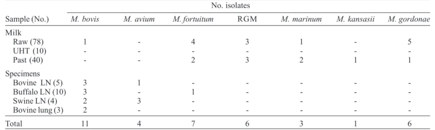
Mycobacteria were isolated from 15 of 22 (68.2%) caseous lesions from livestock and from 23 of 128 (18%) milk samples (Table I). Table II shows the differential characteristics used to identify the environmental myco-bacteria isolated from pathologic specimens and milk samples. Eleven isolates were identified as M. tuberculo-sis complex by PCR identification of IS6110 and were sub-sequently determined to be M. bovis by PCR-RFLP identification of oxyR (Figure). The amplification of a 548-bp region of oxyR by PCR, followed by digestion with
AluI resulted in the generation of 5 DNA bands for M. bovis compared to 4 bands for M. tuberculosis. No M. tuberculosis isolates were found in any sample tested. Of the 11 M. bovis isolates, 3 were found among the 5 bovine lymph nodes and 10 water-buffalo lymph nodes examined, 2 were found among 4 porcine lymph nodes, 2 were found among 3 bovine lung fragments, and 1 was found among the 128 milk samples tested (Table I). Four
M. avium isolates and 1 M. fortuitum isolate were also found in pathologic specimens. Nontuberculous myco-bacteria were found in 9 (22.5%) of the pasteurized milk samples and 14 (16.7%) of the raw milk samples. No my-cobacteria were found in the 10 UHT milk samples tested. Nontuberculous Mycobacterium species isolated from milk samples included M. fortuitum (6 isolates), M. marinum (3 isolates), M. kansasii (1 isolate), M. gordonae
(6 isolates), and 6 unidentified rapidly growing Mycobac-terium isolates.
DISCUSSION
iden-321 321321 321321 Mem Inst Oswaldo Cruz, Rio de Janeiro, Vol. 98(3), April 2003
tified by PCR and RFLP analysis. These molecular meth-ods should be particularly useful for dysgonic slowly growing mycobacterial strains for which biochemical and growth data are difficult to obtain but for which even mini-mal growth can yield sufficient DNA for PCR amplifica-tion. The difference among the growth rates of mycobac-teria species may be justified by differences in key respi-ratory pathways or energy production, in the velocity of oxygen and nutrients diffusion across the cell envelope, in the rate of lipid synthesis and assimilation by cell walls,
or finally in the number of rRNA operon (Goodfellow & Magee 1977). There is clear correlation between slowly growing capacity with disease chronicity and the ability of survive of mycobacteria in a latent state in the host (Goodfellow & Magee 1977). By PCR-RFLP method, we could differentiate M. bovis from M. tuberculosis using specific oxyR nucleotide 285. This method showed rapid, highly sensitive and specific. Algorithms based on re-striction analyses of additional polymorphic gene regions have facilitated the identification of at least 48
Mycobac-TABLE I
Distribution of 38 Mycobacterium isolates from 22 pathologic specimens and 128 milk samples
No. isolates
Sample (No.) M. bovis M. avium M. fortuitum RGM M. marinum M. kansasii M. gordonae
Milk
Raw (78) 1 - 4 3 1 - 5
UHT (10) - - -
-Past (40) - - 2 3 2 1 1
Specimens
Bovine LN (5) 3 1 - - - -
-Buffalo LN (10) 3 - 1 - - -
-Swine LN (4) 2 3 - - - -
-Bovine lung (3) 2 - - -
-Total 11 4 7 6 3 1 6
RGM: unidentified rapidly growing Mycobacterium species; UHT: ultra-high temperature treatment; LN: lymph node; Past: pasteurized
TABLE II
Differential characteristics used to identify mycobacteria from milk and pathology specimens
Mycobacterium RGM M. tuberculosis M.gordonae M. marinum M. kansasii M. avium
Test or property fortuitum complex
Number of isolates 7 6 11 6 3 1 4
Mycolic acids I.V I.VI I.III.IV I.III.IV I.III.IV I.III.IV I.IV.VI
Growth in less than 7 days + + - - - -
-Growth at 25°C + + - + + -
-Growth at 37°C + + + + - + +
Photoreative pigment - - - - + +
-Pigment in dark - - - + - -
-Growth in presence of:
PNB (500 µg/ml) + + - + + + +
TCH (2 µg/ml) + + - + + + +
NaCl (5%) + + - - - -
-Growth on:
McConkey agar + - ND ND ND ND ND
Nutrient agar + + - - + -
-Niacin production - - - - + -
-Nitrate reduction + + - - - +
-Arylsulfatase (3 days) + - - - + -
322 322 322 322
322 Mycobacteria from Livestock Specimens and Milk • Clarice Q Fujimura Leite et al.
terium species (Hernandez et al. 1999, Roth et al. 2000), including those found in our study.
Although M. tuberculosis was not isolated from any sample, M. bovis was isolated from all types of patho-logic samples evaluated and from raw milk. These data support the premise that livestock tissue and milk samples may be a reservoir for M. bovis transmission in these re-gions of Brazil. Concerning the importance of this for the public health, it is necessary and urgent to check if M. bovis is also prevalent in the other regions. Because M. bovis is a primary pathogen causing bovine tuberculosis infections among livestock, it is of particular concern that this species was obtained in 11 of 38 positive cultures (28.9%). Of even greater concern is the presence of M. bovis in one raw milk sample. Since approximately 50% of all milk consumed in Brazil is not pasteurized, consumers are at risk for M. bovis infection.
The additional Mycobacterium species identified from pathologic specimens and milk samples, including M. avium, M. fortuitum, M. marinum and M. kansasii, are considered potentially pathogenic and cause a variety of clinical manifestation in humans (Wolinsky 1992). In a national surveillance of mycobacteriosis, from 500 cul-tures of nontuberculous mycobacteria, M. avium was the major isolates with 44.1% followed by M. kansasii with 13.7%and M. fortuitum with 10.8% (Barreto & Campos 2002). Particularly in immunocompromised patients, the mycobacterial infection caused by M. avium is second only to tuberculosis in prevalence in Brazil (Saad et al. 1997). Although it has been established that pasteuriza-tion kills M. tuberculosis in milk (Hosty & McDurmont 1975), survival of some nontuberculous Mycobacterium
species after simulated laboratory pasteurization has been reported (Harrington & Karison 1965, Grant et al. 1996, Stabel et al. 1997). The organisms found among our pas-teurized samples are among the species known to survive pasteurization. But this does not exclude the possibility
of contamination during the bottling process. Our find-ing, that none of the milk subject to UHT yielded any microorganisms, supports the former possibility. Other mycobacterial species found in this study, particularly M. gordonae and the unidentified rapidly growing organ-isms, have been previously found extensively in the Bra-zilian environment, including natural water systems (Falcão et al. 1993).
The presence of M. bovis, and other potentially patho-genic mycobacteria in livestock tissue and milk suggests that humans may be exposed to these organisms as the result of ingestion. Decreases in food associated myco-bacterial disease in Brazil may be achieved through in-creased surveillance of livestock products, particularly milk, for the presence of mycobacteria and intensification of measures to avoid food contamination.
REFERENCES
Barreto AMW, Campos CED 2000. Micobactérias “não tuberculosas” no Brasil. Bol Pneumol Sanit 8: 23-31. Brasil 1994. Manual de Bacteriologia da Tuberculose, Centro
de Referência Professor Hélio Fraga, Fundação Nacional de Saúde, Ministério da Saúde, Rio de Janeiro, 114 pp. Bryan FL 1969. Infections due to miscellaneous
microorgan-isms. In H Riemann, Food-borne Infections and Intoxica-tions, Academic Press, New York, p. 224-288.
Caffrey JP 1994. Status of bovine tuberculosis eradication programmes in Europe. Vet Microbiol 40: 1-4.
Chapman JS, Speight M 1968. Isolation of atypical mycobac-teria from pasteurized milk. Am Rev Resp Dis 98: 1052-1054.
De los Monteros LE, Galán JC, Gutiérrez M, Samper S, Marín JFG, Martin C, Dominguez L, Rafael L, Baquero F, Gomes-Mampaso E, Blasquez J 1998. Allele-specific PCR method based on pncA and oxyR sequences for distinguishing Myco-bacterium bovis from MycoMyco-bacterium tuberculosis: intraspe-cific M. bovis pncA sequence polymorphism. J Clin Microbiol 36: 239-242.
Eisenach KD, Cave MD, Bates JH,Crawford JT 1992. Poly-merase chain reaction amplification of a repetitive DNA sequence specific for Mycobacterium tuberculosis. J Infect Dis 161: 977-981.
Falcão DP, Valentini SR, Leite CQF 1993. Pathogenic or poten-tially pathogenic bacteria as contaminants of fresh water from different sources in Araraquara, Brazil. Wat Res 27: 1737-1741.
Goodfellow M, Magee JG 1997. Taxonomy of mycobacteria. In PRJ Gangadharam, PA Jenkins (eds), Mycobacteria I Basic Aspects, Chapman and Hall, New York, p. 1-72. Grant IR, Ball HJ,Rowe MT 1996. Thermal inactivation of
several Mycobacterium spp. in milk by pasteurization. Lett Appl Microbiol 22: 253-256.
Gutiérrez M, Samper S, Gavigan JA, Marín JFG, Martin C 1995. Differentiation by molecular typing of Mycobacte-rium bovis strains causing tuberculosis in cattle and goats. J Clin Microbiol 33: 2953-2956.
Harrington R, Karison G 1965. Destruction of various kinds of mycobacteria in milk by pasteurization. Appl Micobrobiol 13: 494-496.
Hernandez SM, Morlock G, Butler WR, Crawford J, Cooksey RC 1999. Identification of Mycobacterium species by PCR-restriction fragment length polymorphism using fluores-cent capillary electrophoresis. J Clin Microbiol 37: 3688-3692.
Hosty TS, McDurmont C 1975. Isolation of acid-fast organ-Differentiation of Mycobacterium tuberculosis from M. bovis by
polymerase chain reaction-restriction fragment lenght polymor-phism analysis of oxy R. Lanes - 1, 6: M. tuberculosis H37Rv; 2: M. bovis AN5; 3, 4, 5: bovine lymph nodes; 7, 8: buffalo lymph nodes; 9, 10: bovine lung fragments; 11, 12: porcine lymph nodes
323 323323 323323 Mem Inst Oswaldo Cruz, Rio de Janeiro, Vol. 98(3), April 2003
isms from milk and oysters. Health Lab Sci 12: 16-19. Kantor IN, Ritacco VB 1994. Bovine tuberculosis in Latin
America and the Caribbean: current status, control and eradi-cation programs. Vet Microbiol 40: 5-14.
Leite CQF, Souza CWO, Leite SR de A 1998. Identification of mycobacteria by thin layer chromatographic analysis of mycolic acids and conventional biochemical method: four years of experience. Mem Inst Oswaldo Cruz 93: 801-805. Roth A, Reischel U, Streubel A, Naumann L, Kroppenstedt RM, Habicht M, Fisher M, Mauch H 2000. Novel diagnos-tic algorithm for identification of mycobacteria using ge-nus-specific amplification of the 16S-23S rRNA gene spacer and restriction endonuclease. J Clin Microbiol 38: 1094-1104.
Roxo E 1997. M. bovis as an agent of zoonoses. Rev Ciênc Farm 18: 101-108.
Saad MHF, Vicent V, Dawson DJ, Pallaci M, Ferraroli L, Fonseca LS 1997. Analysis of Mycobacterium avium complex serovars isolated from AIDS patients from Southeast Bra-zil. Mem Inst Oswaldo Cruz 92: 471-475.
Scorpio A, Collins D, Whipple D, Cave D, Bates J, Zhang Y 1997. Rapid differentiation of bovine and human tubercle bacilli based on a characteristic mutation in the bovine pyrazinamidase gene. J Clin Microbiol 35: 106-110. Sreevatsan S, Escalante P, Xi Pan, Gilles II DA, Siddiqi S, Khalaf
CN, Kreiswirth BN, Bifani P, Adams LG, Ficht T, Perumaalla VS, Cave MD, Van Embden JD, Musser JM 1996. Identification of a polymorphic nucleotide in oxyR specific for M. bovis. J Clin Microbiol 34: 2007-2010. Stabel JR, Steadham EM, Bolin CA 1997. Heat inactivation of
Mycobacterium paratuberculosis in raw milk: Are current pasteurization conditions effective? Appl Environ Microbiol 63: 4975-4977.
Stonebrink B, Duoma J, Manten A, Mulder RJ 1969. A com-parative investigation on the quality of various culture me-dia as used in the Netherlands for the isolation of mycobac-teria. Selected Papers (The Hague) 12: 5-47.
Sweeney RW, Whitlock RH, Rosenberger AE 1992. Mycobac-terium paratuberculosis cultured from milk and supramam-mary lymph nodes of infected symptomatic cows. J Clin Microbiol 30: 166-171.
Vitale F, Capra G, Maxia L, Reale S, Vesco G, Caracappa S 1998. Detection of Mycobacterium tuberculosis complex in cattle by PCR using milk, lymph node aspirates, and nasal swabs. J Clin Microbiol 36: 1050-1055.
Wolinsky E 1992. Mycobacterial diseases other than tubercu-losis. Clin Infect Dis 15: 1-12.