online | memorias.ioc.fiocruz.br
Schistosomiasis control programs have had signifi-cant success controlling the morbidity caused by these infections. However, these programs have not decreased the transmission area in Brazil and in other regions around the world (WHO 2001). Schistosomiasis is prev-alent in 76 countries and affects 200 million individuals, of whom 100 million present some type of clinical occur-rence and 20 million present a more severe form of the disease (WHO 2002). The disease occurs predominantly in the poorest regions of a country and is associated with poor sanitary conditions and adverse social economical conditions. It is estimated that 2.5 million people were infected in Brazil in 2000 and 25 million people are at risk of infection in a 1 million km2area (WHO 2001).
Schistosomal myeloradiculopathy (SMR) is the most severe ectopic form of schistosomiasis (Lambertucci 1993). Three main species of Schistosoma cause lesions in the central nervous system: Schistosoma mansoni, Schistosoma haematobium and Schistosoma japonicum. However, while S. japonicum is frequently associated with clinical cerebral lesions, cases of SMR caused by S. japonicum have only been rarely described.
The pathogenesis of SMR has not been fully eluci-dated, but is accepted that the inflammatory response of the host causes destruction of nervous tissues around the egg that is deposited on the spinal cord and that this inflammation also plays a central role in the medullar le-sion. SMR has been reported more frequently in patients with a chronic intestinal form of schistosomiasis and
appears to be less frequent in acute and hepatosplenic forms of the disease (Pittella 1991, 1997, Gonçalves et al. 1995, Ferrari et al. 2001, Silva et al. 2004a, Lambertucci et al. 2005). Clinically, SMR presents with lower back pain radiating to the lower limbs, followed by a decrease in the muscular strength in these limbs and the loss of sphincter control. Generally, the progression of the dis-ease may take days or weeks. SMR is diagnosed based on the clinical manifestations, the clinical determination of schistosomal infection using immunological or para-sitological methods and the exclusion of other causes of myelopathy (CDC 1984, MS/SVS 2006). The prognosis depends on how soon proper therapy (anti-schistosomal drugs and corticosteroids) can be started.
The prevalence of SMR is unknown and the number of reported cases has increased in the past few years, probably due to the improvement of diagnostic methods. Brazil has had the greater number of reported SMR cases. A series of cases from high-endemic areas have been published (Nobre et al. 2001, Peregrino et al. 2002, Fer-rari et al. 2004) and there have been reports of isolated cases in patients that have been in transmission areas (CDC 1984, Gellido et al. 2000, Chen et al. 2006). As SMR is a form of schistosomiasis that is not related to a high worm burden, it is likely to occur in low-endem-ic areas. The objective of this paper was to determine the occurrence of SMR in a low-endemic area around Campinas, São Paulo (SP), Brazil.
PATIENTS, MATERIALS AND METHODS
Studied area - Campinas is a city of nearly one mil-lion inhabitants (SMS 2007), located in SP (22°50’S 47°00’W), the seat of Campinas metropolitan region, which is composed of 19 cities and towns with ap-proximately 2,620,000 inhabitants distributed over 3,645,666 km2. Campinas is economically character-+ Corresponding author: arrfreitas@yahoo.com.br
Received 9 January 2009 Accepted 16 September 2009
Schistosomal myeloradiculopathy in a low-prevalence area: 27 cases
(14 autochthonous) in Campinas, São Paulo, Brazil
André Ricardo Ribas Freitas/+, Augusto César Penalva Oliveira, Luiz Jacintho Silva
Departamento de Clínica Médica, Faculdade de Ciências Médicas, Universidade Estadual de Campinas, Campinas, SP, Brasil
Schistosomal myeloradiculopathy (SMR) is a form of schistosomiasis that is not linked with a high worm burden but rather is found in patients who have been sporadically exposed to Schistosoma mansoni. This paper aims to determine the occurrence of SMR in a low-endemic area with urban transmission in Campinas, São Paulo, Brazil. A retrospective study was performed, identifying confirmed cases in the two largest public hospitals on the region. Pa-tients were diagnosed with SMR using standardised criteria, common clinical parameters, evidence of schistosomal infection and exclusion of other causes of myelopathy. A total of 27 patients were identified; 19 (85.2%) were men and four (14.8%) were women, ranging from 13-57 years of age (mean = 31.2; standard deviation = 12.8).Patients were classified as autochthonous (n = 14; 51.9%) or allochthonous (n = 11; 40.7%) and epidemiological data could not be obtained for two patients (7.4%). The clinical parameters of these patients were not different from previous studies. The sensitivity of serum immune reactions, cerebrospinal fluid immune reactions and parasitological stool examinations in identifying infected individuals was 87.5%, 93.8% and 40%, respectively. The epidemiological im-portance of these findings and their relationship with the control policies of schistosomiasis are discussed.
ised as an industrial, agroindustrial and service indus-try with many scientific research institutions. It also has a large number of health facilities (SEP/SP 2007). Epidemiologically, SP is characterised as having iso-lated areas of schistosomiasis (Silva 1992, Camargo-Neves 2007). Although schistosomiasis is a disease requiring mandatory notification, it is rarely done. The number of cases of schistosomiasis reported by the city of Campinas averaged 455 per year between the years 2000-2005 and, of these cases, 109 (23.9%) were con-sidered autochthonous (SES/CVE-SP 2008).
Study design - A retrospective study was performed in the Hospital de Clínicas of Universidade Estadual de Campinas (HC-UNICAMP) and in the Hospital e Maternidade Celso Pierro of Pontifícia Universidade Católica de Campinas (HMCP-PUC Campinas). These hospitals were selected because together they support 71% of the patients from Sistema Único de Saúde of Campinas. In addition, these hospitals have a variable proportion of patients from other cities in the metro-politan regions and from other Brazilian regions, in addition to having outpatient clinics, infirmaries and diagnostic centres that are able to treat patients with SMR (SMS 2007). The patients were selected through an active search of the admission data bases, outpatient records, results of lab tests and the National System of Information of Diseases with Mandatory Notifica-tion (SINAN). Patients diagnosed with SMR that were examined in these hospitals between 1995-2005 were included in this study. Patients that did not meet the established diagnostic criteria were excluded. The cri-teria were the following: (i) spinal or myeloradicular
syndrome, (ii) demonstration of schistosomal infection by parasitological or immunological methods and (iii) exclusion of other causes of myeloradiculopathy (CDC 1984, MS/SVS 2006). Clinical, epidemiological and laboratory data, as well as imaging examinations of the patients, were gathered. The patients were classi-fied as “autochthonous” (i.e., those who were in contact with contaminated bodies of water around Campinas region and not in other regions), “allochthonous” (i.e., those who were in contact with contaminated bodies of water in other endemic regions and had no contact with contaminated bodies of water around the region of Campinas), “undetermined” (i.e., those whose previ-ous exposure standard does not fit in any previprevi-ously mentioned) or patients “without epidemiological data” (i.e., those whose information did not allow classifica-tion and could not be located for an interview). All au-tochthonous patients were interviewed for possible data correction about the exposure.
The patients were classified according to the out-come: “complete recovery”, the absence of symptoms and normal neurological examination, “partial recovery with no functional limitations”, marked improvement but also presented mild symptoms that did not hinder daily activities (e.g., patients with paraesthaesia in the lower limbs or slight difficult in urinating), “partial recovery with functional limitations”, symptoms that change the daily routine of the patient (e.g., the use of a bladder probe or a patient who walks with some kind of support) or “absence of recovery”, the absence of a marked recovery or only a slight recovery that has not changed the patient’s functional condition (Peregrino et al. 2002, Ferrari et al. 2004).
TABLE I
Frequency of symptoms and time passed between the start of clinical problems and presentation of each symptom of 27 patients with presumptive diagnosis of schistosomal myeloradiculopathy examined
in hospitals of Campinas, São Paulo, Brazil between 1995-2005
Clinical information Frequency
Time in days from the start of the clinical status until the symptom manifestation
N na % Mean Median SD Minimum Maximum
Lower limbs paresis 27 27 100 35.5 5 94.7 0 360
Sensitivity alteration in lower limbs 27 27 100 83.2 1 215.1 0 730
Bladder dysfunction 25 27 92.6 35.8 7 84.6 0 355
Triadb 25 27 92.6 69.8 7 168.8 0 730
Anal dysfunction 19 25 76 67.1 7 125.1 0 365
Erectile dysfunctionc 9 12 75 149.8 48 278.3 3 730
Back pain 20 27 74.1 5.7 0 20.7 0 90
Pain in lower limbs 18 27 66.7 20.9 0 81 0 335
Other sensitivity changes in lower limbs 25 27 92.6 52.6 4 160.7 0 730
Walking disability 16 27 59.3 32.9 5 88.4 0 350
Result analysis - The statistical program MINITAB Release 14.12.0 Statistical Software was used for data analysis. To describe the findings, the central tenden-cy and variability measures were used in addition to graphic and tabular resources of descriptive statistics. For proportion evaluation, the proportion equality test and Fisher’s exact test were used. The evaluation of the differences between continuous variable distributions was done using Mann-Whitney’s test. To define the geographic coordinates of residence and infection loca-tions and to calculate these distances for inhabitants of Campinas region, the program Google Earth 4.0.2416 (beta) Build Date Oct 31 2006 for Microsoft Windows XP was used. To calculate the distance between the residence of patients living in Campinas and the lakes where these patients were infected, the program Map Info 8.0 was used.
Ethical aspects - This research was authorised by the Ethical Committee of the Faculdade de Ciências Médicas-UNICAMP, respecting the law 196/96 of the Ministry of Health and National Health Council (MS/ CNS 1996). Regarding the guidelines of Declaration of Helsinki, modified in Hong Kong in 1989 to preserve the ethical aspects of research, all patients provided in-formed consent after being provided an explanation of the Informed Consent Form, which was previously ap-proved by the Institutional Ethical Committee.
RESULTS
Epidemiological profile of patients - A total of 30 patients who were diagnosed with SMR were identi-fied. Of these, three were excluded from the study: two because other diseases had not been investigated prop-erly and one who tested positive for human T lympho-tropic virus (HTLV). Therefore, 27 patients remained in the study, 22 from HC-UNICAMP and five from HMCP-PUC Campinas. Of these 27 patients, 11 (41%) were notified by SINAN. The remaining patients were located exclusively through the hospital registry data-base and the active search of lab tests. From these 27 patients, seven (patients 2, 16, 17, 18, 11, 14 and 25) had already been published in national journals by other au-thors as isolated cases (Tedrus et al. 1996, 1999, Lima et al. 1999, Faria et al. 2002).
Twenty-three (85.2%) patients were male and four (14.8%) were female. The age at the time of the diag-nosis ranged from 13-57 years [mean = 31.2; standard deviation (SD) = 12.8 and median = 29] and the time between the first visit and the diagnosis averaged 70.6 days (median = 19; SD = 166.9).
Based on the probable location of infection, the patients were classified as autochthonous (n = 14; 51.9%), with 11 patients from Campinas city and three from other towns in this region, allochthonous (n = 11; 40.7%) or patients without epidemiological data (n = 2; 7.4%). No patient was classified as “undeter-mined”. All patients classified as autochthonous were infected in foci located in urban areas no more than 4.450 m from their homes. Except for patient 9, who was infected during his work activities, the remaining
autochthonous patients were infected during sporadic recreational activities. All allochthonous patients were infected in high-endemic areas in the states of Minas Gerais (MG), Alagoas, Sergipe or Bahia in Brazil.
Clinical presentation- The most frequent symptom was lower limbs paresis (n = 27; 100%), followed by sensitivity changes in the lower limbs and bladder dys-function (n = 25; 92.6%). All three symptoms (blad-der dysfunction, paresis and sensitivity changes in the lower limbs) were observed in 25 (92.6%) patients. Half of the patients that presented these symptoms reported the symptoms seven days after the beginning of the manifestation. Although thoracic and back pain is of- Although thoracic and back pain is of-ten reported as an early symptom, it was not among the most frequently reported symptoms (Table I), af-fecting only 18 patients (66.7%). In contrast, erectile dysfunction, although a late symptom (median of start 48 days), had a higher relative frequency among men 75% (12 patients). As expected, there was no statistical difference in the relative frequency of the symptoms presented by the autochthonous and allochthonous pa-tients (Table II).
Results of complementary examinations - Magnetic resonance imaging (MRI) or computerised tomography were performed in all cases and helped to exclude other causes of myelopathies. Figs 1 and 2 show the MRI of patient 2, with gadolinium contrast and granular aspect.
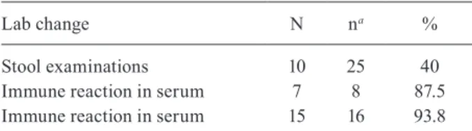
Among non-specified lab results from the initial exam, hyperproteinorachia [proteins in cerebrospi-nal fluid (CSF) > 45 mg/dL] was found in 23 patients (85.2%), pleocitosis in CSF in 22 patients (81.5%) and eosinorachia occurred in 11 patients (40.7%). The most significant change in the blood count was eosinophilia, which occurred in nine patients (36%) (Table III). The time between the beginning of the clinical manifesta-tion and examinamanifesta-tion ranged from 2-1,095 days (mean = 117.3; SD = 255.7).
TABLE II
Frequency of symptoms autochthonous and allochthonous patients with presumptive diagnosis of schistosomal myeloradiculopathy examined in hospitals of Campinas, São Paulo, Brazil between 1995-2005, before treatment
Autochthonous Imported Fisher’s exact test
N na % N n % p
Lower limbs paresis 14 14 100 11 11 100 1
Bladder dysfunction 13 14 93 11 11 100 1
Anal dysfunction 8 14 57 10 11 91 0.09
Erectile dysfunctionb 6 8 75 3 3 100 1
Back pain 12 14 86 8 11 73 0.623
Pain in lower limbs 8 14 57 9 11 82 0.233
Other sensitivity changes in lower limbs 12 14 86 11 11 100 0.486
Walking disability 9 14 64 7 11 64 1
a: because this is a retrospective study, not all patients provided all information. Only patients whose information was available have been included in the calculation (n); b: to calculate the relative frequency of erectile dysfunction, only men whose clinical records presented this information were included.
TABLE III
Frequency of unspecific lab findings altered in 27 patients with presumptive diagnosis of schistosomal myeloradiculopathy examined in hospitals of Campinas, São Paulo, Brazil between 1995-2005
Lab change N na %
Hyperproteinorachia (protein in CSF > 45 mg/dL) 23 27 85.2
Pleocitosis in CSF (white blood cells > 5/mm3) 22 27 81.5
Eosinorachia 11 27 40.7
Eosinophilia (> 500/mm3 in the peripheral blood) 9 25 36
a: because it is a retrospective study not all patients provided all information. For calculations of relative frequency only patients whose information was available have been included (n); CSF: cerebrospinal fluid.
in any of the samples. Patient 4 has also had a rectal biopsy, which showed no presence of S. mansoni eggs. Importantly, in the patients whose stool samples were submitted to quantitative exams, a low quantity of eggs was detected: two patients had eight eggs per gram of faeces (EPG), one had 19 EPG and one had 48 EPG using the Kato-Katz technique. Only two patients had a spinal cord biopsy done because of a suspected neo-plasm (patients 6 and 9); an exudative-necrotic granu-loma was found in both of them. Figs 3 and 4 demon-strate the intense granulomatous reaction around the S. mansoni egg in the nervous tissue of the spinal cord of patient 6, who underwent a surgical procedure to inves-tigate the possibility of a spinal cord tumour, which, in the end, was diagnosed as SMR.
All patients were investigated for syphilis, tuber-culosis, cysticercosis, AIDS, neoplasia, fungal infec-tions, bacterial infections and vitamin B12 deficiency because they are part of the differential diagnosis for non-traumatic spinal cord lesions. Patient 6 was an exception, as his first diagnosis was of a spinal cord
tumour and, with the biopsy, SMR was confirmed. Therefore, the other tests were not required. Diseases such as HTLV infection, autoimmune disease, toxo-plasmosis, cytomegalovirus, herpes virus and Chagas disease were also investigated according to the clinical presentation of patients.
mini-Fig. 1: sagittal magnetic resonance imaging of patient 2 in T2 with granular impregnation pattern of gadolinium magnetic contrast capti-vation in this image of thoracic-lumbar spine.
Fig. 2: axial magnetic resonance imaging of patient 2 in T2. It is ob-served the hypersignal in the medulla on thoracic-lumbar levels.
mum time for diagnosis were also higher for the auto-chthonous patients. This may be because the physicians may not have suspected the existence of autochthonous SMR in SP.
Twenty-three patients (85.2%) were treated with an-ti-schistosomal drugs and corticosteroids. The remain-ing four (14.8%) were treated with anti-schistosomal drugs only.
Table VI shows the outcome distribution presented by the patients. Only four patients (14.8%) demonstrat-ed complete recovery, six patients (22.2%) reportdemonstrat-ed a partial recovery with no functional limitations, 13 pa-tients (48.1%) had a partial recovery with functional limitations and four (14.8%) did not demonstrate a measurable recovery. The worsening of the outcome could be correlated with the delayed introduction of the anti-schistosomal drugs and corticosteroid (Figs 5, 6).
To evaluate the outcome, we compared the clinical status before and after recovery. The interval between
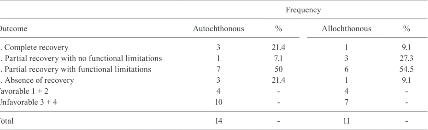
the beginning of treatment and final evaluation ranged from 10-4,380 days (mean ± SD = 1480 ± 1159.3; me-dian = 1460). Table VII shows the relative frequency of each of the related symptoms. There was statisti-cally significant recovery for all symptoms, except for erectile dysfunction. In the final evaluation, the most frequent remaining symptoms were walking difficul-ties (n = 17; 63%), urinary dysfunction (n = 13; 48.1%) and erectile dysfunction among males (n = 11; 33.3%). Table VIII shows the outcome of the autochthonous and allochthonous patients according to the standard classification; the outcome was similar in both groups (autochthonous and allochthonous), but it was not pos-sible to statistically verify the differences between the groups due to the small number of participants. The difference in the frequency of each symptom after the treatment between the two groups was also not statisti-cally significant (Table IX).
Table X shows the outcome of patients in the cur-rent study and other reported cases that used the same classifications. While it is not possible to compare the studies precisely because subjective evaluation meth-odologies were used, the patients examined in Campi-nas city between 1995-2005 appeared to have a worse outcome than those found in the literature. Because the previous studies are from two research groups from high-endemic areas (MG and Pernambuco), they most likely have more expertise and more resources to di-agnose and treat SMR than the professionals in low-endemic areas.
DISCUSSION
The age and gender distribution of patients pre-sented in this paper were very close to those described in the literature: SMR is a disease that predominantly affects male teenagers and young adults (Ferrari 1999, Silva et al. 2004a). Because it affects a young
popu-TABLE IV
Frequency of specific exams for schistosomiasis in 27 patients presumptive diagnosis of schistosomal myeloradiculopathy examined with in hospitals of Campinas,
São Paulo, Brazil between 1995-2005
Lab change N na %
Stool examinations 10 25 40
Immune reaction in serum 7 8 87.5
Immune reaction in serum 15 16 93.8
lation at the beginning of their economically active life and as recovery is often associated with lingering symptoms with serious consequences for life quality of the individual and his family, the morbidity associated with this disease should be reinforced.
As expected, the clinical presentation of SMR in the autochthonous group was not different from the presentation of the non-autochthonous group and it was not different from that described in the literature (Ferrari 1999, 2004, Peregrino et al. 2002, Transverse Myelitis Consortium Working Group 2002, Silva et al. 2004a, b): a spinal cord disease of acute or sub-acute character with sensitive, motor and autonomic symp-tomatology. Importantly, regardless of the level of en-demicity, patient exposure to contaminated bodies of water in transmission areas should suggest this etiolo-gy to healthcare professionals. The majority of patients (51.9%, 14 patients) were classified as autochthonous in a region where the least patients notified in the SINAN are autochthonous (23.9%). This data suggests that the proportion of patients with spinal cord forms among the total of schistosomiasis-carriers is higher among patients from low-endemic areas than among patients from high-endemic areas.
TABLE V
Time in days passed among clinically relevant moments of autochthonous and imported patients with presumptive diagnosis of schistosomal myeloradiculopathy examined in hospitals of Campinas, São Paulo, Brazil between 1995-2005
Autochthonous Allochthonous
Mann- Whitney’s test
Interval in days between Mean Median SD Mean Median SD p
First symptoms and complete clinical condition 42.6 9 88.3 174.6 9 291.3 0.804
First symptoms and the first visit 47.2 10 107.3 213.7 22 369.5 0.336
First visit and diagnosis 112 26 224.3 23.9 9 42.7 0.0247
Time of follow-up 1833.5 2036 1019.4 1077.9 485.5 1231.7 0.381
SD: standard deviation.
Figs 3, 4: optical microscopy with Hematoxin Eosin coloration where it is verified granuloma in medullar nervous tissue of patient 6 (arrows).
us-TABLE VI
Distribution of 27 patients with assumed diagnosis of schistosomal myeloradiculopathy attended in hospitals
of Campinas, São Paulo, Brazil between 1995-2005, according to evolution
Frequency
Outcome N (na = 27) %
Complete recovery 4 14.8
Partial recovery with
no functional limitations 6 22.2
Partial recovery
with functional limitations 13 48.1
Absence of recovery 4 14.8
a: because it is a retrospective study not all patients pro-vided all information. For calculations of relative frequency only patients whose information was available have been included (n).
ing 3-5 stool samples. In the series of 13 patients stud-ied by Carod-Artal et al. (2004) the sensitivity of stool examination was 15.4%; in the thesis by Lima (2004), in which 65 patients were studied, the sensitivity was 44.6%. Silva et al. (2004b) reported a sensitivity of 25% in a series of 16 patients who all had three stool samples analysed, while Peregrino et al. (2002) found a sensitiv-ity of 42.5% in a series of 80 patients. Finally, Peregrino et al. (2002) reported a sensitivity of 42.9% and Andrade (1986) reported a sensitivity of 25%.
The previously reported sensitivity of the CSF im-mune reaction was higher (92.8%) than the sensitivity observed in the current study (87.5%). Because this is a retrospective study, it is possible to rule out other selection biases and other factors, such as methodological differ-ences. However, we can affirm that, in general, the im-mune reactions in the CSF or serum were more sensitive than stool examination. As the parasitological tests are always used as pattern exams for the programs of schis-tosomiasis control, the ability to identify patients that are vulnerable to this form of schistosomiasis is very low.
TABLE VII
Frequency of symptoms observed before and after recovery in 27 patients with presumptive diagnosis of schistosomal myeloradiculopathy examined in hospitals of Campinas, São Paulo, Brazil between 1995-2005
Pre-treatment Post-treatment Fisher’s exact test
N na % N na % p
Walking disability 16 27 59.3 4 27 14.8 0.002
Lower limbs paresis 27 27 100 17 27 63 0.001
Bladder dysfunction 25 27 92.6 13 27 48.1 0.001
Anal dysfunction 19 27 70.4 6 27 22.2 < 0.001
Erectile dysfunctionb 9 12 75 5 15 33.3 0.054c
a: because it is a retrospective study not all patients provided all information. For calculations of relative frequency only patients whose information was available have been included (n); b: for calculation of relative frequency of erectile dysfunction women have been excluded from the calculation; c: the sole variable, which proportion has not shown statistically significant difference before and after recovery was erectile dysfunction.
TABLE VIII
Distribution of outcome of autochthonous and allochthonous patients with presumptive diagnosis of schistosomal myeloradicu-lopathy examined in hospitals of Campinas, São Paulo, Brazil between 1995-2005
Frequency
Outcome Autochthonous % Allochthonous %
1. Complete recovery 3 21.4 1 9.1
2. Partial recovery with no functional limitations 1 7.1 3 27.3
3. Partial recovery with functional limitations 7 50 6 54.5
4. Absence of recovery 3 21.4 1 9.1
Favorable 1 + 2 4 - 4
-Unfavorable 3 + 4 10 - 7
-TABLE IX
Frequency of symptoms in autochthonous and allochthonous patients with presumptive diagnosis of schistosomal myeloradiculopathy examined in hospitals of Campinas, São Paulo, Brazil between 1995-2005, after treatment
Autochthonous Allochthonous Fischer’s exact test
N na % N na % p
Lower limbs paresis 8 14 57 9 11 82 0.233
Bladder dysfunction 7 14 50 5 10 50 1
Anal dysfunction 3 14 21 2 10 20 1
Erectile dysfunctiona 4 10 40 1 5 20 0.600
Thoracic or back pain 12 14 86 8 11 73 0.623
Pain in lower limbs 8 14 57 9 11 82 0.233
Other sensitivity changes in lower limbs 12 14 86 11 11 100 0.486
Walking disability 3 14 21 1 11 9 0.604
a: to calculate the relative frequency of erectile dysfunction, only men whose clinical records presented this information were included.
TABLE X
Comparison between data collected in literature and the evolution of 27 patients with presumptive diagnosis of schistosomal myeloradiculopathy examined in hospitals of Campinas, São Paulo, Brazil between 1995-2005
Current study Ferrari (2004) Peregrino et al. (2002)
N
(na = 27) %
N
(n = 63) %
N
(n = 80) %
1. Complete recovery 4 14.8 20 31.7 31 38.8
2. Partial recovery with no functional limitations 6 22.2 18 28.6 33 41.3
3. Partial recovery with functional limitations 13 48.1 16 25.4 9 11.3
4. Absence of recovery 4 14.8 9 14.3 7 8.8
Favorable 1 + 2 10 37 38 60.3 64 80
Unfavorable 3 + 4 17 63 25 39.7 16 20
a: because it is a retrospective study not all patients provided all information. For calculations of relative frequency only patients whose information was available have been included (n).
Fig. 5: time in days since the beginning of clinical symptoms until the introduction of anti-schistosomal drug according to the outcome.
Considering the fact that most SMR patients have intestinal forms of schistosomiasis, the low sensitivity of the stool exams is not surprising and it makes diag-nosis more difficult. In spite of this fact, however, the low sensitivity does not hinder the treatment. A physi-cian examining a patient suspected of SMR must in-vestigate the etiology with more sensitive exams such as a mucous rectal biopsy or immunological methods. A problem, however, is that immune exams are not always easily or promptly available, particularly in low-endemic areas. The difficulty in performing the immunological methods, as well as the limited knowledge of the disease, may have contributed to the delay in the diagnosis of the autochthonous and allochthonous patients in this study. The time between the first visit and the diagnosis was 70.6 days (median = 19; SD = 166.9). This delay may have contributed to the poor outcome of the 17 patients (62.9%) that had some significant impairment in their daily lives (Table VI). The relationship between the de-lay in the initiation of treatment and unfavourable out-come is shown in Figs 5 and 6.
Another relevant aspect is that the time necessary to diagnose the autochthonous patients was longer than the time to diagnose the allochthonous patients (p = 0.0247) (Table V). One possible explanation for this difference is the fact that Campinas is a region of low endemic-ity. Therefore, not all physicians would consider schis-tosomiasis for a patient who is originally from this area. Additionally, the difficulty in diagnosing this disease stems from errors and misdiagnoses. Therefore, the identified cases could be just a few out of many cases that occurred during this time. In order to decrease the delay in diagnosis and sub-diagnosis, it is necessary to warn neurologists, urologists, physicians who work in emergency rooms and other health care professionals about the occurrence of this disease within this region.
The epidemiological vigilance system (SINAN) has not paid the necessary attention to the identification of SMR cases, with only 41% of the patients notified. Moreover, there is no space in the epidemiological file to register SMR. Therefore, the 11 notified patients have not had the neurological form of schistosomiasis regis-tered at SINAN.
Even though the outcome verified post-treatment was significant, it was not enough to be satisfactory be-cause only four patients (14.8%) had a complete recovery and the remaining symptoms have deeply lowered the quality of life of the remaining patients. The most ob-served remaining symptoms were lower limb weakness (n = 17; 63%), urinary dysfunction (n = 13; 48.1%) and erectile dysfunction (n = 5; 33.3%). Silva et al. (2004a) reported weakness in nine patients (56.3%) in a study that examined 16 patients with another evaluation six months after the treatment. Sexual dysfunction in 40% of male patients (4 out of 10) was also observed and uri-nary dysfunction was observed in 37.5% of the patients (Silva et al. 2004b). The lingering urological symptoms do not seem to respond well to clinical treatment, as ob-served in this paper and other reports from the literature (Lima 2004, Silva et al. 2004b). This is reflected both in symptom maintenance and in changes in the exams to
evaluate urinary function. Lima (2004) has evaluated the occurrence of urological symptoms in patients after re-covery in a prospective study of 65 patients. Even among these patients, who received the current acceptable treat-ment (anti-schistosomal drugs and corticosteroids), the occurrence and changed exams of urinary function were not significantly different before or after treatment, with re-evaluation after nine months. The urological linger-ing symptoms include both urinary function and erec-tile function, but the latter aspect was neglected even in studies performed by urologists (Gomes et al. 2002, 2005, Lima 2004) and this opinion is shared by other researchers (Ferrari 1999, Silva et al. 2004b).
SMR and control policies - Many previous studies have demonstrated the occurrence of SMR in patients from non-endemic areas. Most of these reports detail pa-tients from European or North American countries that have acquired SMR after being infected during travel in endemic countries (CDC 1984, Gellido et al. 2000, Chen et al. 2006). The current paper describes cases of patients from a low-endemic area. Hepatosplenic schistosomiasis is a disease more commonly diagnosed in patients living in high-endemic areas with repeated re-infections and, while SMR can occur in any schistosomiasis carrier, it seems to be more common in those that have a mild intestinal form. Several researchers have warned of the importance of distinguishing SMR from hepatosplenic schistosomiasis regarding the treatment strategy. In the latter, a mass treatment based on a decrease of worm burden in the carriers is enough to control the disease. SMR, on the other hand, will occur whenever intestinal schistosomiasis occurs, that is, as long as the transmis-sion of schistosomiasis continues (WHO 1998, Lamber-tucci et al. 2000, Richter 2003).
areas and other regions of MG, it was not designed to investigate either the likely loci of infection or the stand-ard pattern of exposure. In 2006, the Ministry of Health released a guide exclusively on this topic: Guide of Epi-demiological Surveillance and Control of Schistosomal Myeloradiculopathy (MS/SVS 2006). Therefore, there seems to be an increase in acknowledging the impor-tance of this form of schistosomiasis in the current spec-trum of epidemiology.
The current paper shows the occurrence of SMR in patients in an urban area of low endemicity who had been infected during recreational activities. As the im-portance of SMR is recognised in low endemicity (or low prevalence) settings, a few choices have to be made. The findings of this study reinforce this idea and point to a need for changing the paradigms and concepts used for controlling schistosomiasis, so that the control of the medullary form of the disease can be effective. To elimi-nate SMR, it is necessary to elimielimi-nate schistosomiasis transmission and not to simply decrease the worm load in patients.
There are many factors that may have contributed to the increase in the identification of SMR cases, including diagnostic resource improvements, increased ecotour-ism activities with the exposure of new populations, the maintenance of schistosomiasis global prevalence and an increased area of transmission, including urban areas (Silva 1985, Enk et al. 2003, Agbessi et al. 2006). The instruments for surveillance and control currently used in the programs for schistosomiasis have not been suffi-cient to decrease the SMR problem or to control it. More studies are needed to improve our knowledge of the epi-demiology of the neurological forms of schistosomiasis. Modification in the monitoring and control programs of schistosomiasis is needed for decrease schistosomal morbidity. It is very advisable to introduce more sen-sitive methods within the public health services to di-agnose chronic intestinal schistosomiasis as a means to avoid SMR, particularly in areas of low prevalence. Technology to introduce serology as routine diagnostic tool in basic health services is already available (Dias et al. 1992, Silva et al. 1998, Oliveira et al. 2005). More studies are needed to develop more effective drugs to cure schistosomiasis and more sensitive methods to ver-ify the treatment effectivity. It will be useful to disclose SMR and further educate professionals about examining these patients both in the studied region and in other ar-eas of low and high prevalence. A secondary advantage of disclosing the existence of severe forms of schisto-somiasis is the increase in the control measures taken by the population to eliminate the infection.
There is a need for a detailed study of SMR focus-ing on the epidemiological aspects such as prevalence, incidence and proportion of SMR cases among schisto-somiasis-carriers in areas of high and low endemicity and predisposing factors.
ACKNOWLEDGEMENTS
To Dr Luciano de Souza Queiroz (Queiroz & Zanardi 2006), from the Departamento de Anatomia Patológica, FCM-UNI-CAMP, for providing optical microscopy pictures of patient 6.
REFERENCES
Agbessi CA, Bourvis N, Fromentin M, Jaspard M, Teboul F, Bougnoux ME, Hanslik T 2006. Acute schistosomiasis in French travellers.
Rev Med Interne 27: 595-599.
Andrade AN 1986. Neuroesquistossomose. Arq Neuropsiquiatr 44: 175-179.
Camargo-Neves VLF. Esquistossomose mansônica. Superintendên-cia de Controle de Endemias - SUCEN 2007. [cited in 2007 jan 20]. Available from: http://www.saude.sp.gov.br.
Camargos ST, Dantas FR, Teixeira AL 2005. Schistosomal myelopathy mimicking spinal cord neoplasm. Scand J Infect Dis 37: 365-367.
Carod-Artal FJ, Vargas AP 2004. Myelopathy due to Schistosoma mansoni. A description of two cases and review of the literature.
Rev Neurol 39: 137-141.
Carod-Artal FJ, Vargas AP, Horan TA, Marinho PB, Coelho Costa PH 2004. Schistosoma mansoni myelopathy: clinical and pathologic findings. Neurology 63: 388-391.
CDC - Centers of Disease Control 1984. Acute schistosomiasis with transverse myelitis in American students returning from Kenya.
MMWR Morb Mortal Wkly Rep 33: 445-447.
Chen AW, Alam MH, Williamson JM, Brawn LA 2006. An unusually late presentation of neuroschistosomiasis. J Infect 53: 155-158.
Dias LC, Marçal Júnior O, Glasser CM, Kanamura HY, Hotta LK 1992. Control of schistosomiasis mansoni in a low transmission area. Mem Inst Oswaldo Cruz 87 (Suppl. IV): 233-239.
Drummond SC, Silva LC, Amaral RS, Sousa-Pereira SR, Antunes CM, Lambertucci JR 2006. Morbidity of schistosomiasis man-soni in the state of Minas Gerais, Brazil. Mem Inst Oswaldo Cruz 101 (Suppl. I): 37-44.
Enk MJ, Amorim A, Schall VT 2003. Acute schistosomiasis outbreak in the metropolitan area of Belo Horizonte, Minas Gerais: alert about the risk of unnoticed transmission increased by growing rural tourism. Mem Inst Oswaldo Cruz 98: 745-750.
Faria AV, Cerqueira EMF, Reis F, Pirani C, Damasceno BP, Menezes Neto JR, Zanardi VA 2002. Apresentações da esquistossomose no sistema nervoso central: correlação clinica e radiológica. Ra-diol Bras 35: 47-50.
Ferrari TC 1999. Spinal cord schistosomiasis. A report of 2 cases and re-view emphasizing clinical aspects. Medicine (Baltimore) 78: 176-190.
Ferrari TC 2004. Involvement of central nervous system in the schis-tosomiasis. Mem Inst Oswaldo Cruz 99 (Suppl. I): 59-62.
Ferrari TC, Moreira PR, Cunha AS 2001. Spinal-cord involvement in the hepato-splenic form of Schistosoma mansoni infection. Ann Trop Med Parasitol 95: 633-635.
Ferrari TC, Moreira PR, Cunha AS 2004. Spinal cord schistosomia-sis: a prospective study of 63 cases emphasizing clinical and therapeutic aspects. J Clin Neurosci 11: 246-253.
Gellido CL, Onesti S, Llena J, Suarez M 2000. Spinal schistosomia-sis. Neurology 54: 527.
Gomes CM, Hisano M, Machado LR, Figueiredo JA, Lucon AM, Trigo-Rocha FE 2005. Urological manifestations of chronic schistosomal myeloradiculopathy. BJU Int 96: 853-856.
Gomes CM, Trigo-Rocha F, Arap MA, Gabriel AJ, Alaor de Figueire-do J, Arap S 2002. Schistosomal myelopathy: urologic manifesta-tions and urodynamic findings. Urology 59: 195-200.
Lambertucci JR 1993. Schistosoma mansoni: pathological and clini-cal aspects. In Jordan P, Webbe G, Sturrock RF (eds.), Human Schistosomiasis, Cab International, Wallingford, p. 195-235.
Lambertucci JR, Serufo JC, Gerspacher-Lara R, Rayes AA, Teixeira R, Nobre V, Antunes CM 2000. Schistosoma mansoni: assessment of morbidity before and after control. Acta Trop 77: 101-109.
Lambertucci JR, Sousa-Pereira SR, Silva LC 2005. Myeloradiculopathy in acute schistosomiasis mansoni. Rev Soc Bras Med Trop 38: 277-278.
Lima PR 2004. Análise das alterações vésico-esfincterianas em pa-cientes com mielopatia esquistossomótica mansônica antes e após o tratamento medicamentoso da doença, PhD Thesis, Uni-versidade Federal de São Paulo, São Paulo, 99 pp.
Lima WL, Tose CS, Ferreira RE 1999. Diagnóstico de neuroesquis-tossomose medular após anestesia subaracnóidea: relato de caso.
Rev Bras Anestesiol 49: 338-340.
MS/CNS - Ministério da Saúde Brasil/Conselho Nacional de Saúde 1996. Resolução 196 de 10 de outubro de 1996. [cited 2006 Dec 20]. Avail-able from: http://conselho.saude.gov.br/resolucoes/reso_96.htm.
MS/SVS - Ministério da Saúde Brasil/Secretaria de Vigilância em Saúde 2002. Guia de vigilância epidemiológica, 5th ed., MS, Brasília, 842 pp.
MS/SVS - Ministério da Saúde Brasil/Secretaria de Vigilância em Saúde 2005. Guia de vigilância epidemiológica, 6th ed., MS, Brasília, 816 pp.
MS/SVS - Ministério da Saúde Brasil/Secretaria de Vigilância em Saúde 2006. Guia de vigilância epidemiológica e controle da mielorradiculopatia esquistossomótica, MS, Brasília, 32 pp.
Nobre V, Silva LC, Ribas JG, Rayes A, Serufo JC, Lana-Peixoto MA, Marinho RF, Lambertucci JR 2001. Schistosomal myeloradicul-opathy due to Schistosoma mansoni: report on 23 cases. Mem Inst Oswaldo Cruz 96 (Suppl.): 137-141.
Oliveira EJ, Kanamura HY, Lima DM 2005. Efficacy of an enzyme-linked immunosorbent assay as a diagnostic tool for schistoso-miasis mansoni in individuals with low worm burden. Mem Inst Oswaldo Cruz 100: 421-425.
Peregrino AJ, Puglia PM, Nóbrega JP, Livramento JA, Marques-Dias MJ, Scaff M 2002. Esquistossomose medular: análise de 80 ca-sos. Arq Neuropsiquiatr 60: 603-608.
Pittella JE 1991. The relation between involvement of the central nervous system in schistosomiasis mansoni and the clinical forms of the parasitosis. A review. J Trop Med Hyg 94: 15-21.
Pittella JE 1997. Neuroschistosomiasis. Brain Pathol 7: 649-662.
Queiroz LS, Zanardi VA 2006. Curso de Neuroimagem para gradu-ação em Medicina. Departamento de Anatomia Patológica - FCM - Unicamp. [cited in 2006 dec 20]. Available from: http:// anatpat.unicamp.br/casos.html.
Richter J 2003. The impact of chemotherapy on morbidity due to schistosomiasis. Acta Trop 86: 161-183.
SEP/SP - Secretaria de Economia e Planejamento do Estado de São Paulo. Agência Metropolitana de Campinas. 2007. [cited in 2007 jan. 20]. Available from: http://www.planejamento. sp.gov.br.
SES/CVE-SP - Secretaria de Estado da Saúde/Centro de Vigilân-cia Epidemiológica. São Paulo. Esquistossomose 2008. [cited 2008 dec 01]. Available from: http://cve.saude.sp.gov.br.
Silva LC, Maciel PE, Ribas JG, Pereira SR, Serufo JC, Andrade LM, Antunes CM, Lambertucci JR 2004a. Mielorradiculopatia es-quistossomótica. Rev Soc Bras Med Trop 37: 261-272.
Silva LC, Maciel PE, Ribas JG, Souza-Pereira SR, Antunes CM, Lambertucci JR 2004b. Treatment of schistosomal myelora-diculopathy with praziquantel and corticosteroids and evalua-tion by magnetic resonance imaging: a longitudinal study. Clin Infect Dis 39: 1618-1624.
Silva LJ 1985. Crescimento urbano e doença: a esquistossomose no município de São Paulo (Brazil). Rev Saude Publica 19: 1-7.
Silva LJ 1992. A esquistossomose mansônica no Estado de São Paulo: origens, distribuição, epidemiologia e controle, Associate pro-fessorship Thesis, UNICAMP, Campinas, 140 pp.
Silva RM, Kanamura HY, Camargo ED, Chiodelli SG, Nakamura PM, Gargioni C, Vellosa SA, Antunes JL 1998. A comparative study on IgG-ELISA, IgM-IFT and Kato-Katz methods for epi-demiological purposes in a low endemic area for schistosomiasis.
Mem Inst Oswaldo Cruz 93 (Suppl. I): 279-282.
SMS - Secretaria Municipal de Saúde de Campinas. TabNet Campinas. 2007. [cited in 2007 jan. 20]. Available from: http://tabnet.saude. campinas.sp.gov.br.
Tedrus GM, Fonseca LC, Nogueira-Júnior E 1999. Esquistossomose do sistema nervoso central. Rev Cienc Med Campinas 8: 28-30.
Tedrus GM, Viana MA, Fonseca LC, Araújo CA 1996. Neuroesquis-tossomose. Rev Cienc Med Campinas 5: 21-23.
Transverse Myelitis Consortium Working Group 2002. Proposed di-agnostic criteria and nosology of acute transverse myelitis. Neu-rology 59: 499-505.
WHO - World Health Organization 1998. Report of the WHO infor-mal consultation on schistosomiasis control. Geneva 2-4 Decem-ber. WHO/CDS/CPC/SIP/99.2.
WHO - World Health Organization 2001. Report of informal con-sultation on schistosomiasis in low transmission areas: control strategies and criteria for elimination, WHO, Geneva, 52 pp.