online | memorias.ioc.fiocruz.br The nematodes of the genus Rhabdias Stiles et Hassall,
1905 are parasites of the lungs of amphibians and reptiles in both tropical and temperate regions. The females are parasites and reproduce predominantly by hermaphrodit-ism and rarely by parthenogenesis (Anderson 2000).
More than 70 species of this genus have been described in Lissanphibia and Squamata. Of these, 18 have been found in bufonid anurans (Bursey et al. 2003, Martinez-Salazar 2006, Junker et al. 2010). Eight species of these have a Neotropical distribution: Rhabdias fulleborni Travassos, 1926, Rhabdias androgyna Kloss, 1971, Rhabdias elegans Kloss, 1971, Rhabdias hermaphrodita Kloss, 1971, Rhab-dias americanus Baker, 1978, Rhabdias alabialis Kuzmin, Tkach et Brooks 2007, Rhabdias kuzmini Martínez-Cala-zar et Léon-Règagnon, 2007 and Rhabdias pseudospha-erocephala Kuzmin, Tkach et Brooks 2007 (Bursey et al. 2003, Martinez-Salazar & León-Règagnon 2007).
At least 11 Rhabdias species appear to be specific to the Bufonidae family (Kuzmin et al. 2007). Four species (R. americanus, R. fulleborni, R. alabialis and R. pseudospha-erocephala) have been found parasitizing the lungs of Rhinella marina L. 1758 (Kloss 1974, Kuzmin et al. 2003, 2007, Espinoza-Jiménez et al. 2007). R. marina is a bufonid toad found throughout much of South and Central America and widely-distributed in Brazil (Kewtt et al. 2006).
Kuzmin et al. (2007) concluded that the reports of Rhabdias sphaerocephala being found in R. marina from two localities in Nicaragua and three sites in Costa Rica (Kloss 1971, Kloss 1974) actually corresponded to
the new species R. pseudosphaerocephala. This inter-pretation was supported by both morphological and mo-lecular analyses, which indicated that the distribution of this species is restricted to the Nearctic region.
Kloss (1971) recorded R. sphaerocephala, R. her-maphrodita, R. fuelleborni and R. androgyna parasitiz-ing the lungs of R. marina in Brazil. The present paper describes the morphological and ultrastructural aspects of Rhabdias found in Brazilian amphibians from the Amazon Region using light microscopy and scanning electron microscopy (SEM), which resulted in the diag-nosis of a new species.
MaterialS and MethodS
Twenty specimens of R. marina were collected from the urban area of Belém, in the Brazilian state of Pará (PA), between August 2007-October 2008. The animals were anesthetized with sodium tiopenthal, euthanized by exsanguination and then examined for helminths. Nematodes encountered in the lungs were fixed in AFA (2% glacial acetic acid, 3% formaldehyde and 95% etha-nol 70ºGL), dehydrated in ethaetha-nol and then clarified in Aman lactophenol for examination under a light micro-scope. For SEM, specimens were postfixed in 1% OsO4 and 0.8% K2Fe(CN)6 and analyzed in JEOL JSM 5310. For the histological analysis, parasites were fixed in 2.5% glutaraldehyde in 0.1 M sodium cacodilate at pH 7.4, de-hydrated in ethanol and then embedded in hydroxyethyl methacrylate resin (Leica Historesin Embedding Kit), sectioned at 3 µm intervals and stained with 1% toluidine blue. Illustrations were made using an Olympus BX41 microscope coupled to a camera lucida. Photomicro-graphs were obtained using a Sony Cybershot dsc-5500 camera. Measurements are given in micrometers unless otherwise indicated, with the standard parameter being the mean ± standard deviation, with the range of values followed by holotype measures given in parentheses. Financial support: CAPES PROCAD/2005, PROCAD NF/2009,
PROPESP/FADESP-UFPA
+ Corresponding author: jeannie@ufpa.br Received 28 October 2010
Accepted 2 February 2011
Rhabdias paraensis
sp. nov.: a parasite of the lungs of
Rhinella marina
(amphibia: Bufonidae) from Brazilian amazonia
Jeannie nascimento dos Santos/+, Francisco tiago de Vasconcelos Melo,
luciana de Cássia Silva do nascimento, daisy esther Batista do nascimento, elane Guerreiro Giese, adriano Penha Furtado
Laboratório de Biologia Celular e Helmintologia Profª Drª Reinalda Marisa Lanfredi, Instituto de Ciências Biológicas, Universidade Federal do Pará, Av. Augusto Correa s/n, 66075-110 Belém, PA, Brasil
The nematode parasites of Rhinella marina include species of the genus Rhabdias (Rhabdiasidae: Rhabditoidea). The present study describes Rhabdias paraensis sp. nov., which parasitizes the lungs of R. marina in Brazilian Ama-zonia. Of the more than 70 known species of this genus, 18 are parasites of bufonids, of which, eight are Neotropical. The new species described here is similar to Rhabdias alabialis in the absence of lips is different by the presence of conspicuous cephalic papillae. We describe details of the four rows of pores, which are distributed equally along the whole of the length of the body and connected with hypodermal cells, using histology and scanning electron micros-copy. Other histological aspects of the internal structure of this nematode are also described.
Ethics - The present study was approved by the Animal Research Ethics Committee of the Federal Uni-versity of Pará (UFPA), through authorization CEPAE-UFPA (BIO010-10).
reSultS
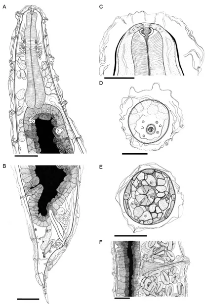
Rhabdias paraensis sp. nov. (Fig. 1A-F)
Description (based on 10 adult specimens) - Total length 7.09 ± 0.66 mm (5.92-8.19 mm, 7.19 mm), body width in the vulvar region 422.88 ± 54.53 (346.15-538.46, 423.08), body width in esophageal dilatation 174.35 ± 9.65 (153.90-180.52, 174.68), buccal capsule 8.90 ± 3.71 (3.90-16.88, 8.44) × 20.65 ± 4.00 (14.93-27.92, 20.78) (Fig. 1A, C), labia or pseudolabia absent (Fig. 1D), cu-ticular swelling with irregular folds starting in the
an-terior portion of the body, esophagus claviform 421.88 ± 11.97 (394.80-439.61, 415.58) × 82.79 ± 5.34 (74.67-90.91, 78.57) (Fig. 1A) with triangular lumen (Fig. 1E), nerve ring situated at 185.88 ± 21.50 (151.30-234.42, 151.30) from anterior end (Fig. 1A), excretory pore not observed, presence of hypodermal gland cells connected with chan-nels that end in a circular opening in the external surface of cuticular swelling (Fig. 1A), anterior portion of intes-tine wide and thick-walled, intestinal lumen starts after the esophagus and filled with a dense black material (Fig. 1A), rectum short, vulva post-equatorial with prominent transversal lips situated 4.12 ± 0.67 mm (3.26-5.20 mm, 3.40 mm) from anterior end, vagina 117.07 ± 44.67 (50.67-213.33, 117.33) in length (Fig. 1F), tail conical, 292 ± 22.47 (266.67-346.67, 293.33) long, ending in a thin tip (Fig. 4A, C), cuticular swelling culminating close to tail tip (Fig.
1B), uteri long, amphidelphic, thin-walled, filled with eggs and larvae, eggs 41.10 ± 3.49 (36.23-43.44, 43.44) × 25.61 ± 2.48 (21.62-28.38, 25.26). Parasite of the lungs of R. marina more numerous in the right lung (mean = 18) than in left lung (mean = 12).
Type host - R. marina (Linneaus, 1758) (Syn. Bufo marinus) (Amphibia: Bufonidae), Cane Toad.
Site of infection - Lungs.
Type locality - Belém, PA, Brazil (01º28’03”S 48º20’18”W).
Type-data and depository - Holotype, hermaphro-dite female. Helminthological Collection of the Instituto Oswaldo Cruz (CHIOC), Fundação Oswaldo Cruz, in Rio de Janeiro, Brazil [CHIOC 35705a (holotype) and CHIOC 35705b (10 paratypes)].
Host-parasite data - Prevalence: 45%; intensity of infection: 27 (3-143).
Etymology - The specific name refers to PA, in which the new species was discovered. In Brazilian Portuguese, paraense is a native of PA.
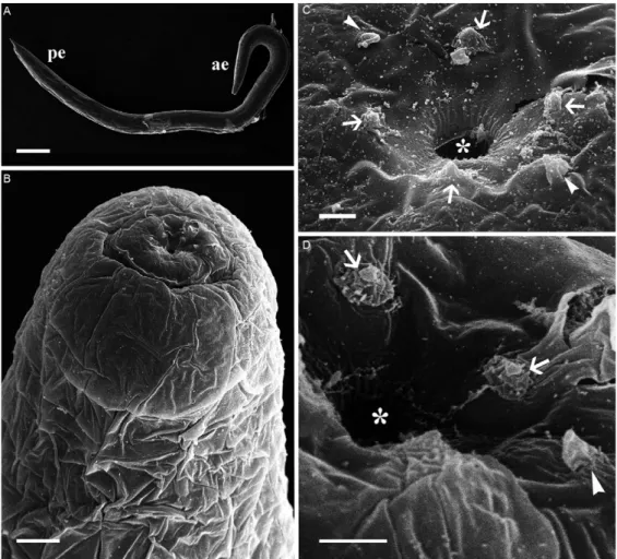
SEM - SEM permitted the observation of the follow-ing superficial characteristics of R. paraensis sp. nov.
The cylindrical body of the parasitic phase of this Rhabdias (Fig. 2A) is coated with a voluminous cuticu-lar swelling which begins close to the oral opening and continues along the body until a discreet connection with the tapered extremity of the tail. This gives the surface of the helminth an irregular appearance (Fig. 2B).
A simple, circular oral opening is found at the ante-rior extremity. Its lipless edges are bordered with tiny cuticular elevations, which radiate outwards from the opening (Fig. 2C, D). Three pairs of head papillae are located around the oral opening. Two of these pairs are large, rounded and submedian, while the third pair are small, lateral papillae (Fig. 2C, D). Both sets of submedi-an papillae are rounded with a wide base submedi-and a small central, spherical structure. The lateral papillae have a flattened conical shape, with a wide base, which tapers progressively towards the extremity (Fig. 2C, D). Am-phids not observed.
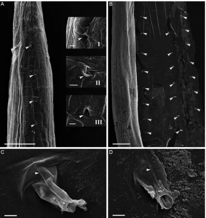
The external surface of the cuticular swelling pres-ents four longitudinal lines of pores: two ventral-lateral and two dorsal-lateral rows equally-spaced, which
tend along the whole length of the body (Fig. 3A, B). These cuticular pores are coated by the invagination of the cuticular swelling, which is projected inwards in the form of a duct (Fig. 3A, B). These conduits are clearly visible when the internal surface of the cuticular swell-ing is analyzed by SEM (Fig. 3C, D).
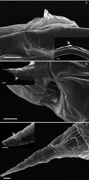
The delicate, threadlike condition of the cuticular swelling along the entire length of the parasite makes this structure vulnerable to fragmentation, which reveals the space between this structure and the cuticular surface (Fig. 4A, B). The material filling this space varies from amorphic to fibrous or filamentous (Fig. 4C, D). In the ar-eas where the cuticular surface is visible, fine and discreet transverse cuticular furrows can be observed (Fig. 4E).
The outermost portion of the vulvar opening pres-ents marked dilation of the cuticular swelling (Fig. 5A), while the anal opening is characterized by a half-moon shaped depression on the body surface (Fig. 5B). The tail tapers abruptly into a “needle-point” tip, with the cuticular swelling, which fades out halfway along this tapering point (Fig. 5C).
Histology - The histological study was based on the analysis of longitudinal sections cut at 3 µm intervals in five specimens of R. paraensis sp. nov. Seven slides were prepared per worm, with 20 sections on each slide.
In the esophageal region, the analysis revealed a simple, thick-walled, lightly basophilic and cylindrical epithelium (Fig. 6A). The interstitial region is more in-tensely basophilic than the esophageal lining, while the amorphic material filling the space within the cuticular swelling presents a subtle metachromatic staining (Fig. 6A, B). The buccal capsule presents a strong affinity with the staining, which revealed that this structure forms a ring around the esophageal opening (Fig. 6A).
The esophagus terminates in the intestinal epitheli-um, in a structure similar to a muscular valve composed of less chromaffin cells than those of the intestine (Fig. 6A). The intestinal epithelium is formed by a single layer of cells, 20-30 µm thick. These cells are cylindrical, tall and voluminous, with widespread microvilli which are highlighted by an intense metachromatic reaction (Fig. 6B). The nuclei of these cells are voluminous and spheri-cal, with a predominance of euchromatin and a promi-nent central nucleolus (Fig. 6B). The whole of the cyto-plasm is filled with different-sized granules, the largest of which are dense and fringed and are found in the middle and lower portions of the cells, while the apical granules are smaller and more acidophilic (Fig. 6B). The fibrous rectal ampulla has an insertion in the intestinal cells, which has the appearance of a valve (Fig. 6C).
The tubular ovary is strongly basophilic, with gametic cells located all along its periphery (Fig. 6D, F). Sequen-tially, embryonated eggs can be observed, some of larvat-ed, and larvae are concentrated close to the vaginal canal, vulvar aperture with small labial projections (Fig. 6E, F).
The numerous and irregularly-shaped hypodermal cells (> 50 µm), which lack basophily, are discreetly aci-dophilic and have cytoplasm filled with small, uniform and achromatic granules (Fig. 7A-D). These cells have a large, spherical nucleus of approximately 10 µm, which are lightly basophilic, and have a prominent central nu-cleolus (Fig. 7C, D). These cells also present an irregular area around the nucleus, which is more intensely baso-philic than the nucleus itself. All these cells commu-nicate with the host environment through a basophilic duct, which begins at the edge of the lateral hypodermal cord, traverses the cuticle and finishes in a pore with an irregular opening in the external surface of the cuticular swelling (Fig. 7E, F).
diSCuSSion
More than 70 species of Rhabdias have been de-scribed in the Lissamphibia and Squamata, including 18 that parasitize bufonids. Eight of these are from the Neotropical regions and can be compared directly with R. paraensis sp. nov. The new species can be easy dis-tinguishable from its congeners by the differences of ce-phalic papillae and some morphological measurement.
The post-equatorial position of the vulva in R. paraen-sis sp. nov. differs from that in R. alabialis, R. kuzmini, R. pseudosphaerocephala, R. americanus and R. fuelle-borni. However, this structure is similar to that found in R. androgyna, R. elegans and R. hermaphrodita, although these species differ from new species in a number of dif-ferent characteristics, such as the length of the esophagus and body and the presence of lips and cephalic papillae.
Few of the studies of Neotropical species have provid-ed data on the dimensions of the buccal capsule, although in R. kuzmini, it measures 31-70 × 35-47 µm, while it is 10 × 12-15 µm in R. americanus, 7-12 × 15-17 µm in R.
pseudosphaerocephala and 10 × 10-15 µm in R. alabialis. These values, and the shape of the structure, are all distinct from those of R. paraensis sp. nov. (8.90 × 20.65 µm).
While most Rhabdias species have labia or pseudola-bia, some species have no labia. Six of these have a post-equatorial vulva and are parasites of amphibians, like R. paraensis sp. nov. However, these species are dis-tinct in terms of bodily dimensions, host species and geographic distribution, being found in the Australian (Rhabdias australiensis Moravec et Sey, 1990), Orien-tal [Rhabdias shortii (Singh et Ratnamala 1977), Baker 1980] and Palearctic biogeographic regions (Rhabdias dossei Hartwich, 1972, Rhabdias niponica Yamaguti, 1943, Rhabdias rhacophori Yamaguti, 1941, and Rhab-dias tokyoensis Wilkie, 1930).
The other nine species have equatorial or pre-equato-rial vulvae and are parasites of a variety of host species, including the order Squamata. This group includes spe-cies from the Ethiopian (Rhabdias collaris Baker, 1987, Rhabdias gemellipara Chabaud, Brygoo et Petter, 1961, Rhabdias madagascariensis Chabaud, Brygoo et Pet-ter, 1961), Oriental (Rhabdias escheri Baer, 1930), and Palearctic regions [Rhabdias bufonis (Schrank, 1788) Stiles et Hassall, 1905, Rhabdias globocephala Kung et Wu, 1945, Rhabdias horiguti Yamaguti, 1943, Rhabdias incerta Wilkie, 1930, Rhabdias martinoi Kurochkin et Guskov, 1963]. The remaining species (R. alabialis) is the only one of the 16 taxa without labia that is found in the Neotropical regions, in according with Martinez-Salazar and León-Règagnon (2007).
Of the Neotropical parasites of R. marina, the ab-sence of labia makes R. paraensis sp. nov. similar to R. alabialis and distinct from R. kuzmini, R. americanus and R. pseudosphaerocephala, which present four sub-median labia and two lateral pseudolabia, and R. fuelle-borni, which has six labia. R. alabialis and R. paraensis sp. nov. differ in the presence of conspicuous papillae on the head of the new species, which were not cited by Kuzmin et al. (2007) in their description of R. alabialis. Similar to R. paraensis sp. nov., a number of different
studies have referred to papillae surrounding the oral opening of the species Rhabdias in both anuran (Baker 1987, Moravec & Kaiser 1995, Tkach et al. 2006, Kuzmin et al. 2007, Martinez-Salazar 2008, Junker et al. 2010) and squamate hosts (Martinez-Salazar 2006, Lhermitte-Vallarino et al. 2009, 2010, Moravec 2010).
R. paraensis sp. nov. also differs from R. alabialis in a number of morphometric parameters, such as the shorter esophagus (340-445 µm) and reduced distance of the nerve ring in relation to the anterior extremity of the body (146.2 µm) in R. alabialis, while in R. paraen-sis sp. nov., the vulva is located closer to the posterior extremity of the helminth. The sum of this evidence in-dicates clearly that R. paraensis sp. nov. is unlike any other known species of the genus.
Kuzmin et al. (2007) compared species of Rhabdias (R. pseudosphaerocephala, R. sphaerocephala and R. fuelleborni) usingrelationship of the relative length of esophagus (percentage of total length) to the total length. In this work these parameters aid in differentiation of species studied. However, those morphometric values in R. paraensis n. sp compared with these other three spe-cies showed similarities with R. pseudosphaerocephala, but the latter species differs by the presence of four labia and two lateral pseudolabia.
R. paraensis sp. nov. is thus a distinct species, which can be differentiated from the others of the genus due to its unique set of morphometric parameters, such as the difference in the location of the vulva, anal opening and nerve ring and the length of the body and the esopha-gus. In particular, it can be distinguished from the most closely-related species, R. alabialis, based on its set of conspicuous head papillae.
The external morphology of some Rhabdias species has been analyzed using SEM, including R. kuzmini (Martinez-Salazar & León-Règagnon 2007), R. ala-bialis, R. pseudosphaerocephala, R. sphaerocephala (Kuzmin et al. 2007), Rhabdias joaquinensis, Rhab-dias eustreptos (Kuzmin et al. 2003), Rhabdias bakeri, Rhabdias ranae (Tkach et al. 2006), Rhabdias leonae (Martinez-Salazar 2006), and Rhabdias manantlanensis (Martinez-Salazar 2008). In R. paraensis sp. nov. there is a conspicuous presence of a cuticular swelling linked to the external surface through caniculi, which traverse the cuticle and are arranged in two ventral-lateral and two dorsal-lateral rows of regularly-spaced pores. These insertion points, which are linked to hypodermal cells, when observed in the histological sections, constitute a pathway of communication between the parasite and the internal environment of the lungs of the host.
Kloss (1974) observed the presence of a number of cells with large nuclei arranged sequentially in the anterior por-tion of R. hermaphodita and R. fuelleborni, although these cells lead into a single excretory canal, which reaches the exterior of the organism through an excretory pore in the cuticle. The author interpreted those cells as belonging to excretory system. In R. paraensis sp. nov., by contrast, the analysis of 202 sequential histological sections and clari-fied specimens found no evidence of an excretory pore, nor of an excretory gland, other than the dozens of pores associated with the hypodermal cells.
Junker et al. (2010) observed no excretory pore in their description of the tropical African species Rhab-dias picardiae and Rhabdias ohlerae, although in R. picardiae, lateral cells impeded observation of the pore, while in R. ohlerae, a long excretory gland was identi-fied. These authors also analyzed Rhabdias vencesi and reported that the excretory pore was difficult to observe in the fixed specimens.
In R. collaris, Baker (1987) observed a cuticle inflat-ed with ducts leading to hypodermal glands all along the body. In R. picardiae, Junker et al. (2010) recorded the cuticle vesicle is attached laterally to the inner layer of the cuticle via conspicuous fibers associated with subcuticu-lar pores. Using histology and SEM, we observed simisubcuticu-lar
features in R. paraensis sp. nov., although the structures and the connections of the hypodermal glandular cells with the outer surface were shown in greater detail.
In the superfamily Rhabditoidea, each lateral and sublateral line of the hypodermal cord is formed by 12-14 hypodermal cells (Chitwood & Chitwood 1974). In R.
Fig. 7: histology of Rhabdias paraensis sp. nov., stained with 1% toluidine blue. Hypodermal cells, canaliculi and the pores. A: mid-portion of the body. Note a voluminous, chromophobic cells between intestine (i) and cuticle (c) with grainy cytoplasm, nucleus and nucleolus (arrows). Asterisk means intestinal lumen. Bar = 100 µm; B: tangential section at the posterior end show chromophobic cells with large nuclei (n), canali-culi (c) and pores (large arrows) in cuticular swelling (cs). Bar = 100 µm; C, D: detail of chromophobic cell, grainy cytoplasm (g), n and nucleoli (nu). Canaliculus (cn) traverses c. Bar = 50 µm; E, F: detail of two cn with pore opening (large arrow) through c in E and the folding of cs at the pore opening; F: note cn enveloped in cytoplasm rich of chromophobic granules (g). Bar = 20 µm.
paraensis sp. nov., we observed (SEM) four lines of 14-16 sublateral pores connected to hypodermal cells. We believe that these pores represent the distribution of the cells of the hypodermal cord.
The ultrastructural characteristics of these special-ized cells of the hypodermis were described by Chit-wood and ChitChit-wood (1974). These cells contain numer-ous small granules and present a strong affinity with alkaline stains. By contrast, R. paraensis sp. nov. is clearly chromophobic.
In R. paraensis sp. nov., in addition, the space be-tween the cuticular swelling and the external surface of the cuticles is filled with an amorphous and basophilic material not found in any other Rhabdias species de-scribed to date. Chitwood and Chitwood (1974) point out that this swelling is caused by the separation of the cortical layer of the cuticle from the remaining layers, which is normally caused by the liquefying of the ma-trix. While the cuticular swelling described by these authors is a specialization of the external layer of the cuticle, it is important to note that we observed delicate transverse striae at uniform intervals on the surface of the helminth - below this swelling - a common feature in many forms of parasitic invertebrates.
The analysis of serial histological sections of samples embedded in methacrylate resin may provide an impor-tant complementary approach and essentials compara-tive parameters for future studies of both microanatomi-cal details and the parasite-host interaction analysis of Rhabdias specimens. This technique, associated with SEM, has provided new insights into the morphology of the genus and contributed to the development of new taxonomic parameters for the characterization of the principal internal structures, which are essential for the development of comparative studies and the understand-ing of host-parasite interactions. This study increases our knowledge of the diversity of the helminths that parasit-ize R. marina in the Amazon Region, expanding on the work of Espinoza-Jiménez et al. (2007), Espinola-Novelo & Guillén-Hernandez (2008) and Santos et al. (2008).
aCknowledGeMentS
To Drs Marcos André Vannier and Adriana Lanfredi (Ul-trastructural Pathology Laboratory of the Gonçalo Muniz Research Center/Fiocruz, Salvador, Bahia), for their techni-cal support for the scanning electron microscopy, to Stephen Ferrari, for revision of the English, and to the anonymous ref-erees, for valuable comments and suggestions.
reFerenCeS
Anderson RC 2000. Nematode parasites of vertebrates. Their devel-opment and transmission, 2nd ed., CAB International, Walling-ford, p. 17-34.
Baker MR 1987. Rhabdias collaris sp. nov. (Nematoda: Rhabdiasi-dae) from frogs of Tanzania. Parasitol 9: 199-201.
Bursey C, Goldberg RS, Telford SR Jr 2003. Rhabdias anolis sp. nov. (Nematoda: Rhabdiasidae) from the lizard, Anolisfrenatus (Sau-ria: Polychrotidae), from Panama. J Parasitol 89: 113-117.
Chitwood BG, Chitwood MB 1974. The external cuticle and hypoder-mis. In BG Chitwood, MB Chitwood, Introduction to nematol-ogy, University Park Press, Baltimore, p. 28-47.
Espínola-Novelo JF, Guillén-Hernández S 2008. Helminth parasites in
Chaunus marinus and Cranopis valliceps (Anura: Bufonidae) from Lagunas Yalahau, Yucatan, México. J Parasitol 94: 672-674.
Espinoza-Jiménez A, García-Prieto L, Osorio-Sarabia D, León-Règag-non V 2007. Checklist of helminth parasites of the cane toad Bufo marinus (Anura: Bufonidae) from Mexico. J Parasitol 93: 937-944.
Junker K, Lhermitte-Vallarino N, Barbuto M, Ineich I, Wanji S, Bain O 2010. New species of Rhabdias (Nematoda: Rhabdiasidae) from Afrotropical anurans, including molecular evidence and notes on biology. Folia Parasitol 57: 47-61.
Kloss GR 1971. Alguns Rhabdias (Nematoda) de Bufo no Brasil.
Papeis Avul Zool 24: 1-52.
Kloss GR 1974. Rhabdias (Nematoda, Rhabditoidea) from the group of
Bufo marinus. The study of sibling species. Arq Zool 25: 61-120.
Kuzmin Y, Tkach VV, Brooks DR 2007. Two new species of Rhab-dias (Nematoda: Rhabdiasidae) from the marine toad, Bufo mari-nus (L.) (Lissamphibia: Anura: Bufonidae) in Central America.
J Parasitol 93: 159-165.
Kuzmin Y, Tkach VV, Snyder SD 2003. The nematode genus Rhab-dias (Nematoda: Rhabdiasidae) from amphibians and reptiles of the Nearctic. ComparativeParasitol 70: 101-114.
Kwett A, Di-Bernardo M, Maneyro R 2006. First record of Chaunus achavali (Anura, Bufonidae) from Rio Grande do Sul, Brazil, with a key for the identification of the species in the Chaunus marinus group. Serie Zoologia96: 479-485.
Lhermitte-Vallarino N, Barbuto M, Junker K, Boistel R, Bain O 2010.
Rhabdias (Nematoda: Rhabdiasidae) from Chamaeleonidae (Sau-ria): two new species from Trioceros ellioti in east Africa and one from Brookesia superciliaris in Madagascar. Parasite 17: 91-105.
Lhermitte-Vallarino N, Barbuto M, Junker K, Boistel R, Ineich I, Wanji S, Bain O 2009. Rhabdiasrhampholeonis sp. nov. and
Rhabdiasmariauxi sp. nov. (Nematoda, Rhabdiasoidea), first lung worms from leaf chameleons: description, molecular evi-dence and notes on biology. Parasitol Int 58: 375-383.
Martínez-Salazar EA 2006. A new rhabdiasid species from Norops megapholidotus (Sauria: Polychrotidae) from Mexico. J Parasi-tol 79: 81-89.
Martínez-Salazar EA 2008. A new rhabdiasid species from Crau-gastoroccidentalis (Anura: Brachycephalidae) from Sierra de Manantlán, Jalisco, Mexico. Rev Mex Biodivers 79: 81-89.
Martínez-Salazar EA, León-Règagnon V 2007. New species of Rhab-dias (Nematoda: Rhabdiasidae) from Bufo occidentalis (Anura: Bufonidae) from Sierra Madre del Sur, Mexico. J Parasitol 93: 1171-1177.
Moravec F 2010. Rhabdias lacertae sp. nov. (Nematoda: Rhabdiasi-dae), the first rhabdiasid species parasitizing lizards in Europe.
J Parasitol 77: 23-27.
Moravec F, Kaiser H 1995. Helminth parasites from west Indian frogs, with descriptions of two new species. J Science 31: 252-268.
Santos JN, Giese EG, Maldonado-Jr A, Lanfredi RM 2008. A new species of Oswaldocruzia in Chaunnus marinus from Brazil.
J Parasitol 94: 264-268.
Tkach VV, Kuzmin Y, Pulis EE 2006. A new species of Rhabdias