Cranial Nerve Injury as an Initial Sign of Wegener Granulomatosis:
Misdiagnosis and Fatal Course in Two Cases
Wegener Granülomatosisin Başlangıç Bulgusu Olarak Kranial Sinir
Tutulumu: Yanlış Tanı ve Ölümcül Seyreden İki Olgu
Kenan tuRgutalp1 tolga köşeci1 Feray tabaKan1 iclal gürses2
Sezer çiçekli turgutalp3 ebru gök oğuz1
arda yılmaz4 Mehmet horoz5 engin KaRa6 ahmet KıyKıM1
1 Mersin University Faculty of Medicine, Department of Internal Medicine, Division of Nephrology, Mersin, Turkey 2 Mersin University Faculty of Medicine,
Department of Pathology, Mersin, Turkey
3 Mersin State Hospital, Department of Dermatology, Mersin, Turkey
4 Mersin University Faculty of Medicine, Department of Neurology,
Mersin, Turkey
5 Harran University Faculty of Medicine, Department of Internal Medicine, Division of Nephrology,
Şanlıurfa, Turkey
6 Mersin University Faculty of Medicine, Department of Radiology,
Mersin, Turkey
Correspondence Address:
Kenan tuRgutalp
Mersin Üniversitesi Tıp Fakültesi, İç Hastalıkları Anabilim Dalı, Nefroloji Bilim Dalı, Mersin, Turkey
Phone : + 90 532 492 68 83 E-mail : [email protected] abStRaCt
Highest prevalence of all antineutrophil cytoplasmic antibody associated systemic vasculitides is the Wegener granulomatosis, and it’s typically characterized by a necrotizing granulomatous vasculitis of the respiratory tracts, kidneys, and skin. Neurologic involvement in Wegener granulomatosis ranges from 22% to 54%, but central nervous system involvement is from 2% to 8%. Cranial nerve involvement as an initial sign with concomitant systemic disease is extremely rare. Involvement of one or more cranial nerves is generally a seemingly isolated inding in Wegener granulomatosis. We present WG cases that presented with treatment-resistant cranial nerve involvement and acute fatal pulmonary-renal syndrome.
key words: Wegener’s granulomatosis, Neurologic manifestation, Pulmonary-renal syndrome
öz
Wegener granülomatosis antinötroil sitoplazmik antikorlarla ilişkili vaskülitlerde yüksek sıklıkla görülmekle beraber cilt, böbrek ve solunum sisteminin nekrotizan granülomatöz vasküliti ile karekterizedir. Wegener granülomatosiste nörolojik tutulum %22 ile %54 arasında değişmektedir. Buna karşılık santral sinir sistemi tutulumu %2 ile % 8 arasında değişmektedir. Sistemik hastalıkların başlangıç bulgusu olarak kranial sinir tutulumu oldukça nadirdir. Bir ya da daha fazla kranial sinir tutulumu Wegener granülomatosisinde nadiren görülür. Bu olguda kranial sinir tutulumlu, tedaviye dirençli, akut pulmoner-renal sendromlu Wegener granülomatosisi tartışmayı amaçladık.
anahtar sözcükler: Wegener granülomatosis, Nörolojik belirti, Pulmoner-renal sendrom
Received : 26.02.2013
Accepted : 12.04.2013 ıntroductıon
Systemic vasculitides is an extremely rare and many physicians still regard of them. However, they don’t prepare for such a surprising come across. The most of organs such as upper and lower respiratory tracts, kidneys, and skin are affected by Wegener’s granulomatosis (WG) which is typically characterized by a necrotizing granulomatous vasculitis (1). The frequency of neurologic involvement in WG ranges from 22 % to 54 %. While peripheral neuropathy is common in patients with WG (43.8 %), cranial nerve involvement is seen in only a small percentage (4.7%) (2). Cranial nerves 2, 6, and 7 are the most frequently involved, either by themselves
or in combination (3). In some prospective studies, the optic nerve was the most commonly affected cranial nerve in patients with WG (2). We present two patients with WG who presented with treatment-resistant cranial nerve involvement and acute fatal pulmonary-renal syndrome.
case 1
with cefuroxime axetil for a week. Her past medical history was unremarkable, and she did not smoke.
All vital indings were normal other than fever of 38.5 0C on admission. Lung examination revealed bilateral basilar late inspiratory crackles. Tympanic membranes and external ear canals were normal. She developed an ulcerated nasolabial skin lesion (Figure 2) and a purpuric eruption together with generalized edema (Figure 3) three days after admission. An audiogram demonstrated bilateral severe sensorineural hearing loss (SNHL), which is deined as both air and bone conduction below 15 dB with no signiicant air-bone gap.
Laboratory indings on admission as follows: white blood cell count 19.000/mm3, hemoglobin concentration 8.6 gr/dl,
platelet count 186.000/mm3, creatinine 3.7 mg/dl, erythrocyte
and progressive dysphagia and right-sided facial nerve paralysis had developed after 5 days of treatment. The patient had been hospitalized in the otorhinolaryngology clinic. Three days after, he had developed microscopic hematuria, generalized skin eruption, and oliguria and acute nephritic syndrome. The patient was referred to our clinic. Physical examination on admission revealed a blood pressure of 100/60 mmHg, pulse 96/min., body temperature 38.7 °C and respiratory rate 18/min. Neurological examination revealed 7th, 8th, and 10th cranial nerve dysfunction.
On admission, laboratory indings were as follows; white blood cell count 24.000/mm3, hemoglobin level 9.6 gr/dl, platelet
count 224.000/mm3, serum creatinine 2.7 mg/dl; serum albumin
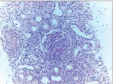
2.9 gr/dl; erythrocyte sedimentation rate 88 mm/h. Urinalysis revealed proteinuria, 150 mg/dl, and microscopic hematuria at 10 red blood cells/hpf. International normalized ratio (INR) was normal. C-reactive protein was 80 mg/L. Rheumatoid factor, anti-nuclear antibody, anti-ds-DNA-antibody, and cryoglobulin were all negative. Serum complement levels were normal. Perinuclear anti-neutrophil cytoplasmic antibody (p-ANCA) was negative but c-ANCA was strongly positive. Decreased tonicity was found in the upper esophageal sphincter on endoscopic examination. Ear canal examination was normal. An audiogram demonstrated bilateral severe sensorineural hearing loss (SNHL). which is deined as both air and bone conduction below 15 dB with no signiicant air-bone gap (>10dB). Echocardiography, chest x-ray, and abdominal ultrasound on admission were normal. A computerized tomography (CT) scan of the temporal bone was normal. Paranasal regions, dura mater thickness, optic canals were all normal as detected by magnetic resonance image of the brain. Renal biopsy was performed because of the acute nephritic syndrome and suspected systemic vasculitic syndrome. Diffuse crescentic glomerulonephritis was found on renal biopsy (Figure 1). The presence of upper respiratory symptoms, acute multiple cranial nerve dysfunction, acute nephritic syndrome and diffuse crescentic glomerulonephritis in renal biopsy, and the high c-ANCA positivity were suggestive of WG. The patient was therefore treated with plasmapheresis and pulse methylprednisolone for 3 days, followed by maintenance prednisolone and cyclophosphamide. The patient needed hemodialysis therapy 3 times during hospitalization. Although aggressive immunosuppressive treatment and plasmapheresis were performed, the patient developed diffuse alveolar hemorrhage and poor pulmonary function on his 10th inpatient day and died due to pulmonary-renal syndrome on 10th day of treatment.
case 2
A 38-year-old-female was admitted to our nephrology service because of tinnitus and progressive hearing loss for two weeks, and oliguria and dyspnea for two days. The patient had been diagnosed with otitis media and a nasolabial skin lesion in an outpatient clinic eight days before the admission, and treated
Figure 1: Diffuse crescentic glomerulonephritis is shown h e original
magniication x200.
scan of the temporal bone was normal. The patient underwent skin and renal biopsies. Renal biopsy revealed diffuse crescentic glomerulonephritis, and skin biopsy was compatible with leukocytoclastic vasculitis. Combined treatment with pulse methylprednisolone (1 gr/d for three days), pulse cyclophosphamide (0.5 gr/m2), and plasmapheresis was used.
Oral corticosteroid therapy at 40 mg/d and cyclophosphamide at 100 mg/d were administered as the maintenance therapy. Hemodialysis was performed 11 times during the hospital stay. Her fever and creatinine level rapidly improved on the seventh day of treatment, but the hearing loss persisted. She developed sudden treatment-resistant massive hemoptysis, and died due to pulmonary-renal syndrome on the 35th day of treatment.
dıscussıon
WG is considered when its choose of the upper and lower respiratory tracts and the kidneys. The most important presenting features of WG are ear, nose, and throat involvement. Numerous patients with WG principally develop the disease by lesions of the upper airway region. Chiely, the ear and nasal mucosa may be the irst region of development disease (20 % to 40 %), and patients with WG frequently apply to otolaryngologists as was seen in our patients (4). The diagnosis of WG is based on the criteria published by the American College of Rheumatology in 1990. The distinctive features of our cases are the normal upper airway tract and external ear canals despite the hearing loss and tinnitus. We observed that these acute complaints had developed due to involvement of the cranial nerves. Nonetheless, the kidneys, skin, and lower respiratory tract were rapidly affected by WG, despite aggressive treatment.
Figure 3: Vasculitic skin eruption on extremities.
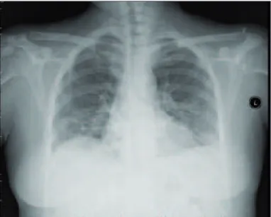
Figure 4: Caviter lesion in right lung lower zone on chest x-ray.
Figure 5: Multiple caviter lesions in lung on high resolution ct hrct.
process that might warrant potent therapy (14). Furthermore, the occurrence of SNHL can be a warning of severe WG and is thought to require initial treatment with cyclophosphamide (14). The hearing loss in WG may progress quickly in days to weeks and can be accompanied by tinnitus (15). The acute onset of SNHL and the poor response to immunosuppressive therapy in our cases are very similar to the patients documented in the Wierzbicka et al. study (9). Furthermore, SNHL can be associated with other severe manifestations including renal disease (15), as seen in our patients.
COnCluSıOn
Lots of physicians still regard WG as uncommon and they don’t prepare themselves for this disease. Physicians will encounter this compulsive disease when cranial neuropathies appear as sole inding of WG, and diagnostic procedure is requirement for not to miss this diagnosis. On the other hand, a multidisciplinary approach with dedicated clinicians is advised since direct histological proof or tests indicating active vasculitis are often not available in these patients.
reFerences
1. Wardyn K-A, Życińska K: Pierwotne systemowe zapalenia naczyń. Pol Arch Med Wewn 2014; 111: 757-767
2. De Groot K, Schimdt DK, Arlt AC, Gross W, Reinhold-Keller E: Standardized neurologic evaluation of 128 patients with Wegener granulomatosis. Arch Neurol 2001; 58: 1215-1221
3. Nishino H, Rubino FA, DeRemee RA, Swanson JW, Parisi JE: Neurological involvement in Wegener’s granulomatosis: An analysis of 324 consecutive patients at the Mayo Clinic. Ann Neurol 1993; 33: 4-9
4. Gottschlich S, Ambrosch P, Kramkowski D, Laudien M, Buchelt T, Gross WL, Hellmich B: Head and neck manifestations of Wegener’s granulomatosis. Rhinology 2006; 44: 227-233
5. Hoffman GS, Kerr GS, Leavitt R-Y, Hallahan CW, Lebovics RS, Travis WD, Rottem M, Fauci AS: Wegener’s granulomatosis: An analysis of 158 patients. Ann Intern Med 1992; 116: 488-498 6. Pamuk ON, Dogutan H, Pamuk GE, Cakir N: Unilateral phrenic
nerve paralysis in a patient with Wegener’s granulomatosis. Rheumatol Int 2003; 23: 201-203
7. Ghilain S, Delreux V, Kevers L, Sindic CJ, Mathurin P, Laterre EC: Multiple cranial nerve involvement with tentorial pachymeningitis of granulomatous type. Acta Neurol Belg 1988; 88: 91-100 8. Nikolaou AC, Vlachtsis KC, Daniilidis MA, Petridis DG, Daniilidis
IC: Wegener’s granulomatosis presenting with bilateral facial nerve palsy. Eur Arch Otorhinolaryngol 2001; 258: 198-202
9. Wierzbicka M, Szyfter W, Puszczewicz M, Borucki L, Bartochowska A: Otologic symptoms as initial manifestation of Wegener’s granulomatosis: Diagnostic dilemma. Otol Neurotol 2011; 32: 996-1000
One of the rare a presenting feature of WG is neurological involvement. This involvement may progress usually while the course of the disease as appeared in 22-50 % of patients (2, 5). Although, multiple neurologic complications are appeared in up to 11 % of patients with WG, the most common single neurologic inding such as peripheral neuropathy is occurred in 16-43 % of patients (2, 5). On the other hand, involvement of the central nervous system has been reported rarely (2% to 8%) in WG patients. One or multiple cranial nerve involvement has been established either simultaneously with apparent vasculitic activity in other organs or as an appeared single manifestation. Another one has been intended to as non-systemic vasculitic neuropathy and the diagnosis is dificult. Cranial nerves 2, 6, and 7 are the most often affected, either singly or in combination (3). Lower cranial nerve palsies are very uncommon and usually occur during advanced stages of the disease (3). Furthermore, phrenic nerve involvement has been reported only once (6) and there are only a few causes of 11th cranial nerve palsy (7). Facial nerve palsy is one of the most important cranial neuropathies in patients with WG. Nikolaou et al. reported that three of their 7 patients showed only facial paresis (8) as was seen in our case 2. Additionally, similar facial nerve palsy indings were reported by Wierzbicka et al. in three of their 7 patients (9). Armani et al. (10) reported a WG case that presented with neurogenic dysphagia due to lower cranial nerve palsy and bilateral denervation of the 9th, 10th, 11th and 12th cranial nerves and right phrenic nerve. Isolated phrenic nerve involvement has also been reported by Pamuk et al. (6). Involvement of optic nerve may appear as exhaustive or ischemic neuropathy, optic nerve edema, and optic neuropathy (11).
Recently, Nishino et al. studied 324 patients in whom the diagnosis was made at the Mayo clinic, and multiple cranial nerve involvement was detected in only 8 patients (3). Groot et al. reported that cranial nerves including the 2nd, 3rd, 5th, and 7th nerve were damaged in only 6 of their 128 cases. Complete remission of CNS involvement was achieved in 6 of the 9 patients and the other 3 patients had a long-standing partial remission with pulse intravenous cyclophosphamide and corticosteroid treatment (2). Likewise, involvement of 7th, 8th, and 9th cranial nerves was found as an initial sign of the disease in our case 1, and involvement of the 8th cranial nerves was found in case 2. However, both of our patients died due to pulmonary-renal syndrome despite aggressive treatment. Some authors believe that cranial pathology without renal and pulmonary involvement suggests a variant of Wegener’s granulomatosis called the “limited” form. We can speculate that combined systemic and cranial involvement may be fatal in patients with WG.
13. Takagi D, Nakamaru Y, Maguchi S, Furuta Y, Fukuda S: Otologic manifestations of Wegener’s granulomatosis. Laryngoscope 2002; 112: 1684-1690
14. Dekker PJ: Wegener’s granulomatosis: Otological aspects. J Otolaryngol 1993; 22: 364-367
15. Bakthavachalam S, Driver MS, Cox C, Spiegel JH, Grundfast KM, Merkel PA: Hearing loss in Wegener’s granulomatosis. Otol Neurotol 2004; 25: 833-837
10. Armani M, Spinazzi M, Andrigo C, Fassina A, Mantovan M, Tavolato B: Severe dysphagia in lower cranial nerve involvement as the initial symptom of Wegener’s granulomatosis. J Neurol Sci 2007; 263: 187-190
11. Pakrou N, Selva D, Leibovitch I: Wegener’s granulomatosis: Ophthalmic manifestations and management. Semin Arthritis Rheum 2006; 35: 284-292