BGD
6, 1267–1316, 2009Element/Ca small-scale variability
in bivalve calcite
P. S. Freitas et al.
Title Page
Abstract Introduction
Conclusions References
Tables Figures
◭ ◮
◭ ◮
Back Close
Full Screen / Esc
Printer-friendly Version
Interactive Discussion Biogeosciences Discuss., 6, 1267–1316, 2009
www.biogeosciences-discuss.net/6/1267/2009/ © Author(s) 2009. This work is distributed under the Creative Commons Attribution 3.0 License.
Biogeosciences Discussions
Biogeosciences Discussionsis the access reviewed discussion forum ofBiogeosciences
Ion microprobe assessment of the
heterogeneity of Mg/Ca, Sr/Ca and Mn/Ca
ratios in
Pecten maximus
and
Mytilus
edulis
(bivalvia) shell calcite precipitated
at constant temperature
P. S. Freitas1,2, L. J. Clarke1, H. Kennedy1, and C. A. Richardson1
1
School of Ocean Sciences, College of Natural Sciences, Bangor University, Askew Street, Menai Bridge, Isle of Anglesey, LL59 5AB, UK
2
Departamento de Geologia Marinha, Laborat ´orio Nacional de Energia e Geologia, Estrada da Portela – Zambujal, 2721-866 Alfragide, Portugal
Received: 19 December 2008 – Accepted: 8 January 2009 – Published: 23 January 2009 Correspondence to: P. S. Freitas ([email protected])
BGD
6, 1267–1316, 2009Element/Ca small-scale variability
in bivalve calcite
P. S. Freitas et al.
Title Page
Abstract Introduction
Conclusions References
Tables Figures
◭ ◮
◭ ◮
Back Close
Full Screen / Esc
Printer-friendly Version
Interactive Discussion
Abstract
Small-scale heterogeneity of biogenic carbonate elemental composition can be a sig-nificant source of error in the accurate use of element/Ca ratios as geochemical prox-ies. In this study ion microprobe (SIMS) profiles showed significant small-scale variabil-ity of Mg/Ca, Sr/Ca and Mn/Ca ratios in new shell calcite of the marine bivalvesP.
max-5
imus andMytilus edulisthat was precipitated during a constant-temperature culturing experiment. Elevated Mg/Ca, Sr/Ca and Mn/Ca ratios were found to be associated with the deposition of elaborate shell features, i.e. a shell surface stria inP. maximus and surface shell disturbance marks in both species, the latter a common occurrence in bivalve shells. In both species the observed small-scale elemental heterogeneity
10
most likely was not controlled by variable transport of ions to the extra-pallial fluid, but by factors such as shell Mg content influencing Sr and Mn heterogeneity, the influence of shell organic content and/or conditions at the shell crystal-solution interface. Invari-ant Mg/Ca ratios observed in the mid and innermost regions of theP. maximus shell suggests a potential application as a palaeotemperature proxy.
15
1 Introduction
The elemental composition of marine biogenic carbonates has been thought to pro-vide a powerful tool to obtain information on Earth’s past climate and oceanographic conditions. The basis of this approach is the observed dependence of the elemen-tal composition of marine biogenic calcite and aragonite minerals on several ambient
20
environmental parameters, such as temperature, salinity, nutrient levels and seawa-ter chemistry (Boyle, 1981, 1988; Lea and Boyle, 1989; Lea et al., 1989; Beck et al., 1992; Delaney et al., 1993; Nurnberg et al., 1996; Martin et al., 1999; McCulloch et al., 1999). However, biological processes and environmental parameters other than the main controlling parameters, e.g. seawater temperature for Mg/Ca, also can influence
25
BGD
6, 1267–1316, 2009Element/Ca small-scale variability
in bivalve calcite
P. S. Freitas et al.
Title Page
Abstract Introduction
Conclusions References
Tables Figures
◭ ◮
◭ ◮
Back Close
Full Screen / Esc
Printer-friendly Version
Interactive Discussion 1977; Lorens and Bender, 1980; Delaney et al., 1985; Rosenberg et al., 1989;
Rosen-berg and Hughes, 1991; Klein et al., 1996b; NurnRosen-berg et al., 1996; Lea et al., 1999; Elderfield et al., 2002; Bentov and Erez, 2005; Gillikin et al., 2005; Lorrain et al., 2005; Freitas et al., 2006). The reliable and accurate use of element/Ca ratios in marine biogenic calcite and aragonite minerals as geochemical proxies thus is dependent on
5
appreciating the occurrence of any secondary influences on elemental incorporation. The environmental and/or biological factor(s) that determine small-scale (<100µm) variability in elemental composition will most likely control, to a large extent, the ob-served variation of element/Ca ratios in bivalve calcite at a larger scale if they are a major influence(s) on shell chemistry. Conventional “bulk” sampling techniques, i.e. that
10
mill to depths of up to a few hundred microns in order to obtain powders for solution ele-mental analyses, could integrate any compositional heterogeneity to variable extents in different samples, thereby reducing spatially resolved records and introducing a signif-icant unknown error. Thus, application of high spatial resolution analytical techniques allows a greater appreciation of the implications that this compositional heterogeneity
15
may have on the limited temperature control and large variability of Mg/Ca ratios re-ported for some bivalves, i.e.Mytilus edulis (Dodd, 1965; Klein et al., 1996a; Vander Putten et al., 2000; Freitas et al., 2008), Crassostrea virginica(Surge and Lohmann, 2008) and P. maximus (Lorrain et al., 2005; Freitas et al., 2006, 2008). A greater knowledge of the extent and causes of any small-scale elemental heterogeneity also
20
has implications for the application of Sr/Ca and Mn/Ca ratios for palaeoceanographic and palaeoenvironmental reconstructions.
Ion microprobe, or secondary ionisation mass spectrometry (SIMS), has proven ex-tremely useful for assessing small-scale heterogeneity (∼10µm) in the distribution of thermodynamically-controlled elements, i.e. Mg in calcite and Sr in aragonite, within the
25
BGD
6, 1267–1316, 2009Element/Ca small-scale variability
in bivalve calcite
P. S. Freitas et al.
Title Page
Abstract Introduction
Conclusions References
Tables Figures
◭ ◮
◭ ◮
Back Close
Full Screen / Esc
Printer-friendly Version
Interactive Discussion shells (Jeffree et al., 1995; Siegele et al., 2001; Markich et al., 2002). Electron
micro-probe (Lorens and Bender, 1980; Lutz, 1981; Rosenberg and Hughes, 1991; Rosen-berg et al., 2001), particle-induced X-ray emission (PIXE) (Swann et al., 1991; Siegele et al., 2001), cathodoluminescence emission (Langlet et al., 2006), proton microprobe (Coote and Trompetter, 1995), synchrotron radiation-based X-ray fluorescence (Thorn
5
et al., 1995; Kurunczi et al., 2001) and laser ablation ICP-MS (e.g. Raith et al., 1996; Price and Pearce, 1997; Leng and Pearce, 1999; Toland et al., 2000; Vander Putten et al., 2000; Lazareth et al., 2003; Langlet et al., 2007) also have been used to determine spatial variability, at various different scales, in the elemental composition of bivalve shells, albeit again related to high resolution reconstructions in animals experiencing
10
time varying environmental conditions.
For archives that retain a potential palaeotemperature proxy, such as Mg/Ca ratios, it is as critical to test the veracity of the proxy under a single constant temperature as it is to appraise the veracity of the proxy under a range of temperatures. In contrast to other biominerals produced by marine organisms, the high spatial resolution distribution of
15
elements within bivalve shell calcite deposited at a constant temperature has not been studied in any detail. The majority of these studies have focussed instead on small-scale (∼10µm) temporal variability in shell chemistry, mainly on Mg/Ca ratios because of their potential application as a palaeotemperature proxy (Lorens and Bender, 1980; Rosenberg and Hughes, 1991; Rosenberg et al., 2001; Dauphin et al., 2003b). Other
20
studies have focused on the small-scale variability of sulphur composition (Rosenberg et al., 2001; Dauphin et al., 2003b, 2005). By comparison, there has been little or no examination of the extent and possible causes of any spatial variability in Sr/Ca or Mn/Ca ratios within marine bivalve shells, despite their potential utility as palaeoprox-ies. At the spatial scale of individual crystals, Dauphin et al. (2003b) observed variation
25
BGD
6, 1267–1316, 2009Element/Ca small-scale variability
in bivalve calcite
P. S. Freitas et al.
Title Page
Abstract Introduction
Conclusions References
Tables Figures
◭ ◮
◭ ◮
Back Close
Full Screen / Esc
Printer-friendly Version
Interactive Discussion new laboratory environment, has been observed in new shell growth fromM. edulis
cul-tured in natural seawater and in semi-artificial “seawater” solutions with varying Mg/Ca and Sr/Ca ratios under controlled conditions at temperatures between 22 and 24◦C (Lorens and Bender, 1980). Furthermore, Rosenberg et al. (2001) demonstrated us-ing digital electron probe microscopy that small scale variations in Mg concentrations
5
inM. edulis calcite were due to Mg being concentrated along the margins of calcite prisms, especially along the terminations of the crystals. InM. edulis, the Mg content of the outer calcite shell layer also was shown to be higher in regions with slow-growth, high shell curvature and with high mantle (the organ that controls calcification) activity, than in shell areas with fast shell growth, low shell curvature and low mantle activity
10
(Rosenberg and Hughes, 1991).
In an initial preliminary assessment of the extent of any small-scale heterogeneity in Mg/Ca, Sr/Ca and Mn/Ca ratios in bivalve shell calcite, new shell material precipi-tated byPecten maximus(king scallop) andMytilus edulis(blue mussel) in a constant-temperature laboratory culturing experiment, during which other seawater parameters
15
also were monitored, has been analysed using the ion microprobe (SIMS) technique. These two bivalve species, as well as closely related taxa, have been proposed pre-viously as archives for palaeoceanographic studies (Krantz et al., 1988; Klein et al., 1996a; Hickson et al., 1999; Chauvaud et al., 2005; Gillikin et al., 2006; Th ´ebault et al., 2007; Wanamaker et al., 2007) and hence are suitable materials for such an
investi-20
gation of the extent of spatial variability in bivalve shell elemental concentrations. This methodological approach is especially valid for Mg/Ca ratios in bivalve calcite, which should be invariant within the new shell growth if a simple thermodynamic influence is the predominant control on this geochemical proxy in these archival materials, and also may provide further insights into explaining the reported limited temperature
con-25
BGD
6, 1267–1316, 2009Element/Ca small-scale variability
in bivalve calcite
P. S. Freitas et al.
Title Page
Abstract Introduction
Conclusions References
Tables Figures
◭ ◮
◭ ◮
Back Close
Full Screen / Esc
Printer-friendly Version
Interactive Discussion
2 Materials and methods
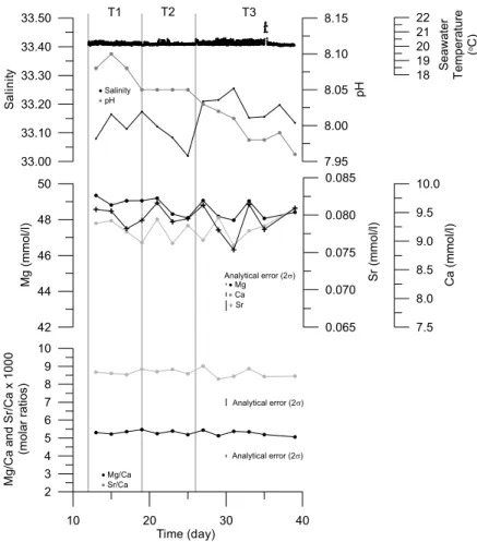
Seawater temperature in the aquarium was measured every 15 min by in-situ log-ger (Gemini Data Loglog-gers TinyTag Aquatic – TGI 3080) as 20.33±0.13◦C (N=2952) and 20.21±0.13◦C (N=2304) for the duration of the two culturing periods (Fig. 1). Aquarium seawater samples for measurement of pH, element/Ca ratios and δ18O,
5
were collected every other day, whereas salinity was measured on samples every fourth to eighth day (Fig. 1). Seawater pH measurements were taken manually with a commercial glass electrode (Mettler Toledo Inlab 412). For determination of element/Ca ratios seawater samples were diluted 10 fold and acidified to 3% v/v with Merck Ultrapur HNO3 and measured on a Perkin Elmer Optima 3300RL
ICP-10
AES instrument housed at the NERC ICP Facility, Royal Holloway University of Lon-don. Instrumental drift, for the larger batch of solutions that included these sea-waters, was monitored by running an independent solution every 10 samples. Fol-lowing ICP-AES analyses signal intensity drift was checked and corrected for. Pre-cision of seawater elemental analyses, expressed as relative standard deviation or
15
RSD, was obtained from repeat measurements of the CASS-4 seawater certified ref-erence material (Mg=42.975±0.469µmol/ml, 1.09 %RSD; Ca=8.635±0.069µmol/ml, 0.80%RSD; Sr=0.071±0.002µmol/ml, 2.18%RSD; Mg/Ca (molar)=4.977±0.079, 1.58 %RSD; Sr/Ca (molar)=0.0082±0.0002, 2.66%RSD; N=14). Aquarium seawater Mn concentrations were below the detection limits of the ICP-AES. Seawater Mg, Ca and
20
Sr concentrations, as well as Mg/Ca and Sr/Ca molar ratios are shown in Fig. 1. Seawaterδ18O and salinity samples were collected in sealed Winchester glass bot-tles. Seawaterδ18O was measured at the School of Environmental Sciences, Univer-sity of East Anglia, by off-line equilibration with CO2 and subsequent measurement
of isotope ratios using a Europa-PDZ Geo 20/20 isotope-ratio mass spectrometer,
25
with normalisation relative to a laboratory standard, North Sea Water (accepted value of+0.13‰ VSMOW). The precision of replicateδ18Oseawater analyses is 0.05‰ (1σ;
cal-BGD
6, 1267–1316, 2009Element/Ca small-scale variability
in bivalve calcite
P. S. Freitas et al.
Title Page
Abstract Introduction
Conclusions References
Tables Figures
◭ ◮
◭ ◮
Back Close
Full Screen / Esc
Printer-friendly Version
Interactive Discussion ibrated with International Association for Physical Sciences of the Ocean (I.A.P.S.O.)
standard seawater (analytical accuracy and resolution of±0.003 equivalent PSU). To obtain a larger temporal coverage of the salinity variation during the experiment the fol-lowing relationship, with 95% confidence intervals, between salinity and seawaterδ18O was obtained: salinity=33.05(±0.04)×2.53(±0.46)δ18O (r2=0.96;p<0.001;N=7) and
5
used to estimate salinity for those dates during the experimental period when only sea-waterδ18O data were available (N=16). Salinity data are shown in Fig. 1. A full and detailed description of the culturing experiment set-up is reported elsewhere (Freitas, 2007; Freitas et al., 2008).
Single specimens of P. maximus (shell height 34.5 mm) andM. edulis (shell height
10
29.3 mm) were selected from a group of individuals that had been cultured for 27 and 24 days, respectively, in the laboratory constant-temperature aquarium. For both species studied, approximately 5–6 mm of new shell was precipitated during the total experimental period; emergence (M. edulis) or handling (P. maximus) of each animal occurred at the beginning of the culturing period and twice more during the experiment
15
and resulted in three “growth intervals” (denoted as T1, T2 and T3 on Figs. 2 and 3), separated from one another by disturbance marks on the surface of the shell (denoted by the black vertical lines on Figs. 2 and 3), with each “growth interval” representing ca. one weeks shell growth. A combination of disturbance marks and photographs was used to identify and measure all shell growth between emersions and provided a time
20
control on the new shell growth laid down throughout the experiments.
One shell of each bivalve species was mounted in blocks usingRobnor resinsepoxy resin (direct equivalent of araldite CY1301 and MY778) and Aradur hardener (HY951) and subsequently sectioned parallel to the main growth axis. Polished sections were digitally photographed under a light microscope and then sputter-coated with gold to
in-25
BGD
6, 1267–1316, 2009Element/Ca small-scale variability
in bivalve calcite
P. S. Freitas et al.
Title Page
Abstract Introduction
Conclusions References
Tables Figures
◭ ◮
◭ ◮
Back Close
Full Screen / Esc
Printer-friendly Version
Interactive Discussion The general shell structure ofP. maximusconsists of both outer and inner irregularly
oriented foliated calcite layers (Taylor et al., 1969; Carter, 1990b), with some pectinid species also having a very thin aragonite prismatic pallial myostracum (Taylor et al., 1969). Neither the inner layer nor the myostracum were expected or observed to occur in the new growth region of the P. maximus specimen subjected to SIMS analyses.
5
The general structural characteristics of M. edulis bivalve shells are reported to be two primary calcium carbonate layers and an outer organic layer, the periostracum, which covers the outer surface of the shell. The outer shell layer is finely prismatic calcite with the inner layer being a nacreous aragonite, these separated by a thin pal-lial myostracum made up of irregular simple prismatic aragonite (Taylor et al., 1969;
10
Carter, 1990a). In the new growth region of theM. edulisspecimen subjected to SIMS analyses, the inner nacreous aragonite layer was observed on the inner shell surface between the pallial line, a mark observed on the inner shell surface that is caused by attachment of the animal’s mantle organ, and the shell umbo (Fig. 3a). This layer was, however, extremely thin and if sampled by SIMS analyses was restricted to the
deep-15
est sample(s) in profiles P4 and P5. In the other profiles (P6, P7, P8 and P9) only the periostracum and outer prismatic calcite layer were sampled by SIMS analyses. Therefore, for both species the SIMS profiles predominantly only spanned a single, outer, structural layer of calcite mineralogy.
Ion microprobe analyses were completed at the NERC Ion Microprobe Facility, at
20
Edinburgh University, UK, using a Cam ´eca ims-4f ion microprobe instrument. The op-timisation of this instrument and application of the SIMS technique (Hinton, 1995) for the determination of element/Ca ratios in biogenic carbonates is described in more detail elsewhere (Allison, 1996; Allison et al., 2007). The samples were analysed us-ing an 8–10 nA 16O− primary ion beam, accelerated at −10.7 keV. The sample was
25
BGD
6, 1267–1316, 2009Element/Ca small-scale variability
in bivalve calcite
P. S. Freitas et al.
Title Page
Abstract Introduction
Conclusions References
Tables Figures
◭ ◮
◭ ◮
Back Close
Full Screen / Esc
Printer-friendly Version
Interactive Discussion from the outer to inner surfaces of the two shells were undertaken in the new shell
growth only using a step-size of 10µm resolution (Supplementary Material 1). Three profiles were completed on theP. maximus shell (profiles P1 to P3 in Fig. 2) and six profiles on the M. edulis shell (profiles P4 to P9 in Fig. 3); for both shells individual profiles traversed the full shell thickness, but in some cases also included other shell
5
features, i.e. shell surface disturbance marks in both species (Figs. 2 and 3) and a shell surface stria (growth ridge present on the surface of the left valve) inP. maximus (Fig. 2). An energy offset of−75 eV was applied to the sample and data collected with a±20 eV energy window in order to minimise measurement of interferences caused by molecular ions. The ims-4f was operated in low mass resolution (M/∆M=400–500) and
10
secondary positive ions were counted by an electron multiplier at the following masses, for counting times appropriate to the expected relative elemental concentrations: mass 22.5 (average background counts of<1/s; 10 s);26Mg (5 s);30Si (2 s);44Ca (2 s);55Mn (5 s); 88Sr (2 s); 138Ba (10 s). Mass 22.5 was selected to monitor background counts and also to allow settling of the Cam ´eca ims-4f magnet prior to commencement of each
15
data collection cycle. Prior to data collection, initial pre-spluttering was completed for 20 s. The88Sr signal was corrected for interference from the Ca2+ dimer (principally
48
Ca40Ca+) using a constant Sr/Ca ratio of 0.0001, and the44Ca signal was used to identify where the individual line profiles crossed from the resin to the shell and vice versa. Silicon and barium were not above detection limits throughout the two samples.
20
A single-point calibration was completed using the OKA carbonatite standard, includ-ing application of standardisation factors to compensate for offsets observed between SIMS and ICP-MS derived Mg/Ca and Sr/Ca ratios (Allison et al., 2007), assuming that those offsets observed for their fossilPorites coral aragonite are applicable to the calcite measured in this study. Nine repeat analyses of the OKA carbonatite standard
25
BGD
6, 1267–1316, 2009Element/Ca small-scale variability
in bivalve calcite
P. S. Freitas et al.
Title Page
Abstract Introduction
Conclusions References
Tables Figures
◭ ◮
◭ ◮
Back Close
Full Screen / Esc
Printer-friendly Version
Interactive Discussion
3 Intra-shell spatial heterogeneity of element/Ca ratios
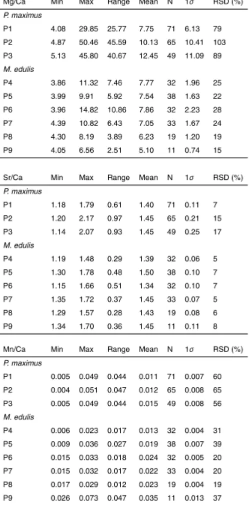
Significant small-scale Mg/Ca, Sr/Ca and Mn/Ca heterogeneity was observed in both P. maximus and M. edulis shells (Figs. 2 and 3; Table 1; Supplementary Material 1 http://www.biogeosciences-discuss.net/6/1267/2009/bgd-6-1267-2009-supplement. pdf). InP. maximusthe range of Mg/Ca ratios (4.08 to 50.46 mmol/mol) was larger than
5
that observed inM. edulis(3.86 to 14.82 mmol/mol). The range of Sr/Ca and Mn/Ca ratios was similar in the two species (Table 1), albeit with higher maximum Sr/Ca ratios inP. maximus(2.17 mmol/mol) than inM. edulis(1.78 mmol/mol) and lower maximum Mn/Ca ratios inP. maximus(0.051 mmol/mol) than inM. edulis(0.073 mmol/mol).
3.1 Pecten maximus
10
InP. maximus(Fig. 2b), Mg/Ca ratios in all profiles decrease from maxima in the outer-most shell to a minimum value at depths of ca. 110–170µm, below which Mg/Ca ratios were relatively invariant, both within (P1: depths of 110–700µm, 5.46±0.52 mmol/mol,
N=60; P2: depths of 160–640µm, 5.54±0.32 mmol/mol, N=49; and P3: depths of 170–480µm, 5.58±0.34 mmol/mol, N=32) and between each of the three profiles,
15
which showed no significant difference in mean Mg/Ca ratios between each profile (t-test, p>0.05 for all comparisons). The highest Mg/Ca ratios are the highest ever reported forP. maximus(Lorrain et al., 2005; Freitas et al., 2008) or any other bivalve shell calcite (Lazareth et al., 2007) and were observed in regions of modified shell structures (the two shaded areas in Fig. 2b), i.e. the shell surface stria (profile P2) and
20
disturbance growth mark (profile P3).
Shell Sr/Ca ratios were highest in the 100–120µm of the P. maximus profiles prox-imal to the outer shell surface (1.7 to 2.2 mmol/mol), especially in the modified shell structures sampled in profiles P2 and P3 (shaded areas in Fig. 2b), i.e. the shell surface stria (up to 2.2 mmol/mol) and disturbance growth mark (up to 2.1 mmol/mol). Sr/Ca
ra-25
inter-BGD
6, 1267–1316, 2009Element/Ca small-scale variability
in bivalve calcite
P. S. Freitas et al.
Title Page
Abstract Introduction
Conclusions References
Tables Figures
◭ ◮
◭ ◮
Back Close
Full Screen / Esc
Printer-friendly Version
Interactive Discussion mediate ratios below the outer shell Sr/Ca maxima. Shell Sr/Ca ratios then increased
from these minima to higher values towards the inner shell surface.
Shell Mn/Ca ratios were high in the ca. 100µm proximal to the outer shell surface, particularly in the shell features sampled in profiles P2 and P3, i.e. the shell surface stria (up to 0.023 mmol/mol) and disturbance growth mark (up to 0.050 mmol/mol).
5
Mn/Ca ratios then were lowest (0.004 to 0.016 mmol/mol), but also variable, at the mid-depths within the profiles, i.e. from the ca. 100µm proximal to the outer shell sur-face to 200–300µm above the inner shell surface. Shell Mn/Ca ratios increased to higher values towards the inner shell surface (0.030 to 0.051 mmol/mol).
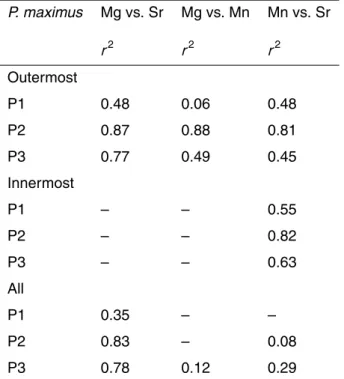
Taking the whole of each profile, the Sr/Ca and Mg/Ca ratios were significantly
cor-10
related (Table 2; allp<0.001) in profiles P1, P2 and P3. The greatest degree of cor-relation was found in the 110–180µm proximal to the outer shell surface where both Sr/Ca and Mg/Ca reach maxima (Table 2; all p<0.001), and which correspond to a brighter area in the light microscope image (Fig. 2a). Between this outermost section (i.e. to depths of 110–180µm) and the inner shell surface no significant relationship
15
was observed between Sr/Ca and Mg/Ca ratios in any of the profiles.
Taking the whole of each profile, the Mn/Ca and Mg/Ca ratios were not significantly correlated (Table 2). However, shell Mn/Ca and Mg/Ca ratios were significantly corre-lated in the shell features sampled in profiles P2 and P3, i.e. the shell surface stria and disturbance growth mark (Table 2; bothp<0.001). Shell Mn/Ca ratios were significantly
20
correlated to Sr/Ca ratios in profiles P1, P2 and P3 in the 110–200µm closest to the outer shell surface and also in the 170–490µm proximal to the inner shell surface of P. maximus(Table 2; allp<0.001).
3.2 Mytilus edulis
Spatial variability of Mg/Ca, Sr/Ca and Mn/Ca ratios within theM. edulis shell (Fig. 3)
25
BGD
6, 1267–1316, 2009Element/Ca small-scale variability
in bivalve calcite
P. S. Freitas et al.
Title Page
Abstract Introduction
Conclusions References
Tables Figures
◭ ◮
◭ ◮
Back Close
Full Screen / Esc
Printer-friendly Version
Interactive Discussion T3, to maximum values at depths of ca. 240–270µm. Shell Mg/Ca ratios then usually
decreased to lower (but not always the lowest in each profile) values at the inner shell surface. The remaining profile P9 exhibited no particular trend in Mg/Ca ratios with profile depth. Compared to Mg/Ca ratios, shell Sr/Ca and Mn/Ca ratios showed no consistent pattern with profile depth. In addition, shell Mg/Ca, Sr/Ca and Mn/Ca ratios
5
were not significantly correlated within any of theM. edulisprofiles (p>0.05).
3.3 Relative importance of intra- and inter-profile variability in element/Ca ratios
Spatial variability of element/Ca ratios was evident both between and within each ion microprobe profile for each bivalve species (Figs. 2 and 3; Table 1). To evaluate the relative significance of the inter- and intra-profile heterogeneity in element/Ca ratios
10
observed in each species, ANOVA was used to determine the percentage of the total variability that the inter- and intra-profile variation in elemental concentrations repre-sents. Small-scale variability of element/Ca ratios in both bivalve species, with the exception of Mn/Ca ratios inM. edulis, was found to be dominated by intra-profile vari-ability, i.e. between the outer and the inner shell surfaces. In both species, the majority
15
of the variation of Mg/Ca ratios (95.6% for P. maximusand 83.8% for M. edulis) and Sr/Ca ratios (99.9% for P. maximus and 69.5% for M. edulis) occurs as intra-profile variability. A similar observation is evident for the variability of shell Mn/Ca ratios in P. maximus(95.5%), whereas the relative importance of intra- and inter-profile variabil-ity of shell Mn/Ca ratios is similar inM. edulis (47.5 and 52.5%, respectively). In M.
20
edulis, the high inter-profile variability of shell Mn/Ca ratios is evident from mean profile Mn/Ca ratios which decreased from profile P9 towards profile P4, i.e. from the shell margin towards the umbo region (Fig. 3b and Table 1).
3.4 Shell element/Ca ratios variability between growth intervals
Each individual SIMS profile sampled, at different depths, one or more growth intervals
25
BGD
6, 1267–1316, 2009Element/Ca small-scale variability
in bivalve calcite
P. S. Freitas et al.
Title Page
Abstract Introduction
Conclusions References
Tables Figures
◭ ◮
◭ ◮
Back Close
Full Screen / Esc
Printer-friendly Version
Interactive Discussion profile that correspond to individual growth intervals (Table 3). However, due to the
influence of intra-profile variability on element/Ca ratios, particularly in P. maximus, mean elemental/Ca ratios for each growth interval can not be interpreted solely as representing inter-profile variability.
Mean Mg/Ca ratios in P. maximuswere higher in shell deposited during growth
pe-5
riods T1 and T2, as sampled by profiles P1, P2 and P3 in the outermost shell, and lower in shell deposited during growth periods T2 and T3 that were sampled by profiles P1, P2 and P3 (Fig. 2 and Table 3). Mean Sr/Ca and Mn/Ca ratios inP. maximuswere variable in shell deposited during growth periods T1 and T2, as sampled by profiles P1, P2 and P3 in the outermost shell, but similar in shell deposited during growth period
10
T3, as sampled by profiles P1, P2 and P3 (Fig. 2 and Table 3).
Mean profile Mn/Ca ratios inM. eduliswere higher in profiles P5, P6, P7, P8 and P9 that sampled shell deposited during growth period T3 and lower in profiles P4 and P5 that sampled shell deposited during growth periods T2 and T1, respectively (Fig. 2 and Table 3).
15
4 Relationships between element/Ca ratios and shell features and structure
4.1 Pecten maximus
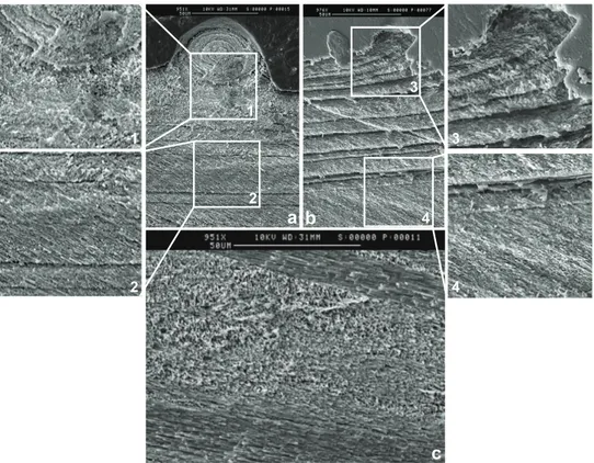
SEM images (Figs. 2 and 4) illustrate that the threeP. maximusion microprobe profiles, situated in the new growth region of the shell, traversed only one layer of irregularly ori-ented foliated calcite. Nevertheless, high and variable Mg/Ca, Sr/Ca and Mn/Ca ratios
20
are associated with some differences in the crystal arrangement, particularly within the shell surface stria (profile P2) and to a lesser extent within the region of the sur-face disturbance mark (profile P3), which are comprised of a relatively unorganized arrangement of calcite crystals. Clearly, shell features, such as the shell surface stria and disturbance growth marks, and associated variations in crystal arrangement may
25
thermo-BGD
6, 1267–1316, 2009Element/Ca small-scale variability
in bivalve calcite
P. S. Freitas et al.
Title Page
Abstract Introduction
Conclusions References
Tables Figures
◭ ◮
◭ ◮
Back Close
Full Screen / Esc
Printer-friendly Version
Interactive Discussion dynamic control. By comparison, Mg/Ca ratios are of lower magnitude and relatively
invariant in the mid region to innermost shell, whereas Sr/Ca and Mn/Ca ratios vary sig-nificantly in this region of the shell and must be controlled by some other factor other than by variable crystal arrangement. Nevertheless, the coherent variation of Sr/Ca and Mn/Ca ratios in the mid region to innermost shell for shell deposited
contempo-5
raneously during growth intervals T2 and T3, but sampled by different SIMS profiles (Fig. 2), suggests that a common process, or processes, could control both Sr and Mn incorporation in this region of the shell.
4.2 Mytilus edulis
All of theM. edulisSIMS analyses sampled the outer prismatic calcite layer (Figs. 3 and
10
5), and only the deepest sample(s) closest to the inner shell surface in profiles P4 and P5 may have potentially sampled the inner nacreous aragonitic layer (see Fig. 3a and Sect. 2). Internal disturbance/growth lines are easily identifiable in the SEM images of theM. edulis shell (Figs. 3a and 5) and these run obliquely between the outer and inner shell surfaces. The disturbance marks on the surface of the shell (“hump-like”
15
features where profiles P4, P5 and P6 are directly or proximally situated; Figs. 3a and 5) and their associated internal disturbance lines (the most prominent lines labelled a, b and c on Figs. 3a and 5) were formed during emersion of the animals between growth intervals, whereas the other internal growth lines formed while the animal remained immersed. Because of the nature of the incremental growth pattern in bivalve shells
20
(Lutz and Rhoads, 1980), internal growth lines can be used to identify shell deposited contemporaneously at different locations within the shell. This incremental growth pat-tern of bivalve shells is such that within an individual SIMS profile the uppermost data points are representative of shell material deposited at the shell margin at one point in time, with data points lower in that profile representing shell material precipitated
25
BGD
6, 1267–1316, 2009Element/Ca small-scale variability
in bivalve calcite
P. S. Freitas et al.
Title Page
Abstract Introduction
Conclusions References
Tables Figures
◭ ◮
◭ ◮
Back Close
Full Screen / Esc
Printer-friendly Version
Interactive Discussion time line, can be traced between SIMS profiles. For example, the internal disturbance
line labelled b in Figs. 3 and 5 that runs from near the base of SIMS profile P4 to the disturbance mark on the shell surface at the top of profile P5 delineates a break in shell deposition during emersion. A similar structural relationship and internal disturbance line (labelled c) can be seen running from near the base of profile P5 and the
distur-5
bance mark on the shell surface at the top of profile P6 (Figs. 3 and 5). As in all bivalve shells, the incremental growth pattern is such that the individual internal growth bands, defined by two growth lines, also thicken towards the margin of the shell (Fig. 5), indi-cating variable mineral crystallisation rates within individual growth bands. Therefore, it is evident that the fastest shell precipitation rates occurred at the shell margin.
10
Given that the shell was deposited during a period of constant seawater temperature and Mg/Ca ratio, it would be expected that shell deposited between any two growth lines would have a constant Mg/Ca ratio due to contemporaneous precipitation, inde-pendent of the location of new mineral crystallisation within the shell. Calcite Sr/Ca and Mn/Ca ratios, on the other hand, are influenced by mineral precipitation rate, the former
15
being positively correlated with precipitation rate (Lorens, 1981; Morse and Bender, 1990; Tesoriero and Pankow, 1996) and the latter inversely correlated to precipitation rate (Lorens, 1981; Mucci, 1988; Pingitore et al., 1988; Dromgoole and Walter, 1990). Sr/Ca and Mn/Ca ratios, thus, are not expected to be constant throughout an individual internal band due to the variable mineral precipitation rates. Whereas seawater Sr/Ca
20
ratios remained constant, due to the conservative behaviour of these two elements, it is impossible to determine whether variable concentrations of non-conservative seawater Mn also could have contributed to the observed heterogeneity in shell Mn content since temporal changes in seawater Mn concentration were not monitored.
Interestingly, Mg/Ca ratios differ more strongly than Sr/Ca ratios depending on where
25
BGD
6, 1267–1316, 2009Element/Ca small-scale variability
in bivalve calcite
P. S. Freitas et al.
Title Page
Abstract Introduction
Conclusions References
Tables Figures
◭ ◮
◭ ◮
Back Close
Full Screen / Esc
Printer-friendly Version
Interactive Discussion the initial part of growth increments T2 and T3 (Fig. 5b, blue and red areas at the top
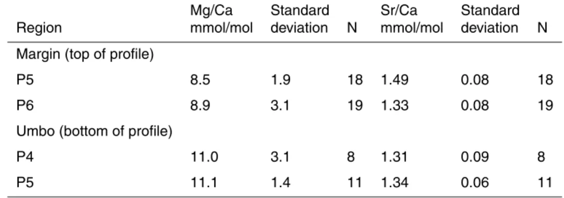
of profiles P5 and P6, respectively) exhibit a decrease and then general increase in Mg/Ca and lower mean ratios (Table 4) than shell precipitated contemporaneously in the innermost shell away from the shell margin (Fig. 5b, blue and red areas near the base of profiles P4 and P5, respectively). In contrast, during the initial part of growth
5
increments T2 and T3, Sr/Ca ratios were similar (Table 4) in shell precipitated at the shell margin (Fig. 5b, blue and red areas at the top of profiles P5 and P6, respectively) and shell precipitated contemporaneously in the innermost shell away from the shell margin (Fig. 5b, blue and red areas near the base of profiles P4 and P5, respectively). Lorens and Bender (1980) have described previously an influence of disturbance on
10
M. edulisshell Mg/Ca and Sr/Ca ratios, whereby the stress of capture and adaptation to a new laboratory environment induced the deposition of a shell region (termed “tran-sition zone calcite” by those authors) with high organic matrix content and high Mg/Ca (>40 mmol/mol) and Sr/Ca (>1.4 mmol/mol) ratios. Subsequent shell growth then ex-hibited decreasing Mg/Ca (to<10 mmol/mol) and Sr/Ca (to<0.6 mmol/mol) ratios as
15
the animals adapted to their new environment. However, Lorens and Bender (1980) only inferred the “transition zone” between new and old calcite using spot chemical compositions measured by electron microprobe, and their transition zone, where higher Mg/Ca and Sr/Ca ratios were observed, occurs perpendicular to the shell surface, an observation that is inconsistent with the incremental growth pattern ofM. edulis,
20
i.e. growth lines actually run oblique to the shell surface. In this study the Mg/Ca and Sr/Ca ratios have been compared directly to SEM images of the shell’s internal growth banding and indicate a more complex relationship between elemental composition and shell structure. Furthermore, the pattern of Mg/Ca ratio variability observed within the M. edulis shell calcite layer in this study is similar to that recognised by Dalbeck et
25
BGD
6, 1267–1316, 2009Element/Ca small-scale variability
in bivalve calcite
P. S. Freitas et al.
Title Page
Abstract Introduction
Conclusions References
Tables Figures
◭ ◮
◭ ◮
Back Close
Full Screen / Esc
Printer-friendly Version
Interactive Discussion
5 Potential causes of the observed small scale element/Ca ratio heterogeneity
withinP. maximusandM. edulisshell calcite
The extent of the small-scale heterogeneity of element/Ca ratios differs significantly be-tweenP. maximus andM. edulis, suggesting that the processes controlling elemental incorporation into shell calcite also differ between the two bivalve species investigated
5
in this study. For instance, P. maximus produces elaborate shell features, such as shell surface striae, whileM. edulis has a smoother shell surface that is also covered by an organic periostracum, a shell component which is absent in the former species. Several processes can be considered as potential explanations for the differential in-corporation of elements into bivalve shells and may explain the significant small-scale
10
heterogeneity of Mg, Sr and Mn contents inP. maximusandM. edulisshell calcite that has been observed in the present study. Of all the elements investigated in this study, Mg in the outermost parts of theP. maximusshell provides the strongest indication for the likely presence of control(s) other than temperature on the elemental composition of bivalve calcite.
15
5.1 Environmental variables: seawater salinity, pH, Mg/Ca and Sr/Ca ratios, Mg and Sr concentrations
Deposition of new shell material in P. maximus and M. edulis during the laboratory-culturing experiment occurred at constant temperature and constant seawater Mg/Ca and Sr/Ca ratios (Fig. 1). However, other environmental parameters that are known to
20
influence the elemental composition of biogenic carbonates, such as salinity and pH (e.g. Nurnberg et al., 1996; Lea et al., 1999), were not constant for the duration of the experiment (Fig. 1). Higher salinities have been reported to increase Mg/Ca and Sr/Ca ratios and higher pH appear to decrease Mg/Ca ratios and to increase Sr/Ca ratios (e.g. Nurnberg et al., 1996; Lea et al., 1999). Nevertheless, the range of salinity (33.02
25
BGD
6, 1267–1316, 2009Element/Ca small-scale variability
in bivalve calcite
P. S. Freitas et al.
Title Page
Abstract Introduction
Conclusions References
Tables Figures
◭ ◮
◭ ◮
Back Close
Full Screen / Esc
Printer-friendly Version
Interactive Discussion ratios observed in the SIMS profiles of P. maximus and M. edulis (Figs. 2 and 3).
Furthermore, seawater Mg/Ca and Sr/Ca ratios did not vary over the salinity variation observed during the experiment (Fig. 1). Seawater Mg and Sr concentrations exhibited no significant variation during the culturing period and thus also cannot be associated with the observed variation in shell Mg/Ca and Sr/Ca ratios (Fig. 1). In contrast, an
5
increase in seawater Mn concentration (not measured) during the culturing experiment could potentially explain the observed increase in mean profile Mn/Ca ratios observed in SIMS profiles that sampledM. edulis shell deposited from growth interval T1 to T3 (Table 1), but such a response would appear to be restricted toM. edulissince a similar pattern was not observed in theP. maximusshell Mn/Ca data.
10
5.2 Shell growth and mineral precipitation rates
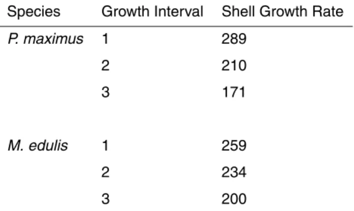
No data on the metabolic activity and food uptake of the animals were collected during the experimental period. However, shell growth rate (SGR) was measured and should reflect changes in the physiological condition, food uptake and metabolic activity of the animals during the experiment, i.e. assuming that an increase in food uptake or
15
metabolic activity of the animals will correspond in an increase in SGR. For both the P. maximusand M. edulis specimens, at the whole shell level, SGR decreased from growth interval T1 to T3 (Table 5) and, from the comparison to mean Mg/Ca, Sr/Ca and Mn/Ca ratios for each growth interval sampled by different SIMS profile (Table 3), SGR did not influence the variation of Mg/Ca, Sr/Ca and Mn/Ca observed in the SIMS
20
profiles ofP. maximusandM. edulis (Figs. 2 and 3).
In calcite Mg incorporation is not thought to be influenced by mineral precipitation rate, both (in synthetic e.g. Morse and Bender, 1990) and in bivalve calcite (Lorens and Bender, 1980). Increasing precipitation rate has, however, been shown to cause an increase in Sr/Ca ratios and a reduction in Mn/Ca ratios in calcite (Lorens, 1981;
25
BGD
6, 1267–1316, 2009Element/Ca small-scale variability
in bivalve calcite
P. S. Freitas et al.
Title Page
Abstract Introduction
Conclusions References
Tables Figures
◭ ◮
◭ ◮
Back Close
Full Screen / Esc
Printer-friendly Version
Interactive Discussion In the M. edulis shell the individual internal growth bands, defined by two growth
lines representing contemporaneous periods of shell deposition, thicken towards the margin of the shell (Fig. 5). This observation indicates variable mineral precipitation rates with the highest shell precipitation rates occurring at the shell margin. If Sr/Ca and Mn/Ca ratios inM. eduliswere controlled by mineral precipitation rate, Sr/Ca ratios
5
should be higher and Mn/Ca ratios lower in the shell deposited at the shell margin compared to shell deposited away from the shell margin. In addition, Sr/Ca ratios also should decrease and Mn/Ca ratios increase from the outer shell surface towards the inner shell surface, in accordance with the gradient in shell mineralization rate that is highest in the former and lowest in the latter parts of the shell. However, such variation
10
of Sr/Ca and Mn/Ca ratios was not observed in any of the individual SIMS profiles, albeit such a pattern can be identified in one of two growth bands sampled at different locations within the shell in profiles P4, P5 and P6 (Fig. 5). Mean Sr/Ca ratios were higher in shell deposited closer to the shell margin than in shell deposited away from the shell margin (Table 4 and Fig. 5b; blue areas shaded on the top of profile P5 and
15
at the bottom of profile P4, respectively). Nevertheless, mineral precipitation rate was not a significant general influence on the incorporation of Sr and Mn in the shell calcite ofM. edulis.
A similar comparison cannot be made forP. maximus due to the lack of visible in-ternal growth lines. It can be seen, however, that Sr and Mn positively co-vary in the
20
innermost shell regions (Fig. 2 and Table 2) and thus Sr and Mn incorporation in P. maximus calcite should be controlled by a common process(es) other than precipita-tion rate.
5.3 Composition of the extra-pallial fluid (EPF), the precipitating solution in bivalves
The elemental composition of the solution from which calcification occurs is known to
25
BGD
6, 1267–1316, 2009Element/Ca small-scale variability
in bivalve calcite
P. S. Freitas et al.
Title Page
Abstract Introduction
Conclusions References
Tables Figures
◭ ◮
◭ ◮
Back Close
Full Screen / Esc
Printer-friendly Version
Interactive Discussion oysters, shell deposition at the shell margin occurs from an extra-pallial space (EPS)
that is periodically exposed to the ambient seawater medium, while inM. edulis biomin-eralization occurs from a continuously isolated EPS (Clark II, 1974; Carriker, 1992). In bivalve species with periodically exposed EPS the margins of the mantle (the soft-tissue organ that controls calcification) are frequently withdrawn into the mantle cavity,
5
exposing those crystals along the inner shell surface between the shell margin and the attachment of the mantle on the inner shell surface at the pallial line to a more seawater-like fluid (Clark II, 1974; Carriker, 1992). The periodical exposure of the marginal EPS to seawater, if it causes higher Mg content of the marginal EPF, may thus also help to explain the high Mg/Ca ratios previously observed inP. maximus(Freitas et al., 2006,
10
2008), namely in shell deposited close to the shell margin, i.e. the outermost shell (top ca. 170µm), shel surface stria and surface disturbance mark (Fig. 2), and in another species with periodically exposed EPS,Pinna nobilis(Freitas et al., 2005).
In both P. maximus and M. edulis, Mg/Ca, Sr/Ca and Mn/Ca ratios varied signifi-cantly in shell deposited from the same marginal EPF at the same time, but at different
15
locations on the inner shell surface. Transport to the EPF likely is not a major control on the observed heterogeneity of shell elemental composition, unless variation of ion transport along the mantle could lead to elemental concentration gradients within the EPF. Other smaller-scale mechanism(s) thus must control incorporation of elements in the shell, e.g. processes that act at the shell crystal-solution interface. Furthermore,
20
the close proximity of the mantle epithelium to the shell surface, which are separated by a small distance and may be in contact with each other, allows the transfer of ions and organic molecules to occur virtually by direct contact (Simkiss and Wilbur, 1989; Addadi et al., 2006), and thus could provide the potential for small-scale variations in the chemical and physical conditions at different precipitation sites on the inner shell
25
BGD
6, 1267–1316, 2009Element/Ca small-scale variability
in bivalve calcite
P. S. Freitas et al.
Title Page
Abstract Introduction
Conclusions References
Tables Figures
◭ ◮
◭ ◮
Back Close
Full Screen / Esc
Printer-friendly Version
Interactive Discussion 5.4 Elemental composition of shell calcite
The Mg content of calcite is known to influence the incorporation of other elements during calcite precipitation, whereby substitution of Ca2+ by Mg2+ distorts the crystal lattice and favours the incorporation of other elements, namely ones with large ionic radii, such as Sr2+ and Mn2+ (Mucci and Morse, 1983; Ohde and Kitano, 1984; Morse
5
and Bender, 1990). InP. maximus, significant positive correlations were observed be-tween Mg/Ca and both Sr/Ca and Mn/Ca ratios in the 100–200µm proximal to the outer shell surface, particularly in the shell surface stria and disturbance mark. Therefore, shell Mg content could influence Sr and, to a lesser extent, Mn incorporation in the outermost part of the shell of this species (Table 2), where Mg/Ca ratios are highest
10
(10–50 mmol/mol, Fig. 2). However, such a lattice distortion control is not evident in the mid region and innermost part of theP. maximus shell. In this same part of the shell Mg does not correlate with Sr or Mn, but Sr and Mn are significantly correlated to each other (Table 2), suggesting that a common process, or processes, could control both Sr and Mn incorporation. InM. edulis, Mg/Ca ratios were not significantly correlated
15
to Sr/Ca and Mn/Ca, and thus shell Mg content does not appear to have influenced Sr and Mn incorporation in the calcite shell component of this species.
5.5 Sector zoning and calcite crystal orientation and size
Sector zoning is a phenomenon recognised in both synthetic and natural calcite miner-als, whereby differences in elemental composition occur both within a particular
crys-20
tal sector type (intra-sectoral) and between crystallographically non-equivalent coeval growth sectors (inter-sectoral) in any one crystal (Reeder and Grams, 1987; Reeder and Paquette, 1989; Paquette and Reeder, 1990, 1995). Elemental zoning patterns reflect non-equilibrium elemental incorporation due to differences in the intrinsic char-acteristics of crystal surfaces and their underlying structure, the specific nature of
25
BGD
6, 1267–1316, 2009Element/Ca small-scale variability
in bivalve calcite
P. S. Freitas et al.
Title Page
Abstract Introduction
Conclusions References
Tables Figures
◭ ◮
◭ ◮
Back Close
Full Screen / Esc
Printer-friendly Version
Interactive Discussion Reeder, 1990, 1995). Paquette and Reeder (1995) describe the mechanisms of
trace-element incorporation in calcite in more detail.
Unlike laser-ablation or powder milling sampling, SIMS instrumentation samples only about 0.1–0.2 microns of the polished shell section (R. Hinton, personal communica-tion, 2007). Therefore, if those calcite crystals comprising the sectioned bivalve shells
5
were orientated along different axes within different parts of shells then the observed variability in the SIMS element/Ca ratio data could be explained by inter-sectoral zon-ing, as described above, and the SIMS sampling of different crystal faces.
Dalbeck et al. (2006) used Electron Backscatter Diffraction (EBSD) and electron probe microanalysis (EPMA) to investigate the relative crystallographic orientations of
10
crystals and chemistry, respectively, ofM. edulis calcite and aragonite layers. For the entire calcite layer of this species, with the exception of the outermost 40µm, Dal-beck et al. (2006) demonstrated uniformity of the axes of the individual calcite crystals, which differ in orientation to one another by less than 15◦. Calcite crystals produced early during formation of theM. edulisshell, i.e. the outermost 40µm, are
morphologi-15
cally more varied than the remainder of the calcite layer, with a progressive refinement in the orientation of the planes of adjacent crystals during shell growth (Dalbeck et al., 2006). Consequently, SIMS sampling of sector zoning cannot be used as an explana-tion for the observed variability of Mg/Ca, Sr/Ca and Mn/Ca ratios within the majority ofM. eduliscalcite sampled herein (Fig. 3), but cannot be discounted in the outermost
20
40µm of theM. edulisshell. A similar conclusion was reached by Dalbeck et al. (2006) in terms of the compositional variability evident in their EPMA elemental dataset. In ad-dition, Dalbeck et al. (2006) have suggested that the smaller size of the calcite crystals in the outermost part of theM. edulis shell may allow a greater adsorption of cations due to a greater surface area to volume ratio. Rosenberg et al. (2001) also observed in
25
BGD
6, 1267–1316, 2009Element/Ca small-scale variability
in bivalve calcite
P. S. Freitas et al.
Title Page
Abstract Introduction
Conclusions References
Tables Figures
◭ ◮
◭ ◮
Back Close
Full Screen / Esc
Printer-friendly Version
Interactive Discussion ForP. maximus, Checa et al. (2007) have shown that the foliated calcite layer of this
bivalve species is made up of highly organised folia that display a coherent crystal-lographic pattern, i.e. a sheet texture; several co-existing sets of folia run in different directions in the shell of this species (see also Fig. 4c). As withM. edulis, the uniformity of crystal orientation withinP. maximusfolia means that SIMS sampling of sector
zon-5
ing cannot be invoked as an explanation for variable Mg/Ca, Sr/Ca and Mn/Ca within a folia. However, SIMS sampling of co-existing sets of folia running in different directions, and thus potentially of different crystal faces, could contribute to some of the observed variability in Mg/Ca, Sr/Ca and Mn/Ca ratios inP. maximus. Furthermore, it is impossi-ble to discount the possibility that the more random crystal orientations (Fig. 4) within
10
the shell surface stria (profile P2) and surface disturbance mark (profile P3) contribute to the high Mg/Ca, Sr/Ca and Mn/Ca ratios observed in these regions as the result of SIMS sampling of inter-sectoral compositional zoning, especially when considering the strong correlations observed between the Mg/Ca, Sr/Ca and Mn/Ca ratios measured in this study within the shell surface stria and disturbance mark and the evidence for
15
positive correlations between Mg, Mn and Sr concentrations in different crystal growth sectors, i.e. elevated within the same sector relative to another sector (Reeder and Grams, 1987; Reeder and Paquette, 1989). However, without detailed EBSD studies, further speculation regarding the effect of crystal orientation on the Mg/Ca, Sr/Ca and Mn/Ca ratios of bivalve shell calcite is unwarranted.
20
5.6 Role of the shell organic matrix
Biogenic minerals are composed of both inorganic and organic fractions, the latter controlling crystal nucleation and growth (e.g., Mann, 2001; Weiner and Dove, 2003; Addadi et al., 2006). Within and between shells the organic matrix varies both in con-centration and composition (e.g. Dauphin, 2003; e.g. Dauphin et al., 2003a, 2005) and
25
BGD
6, 1267–1316, 2009Element/Ca small-scale variability
in bivalve calcite
P. S. Freitas et al.
Title Page
Abstract Introduction
Conclusions References
Tables Figures
◭ ◮
◭ ◮
Back Close
Full Screen / Esc
Printer-friendly Version
Interactive Discussion Sulphur has been proposed to represent the shell organic matrix, either as sulphated
polysaccharides or S-rich aminoacids, in biogenic carbonates such as bivalves (Lorens and Bender, 1980; Rosenberg et al., 2001; Dauphin et al., 2003a, 2003b, 2005), gas-tropods (Lazareth et al., 2007), brachiopods (Cusack et al., 2008a) and corals (Cuif and Dauphin, 2005). Observations that the distribution of Mg correlates with S
variabil-5
ity has led to the suggestion that a proportion of the measured Mg content in biogenic calcites is non-lattice bound, being associated with the shell organic matrix and not just the inorganic calcite mineral (Lorens and Bender, 1980; Rosenberg and Hughes, 1991; Rosenberg et al., 2001; England et al., 2007).
The role of the organic component on elemental incorporation in biogenic
carbon-10
ates, however, is far from clear and may likely vary between specific taxa. For instance, removal of the organic component of the aragonitc shell of the estuarine bivalve Cor-bula amurensisresults in a lowering of shell Mg, and Mn content but not of Sr content (Takesue et al., 2008). In the shell calcite of the gastropodConcholepas concholepas lower Mg content occurs in areas with higher organic content (Lazareth et al., 2007). In
15
two species of brachiopods,Terbratulina retusaandNotosaria nigricans, Mg has been shown not to be hosted by organic components (Cusack et al., 2008b). Furthermore, sulphur can also occur in the calcite structure as sulphate that substitutes for carbonate groups (Kontrec et al., 2004), and thus the use of S as a tracer for the organic matrix in biogenic carbonates is not straightforward. For instance, in the calcitic shells of the
20
bivalvesP. nobilis andP. margaritiferano correlation was observed between Mg and S (Dauphin et al., 2003b), whereas in the shell calcite of the gastropodC. concholepas Mg and S were observed to be inversely correlated (Lazareth et al., 2007).
Lorens and Bender (1980) observed that high S/Ca and Mg/Ca ratios were both found inM. edulis shell regions deposited under stress conditions and suggested that
25
BGD
6, 1267–1316, 2009Element/Ca small-scale variability
in bivalve calcite
P. S. Freitas et al.
Title Page
Abstract Introduction
Conclusions References
Tables Figures
◭ ◮
◭ ◮
Back Close
Full Screen / Esc
Printer-friendly Version
Interactive Discussion crystal elongation and hence shell form along different axes (Rosenberg et al., 2001).
InM. edulis, shell regions with high curvature are organic-rich and have high Mg and S content in contrast to shell regions with low curvature that are mineral-rich and have low Mg and S content (Rosenberg et al., 1989, 2001 Rosenberg and Hughes, 1991).The deposition of complex features such as the surface ridge and disturbance growth marks
5
in P. maximus involves a marked disruption of shell deposition, with changes in the orientation of the shell surface and also of calcite crystal orientation (Figs. 2 and 4). Such disruption of shell deposition appears to be of smaller intensity in theM. edulis shell (Fig. 5). Therefore, considering the observations of Lorens and Bender (1980), Rosenberg et al. (1989, 2001) and Rosenberg and Hughes (1991), it is plausible that
10
deposition of the surface ridge and disturbance growth marks inP. maximus involves an increase in the organic matrix relative to the remainder of the shell and that such an increase is associated with higher Mg contents, although not necessarily bound to the organic matrix.
Variations in both the amount and type of the shell organic matrix remain plausible
15
causes of the SIMS element/Ca ratio heterogeneity observed in this study, especially with regards to the shell surface stria inP. maximusand the disturbance growth marks evident inP. maximusandM. edulis.
6 The potential of Mg/Ca ratios in the innermost shell region ofP. maximusas
a palaeotemperature proxy
20
The relatively invariant Mg/Ca ratios in the mid region to innermost shell ofP. maximus suggest a region of shell deposition where temperature was the main control on Mg incorporation with potential to use measurements of the Mg/Ca ratio in this part of the shells of this bivalve species as a palaeotemperature proxy. Such a hypothesis is strengthened by the evidence that the Mg/Ca ratios were relatively invariant both within
25
BGD
6, 1267–1316, 2009Element/Ca small-scale variability
in bivalve calcite
P. S. Freitas et al.
Title Page
Abstract Introduction
Conclusions References
Tables Figures
◭ ◮
◭ ◮
Back Close
Full Screen / Esc
Printer-friendly Version
Interactive Discussion surface (Fig. 2).
7 Small-scale element heterogeneity and implications for the use of
geochemical proxies in bivalves
It is clear from the ion microprobe elemental data collected in this study that for both bivalve species investigated herein highly variable Mg/Ca, Sr/Ca and Mn/Ca ratios can
5
occur within one structural layer of shell calcite precipitated at a single and constant seawater temperature and via deposition from the same marginal extra-pallial fluid (EPF), albeit a variably non-isolated EPF inP. maximus. Such significant small-scale heterogeneity of the Mg, Sr and Mn content in the shells ofM. edulisandP. maximus has profound implications for the application of these geochemical proxies in bivalve
10
calcite. First, the deposition of elaborate shell features and surface disturbance growth marks, the latter ubiquitous features of shells of many bivalve shells, is associated with highly variable element/Ca ratios. Second, factors/processes that may influence el-emental incorporation into bivalve calcite potentially do not just operate at the scale of whole shells, or even within a single shell structural layer, but can vary at tens of
15
microns scale. Third, users of large-scale “bulk” shell sampling methods, and even micro-sampling methods such as micro-milling or laser ablation sampling, need to con-sider carefully which section, or sections, of the shell are sampled, otherwise they risk obtaining a large variability in element/Ca ratio measurements that do not relate to any change in environmental conditions. Finally, Mg, Sr and Mn incorporation into bivalve
20
calcite most likely is under the control of multiple factors and that the relative influ-ence of any one factor most likely varies with time and with the location of the shell deposition.
In particular, significant small-scale heterogeneity (i.e. tens of microns) in bivalve shell Mg/Ca ratios is potentially a significant source of error when attempting to use
25
BGD
6, 1267–1316, 2009Element/Ca small-scale variability
in bivalve calcite
P. S. Freitas et al.
Title Page
Abstract Introduction
Conclusions References
Tables Figures
◭ ◮
◭ ◮
Back Close
Full Screen / Esc
Printer-friendly Version
Interactive Discussion ones analysed by SIMS in the present study, but sampled by surface milling, displayed
a large variability (up to ca. 6 mmol/mol and up to ca. 16 mmol/mol, respectively) in Mg/Ca ratios at constant temperature and a weak correlation between shell Mg/Ca ra-tios and seawater temperature (r2=0.37,p<0.001 andr2=0.21,p<0.001, respectively) was observed over a range from 10 to 20◦C (Freitas et al., 2008). It is clear from the ion
5
microprobe data obtained in this study that small-scale variability in the Mg content is one possible reason why such a weak relationship was observed between shell Mg/Ca ratios and seawater temperature inP. maximus(Lorrain et al., 2005; Freitas et al., 2006, 2008). The inclusion of variable amounts of material from parts of the shell structure with different Mg/Ca ratios, as well as the sampling of shell areas with shell surface
10
striae and/or surface disturbance marks, may provides an explanation for the higher and variable large variability observed in Mg/Ca ratios from observed in the upper (top ca. 200µm) shell calcite grown at constant temperatures relative to the lower and less variable Mg/Ca ratios of the mid and innermost regions in the sameP. maximusshells grown at constant temperatures. In another field culturing experiment study, the
un-15
expected increase in Mg/Ca ratios of P. maximus specimens grown in a field-based culturing experiment at low winter temperatures reported by Freitas et al. (2006) could now be explained by a higher number of shell surface striae (ca. 12 striae/mm) be-ing milled and included in each “winter” powder sample, compared to Mg/Ca ratios in spring and summer samples with a lower number of shell surface striae (ca. 4–6
20
striae/mm), which had a more robust relationship to water temperatures.
InM. edulis there is some evidence that Mg/Ca ratios vary significantly depending on whether shell deposition occurs at the shell margin or on the inner shell surface. The observation thatM. edulisshell Mg/Ca ratios are influenced by disturbance marks formed during emersion could be particularly significant since in a natural inter-tidal
en-25
BGD
6, 1267–1316, 2009Element/Ca small-scale variability
in bivalve calcite
P. S. Freitas et al.
Title Page
Abstract Introduction
Conclusions References
Tables Figures
◭ ◮
◭ ◮
Back Close
Full Screen / Esc
Printer-friendly Version
Interactive Discussion 2001). Such observations further question the validity, and indicate the difficulty, of
us-ing Mg/Ca ratios inM. edulisas a palaeotemperature proxy, irrespective of a previous study that demonstrated a Mg/Ca ratio temperature relationship in the related species M. trossulus(Klein et al., 1996a).
8 Summary
5
In the present study, significant heterogeneity in Mg/Ca, Sr/Ca and Mn/Ca ratios within the new growth of shells ofP. maximusandM. edulisdeposited at a constant temper-ature of 20◦C has been determined using SIMS. Differences in the relative contribution of specific shell features, i.e. the number and size of shell surface striae (inP. maximus) and surface disturbance growth marks (in both species) milled, as well as the depth of
10
milling through regions of shell with variable Mg/Ca, Sr/Ca and Mn/Ca ratios, likely ex-plains some of the variability in element/Ca ratios observed previously forM. edulisand P. maximusshells. Most importantly, the observed variation in Mg/Ca ratios, as well as in Sr/Ca and Mn/Ca ratios, cannot be attributed to particular structural shell layers that are recognised forP. maximus orM. edulis shells, since only one structural layer was
15
sampled by the SIMS analyses. InP. maximus, Mg appears to strongly influence the incorporation of Sr and Mn in the uppermost shell, while in the innermost shell of this species Sr and Mn co-vary, an observation that suggests incorporation of these two elements may be controlled by (a) common process or processes.
The present study clearly confirms that processes that control shell structure and
fea-20
tures can exert a strong influence on the elemental composition of bivalve shell calcite, even in shell deposited from the same solution. Therefore, it is evident that disruption of “normal” shell deposition significantly perturbs the elemental composition of shell calcite in these species. The common presence of shell features and growth marks in bivalve shells thus further questions the use of Mg/Ca, Sr/Ca and Mn/Ca from bivalve
25
BGD
6, 1267–1316, 2009Element/Ca small-scale variability
in bivalve calcite
P. S. Freitas et al.
Title Page
Abstract Introduction
Conclusions References
Tables Figures
◭ ◮
◭ ◮
Back Close
Full Screen / Esc
Printer-friendly Version
Interactive Discussion thermodynamic control, and therefore the potential to use measurements of the Mg/Ca
ratio in this part of the shells of this animal as a palaeotemperature proxy is confirmed.
Acknowledgements. The authors thank Berwyn Roberts and Gwyn Hughes for their
ever-present technical support in relation to the maintenance of the laboratory aquaria and supply of algal cultures during the laboratory culturing experiment, John Rowlands for technical support
5
during use of the scanning electron microscope and David Oakes from Ramsay Sound Shell-fish Ltd. for supplying the scallop specimens. Access to the UK. Natural Environment Research Council Ion Microprobe Facility at Edinburgh University and the assistance of Richard Hinton and John Craven is acknowledged gratefully. We thank Claire Lazareth, Vincent Salters and an anonymous reviewer for their comments that substantially improved an earlier version of
10
this manuscript. This research was partially funded by Fundac¸ ˜ao para a Ci ˆencia e Tecnologia (FCT), Portugal, through a scholarship to Pedro Freitas, Contract No. SFRH/BD/10370/2002.
References
Addadi, L., Joester, D., Nudelman, F., and Weiner, S.: Mollusk shell formation: A source of new concepts for understanding biomineralization processes, Chem. Eur. J., 12, 980–987, 2006.
15
Allison, N.: Comparative determination of trace and minor elements in coral aragonite by ion mi-croprobe analysis, with preliminary results from Phuket, southern Thailand, Geochim. Cos-mochim. Ac., 60, 3457–3470, 1996.
Allison, N. and Austin, W.: The potential of ion microprobe analysis in detecting geochemi-cal variations across individual foraminifera tests, Geochem. Geophys. Geosyst., 4, 8403,
20
doi:8410.1029/2002GC000430, 2003.
Allison, N., Finch, A. A., Webster, J. M., and Clague, D. A.: Palaeoenvironmental records from fossil corals: The effects of submarine diagenesis on temperature and climate estimates, Geochim. Cosmochim. Ac., 71, 4693, 2007.
Beck, J., Edwards, R., Ito, E., Taylor, F., Recy, J., Rougerie, F., Joannot, P., and Henin, C.: Sea
25
surface temperature from skeletal Sr/Ca ratios, Science, 257, 644–647, 1992.
Bentov, S. and Erez, J.: Novel observations on biomineralization processes in foraminifera and implications for Mg/Ca ratio in the shells, Geology, 33, 841–844, 2005.
Berner, R.: The role of magnesium on the crystal growth of calcite and aragonite in seawater, Geochim. Cosmochim. Ac., 39, 489–504, 1975.