A
rt
ic
le
J. Braz. Chem. Soc., Vol. 20, No. 7, 1327-1333, 2009. Printed in Brazil - ©2009 Sociedade Brasileira de Química 0103 - 5053 $6.00+0.00
*e-mail: [email protected]
# Present address: Laboratório de Farmacognosia, Departamento de Ciências Farmacêuticas, Centro de Ciências da Saúde, Universidade Federal de Santa Catarina, Trindade, 88040-900 Florianopolis-SC, Brazil
A New Polyacetylene from Vernonia scorpioides (Lam.) Pers. (Asteraceae)
and its in vitro Antitumoral Activity
Humberto Buskuhl,a Rilton A. Freitas,a Franco Delle Monache,b Andersson Barison,c Francinete R. Campos,c Yuri E. Corilo,d Marcos N. Eberlind and Maique W. Biavatti*,a,#
aCentro de Ciências da Saúde, Universidade do Vale do Itajaí, 88302-202 Itajaí-SC, Brazil
bDipartimento di Chimica e Tecnologia delle Sostanze Biologicamente Attive, Università “La Sapienza”, Roma, Italy
cDepartamento de Química, Centro Politécnico, Universidade Federal do Paraná, 81530-900 Curitiba-PR, Brazil
dInstituto de Química, Universidade Estadual de Campinas, 13083-970 Campinas-SP, Brazil
A fração diclorometano obtida pelo particionamento do extrato hidroalcoólico obtido das
folhas e flores de Vernonia scorpioides (Asteraceae) resultou no isolamento e caracterização de
um novo poliacetileno, 5-octa-2,4,6-triinil-furan-2(5H)-ona. A determinação estrutural foi baseada em dados de espectrometria de Ressonância Magnética Nuclear mono e bidimensionais, de Massas e de Infravermelho. Foi investigado o potencial antitumoral e genotóxico do novo poliacetileno e os resultados encontrados sugerem genotoxicidade e citotoxicidade, onde a morte celular ocorreu
via apoptose, envolvendo um mecanismo dependente de caspase.
The dichloromethane fraction obtained from hydroalcoholic crude extract of leaves and flowers of Vernonia scorpioides (Asteraceae) was investigated, resulting in the isolation and structure elucidation of a new polyacetylene namely 5-octa-2,4,6-triynyl-furan-2(5H)-one. The structure of the isolated compound was determined based on IR, NMR (1D and 2D) and MS spectrometric data. The antitumor potential, including cytotoxicity to tumor cells and genotoxicity, was investigated.
The results suggest that apoptotic cell death may have occurred, at least in part, via a
caspase-dependent mechanism.
Keywords: polyacetylene, Vernonia scorpioides, cytotoxicity, genotoxicity, Asteraceae
Introduction
The widespread genus Vernonia (Asteraceae)
is characterized by the fact that it produces highly oxygenated sesquiterpene lactones, such as glaucolides and hirsutinolides, some of which are allenic lactones. Many other compounds have also been isolated, such as thiophenes and dithio compounds.1,2 Among the species
of Vernonia, cytotoxicity has been described for isolated sesquiterpene lactones from V. cinerea,3V. lasiopus,4 and
V. amygdalina.5 Besides sesquiterpene lactones, polar
compounds from water-soluble extracts of Vernonia have also been found to have cytotoxic and imunomodulatory activities such as those from the edible leaves of V.
amygdalina6 and from the roots of V.kotschyana7 and V.
anthelmintica.8
The species Vernonia scorpioides (Lam.) Pers., which is very common in Brazil, is popularly known as “Piracá”, “Enxuga” or “Erva-de-São-Simão” and usually grows in poor deforested neotropical soils.9 Alcoholic extract from
the fresh leaves is widely used in topical applications, to treat a variety of skin disorders, including chronic wounds, such as ulcers of the lower limbs. In previous studies, chloroform and hexane crude extracts of V.scorpioides leaves have shown fungicide activity,10 moderate bactericide
activity and mild wound healing effects.11 In addition, in
mice treated with dichloromethane extract of the leaves, the Ehrlich tumor disappeared completely.12 Therefore,
A New Polyacetylene from Vernonia scorpioides (Lam.) Pers. (Asteraceae) J. Braz. Chem. Soc.
1328
Results and Discussion
The crude ethanol extract of V. scorpioides was successively partitioned with hexane, dichloromethane and ethyl acetate. Successive chromatographic separations of dichloromethane fraction as described in the Experimental section resulted in the isolation of a new polyacetylene (1).
Polyacetylene 1 was obtained in the form of colorless crystals with the molecular formula C12H8O2, as deduced from its HR-TOF-MS (observed m/z 185.0597 [M + H]+) and
NMR data. Additionally, the ESI-MS/MS (25%) experiment of deprotonated molecule 1 gave a predominant ion product at m/z 139 [(M - H - 44)−], corresponding to the loss of
carbon dioxide, suggesting the presence of a lactone carbonyl group. The IR spectrum exhibited strong absorption bands at 1289 and 1786 cm-1, which were attributed to the α,β
-unsaturated five member conjugated lactone, supported by a 13C NMR chemical shift at 171.9 ppm. On the other hand,
the expected alkyne stretching in the 2100-2250 cm-1 range
was not observed. This is because the internal alkynes had very small bond dipole moments and as a result, symmetrical alkynes showed little or no absorption. The UV spectrum revealed only one band at 207 nm. The 1H NMR spectrum
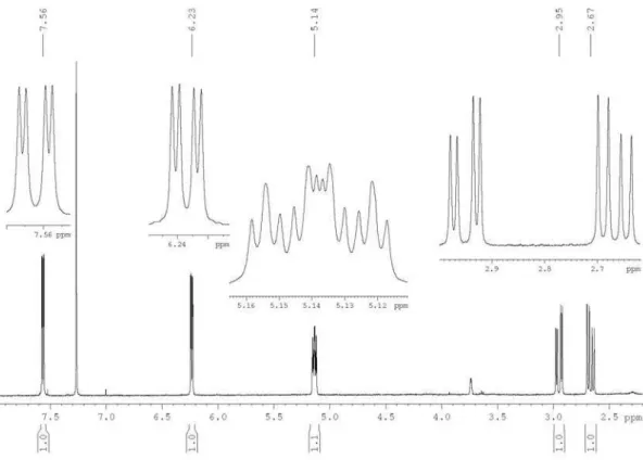
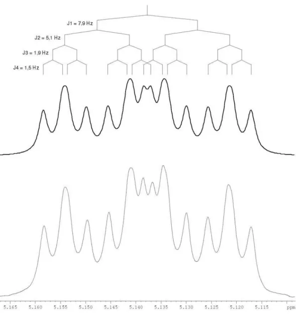
showed only two spin systems, one containing an isolated methyl group (d1.97 s, H-8’) and the other consisting of one methyne [d 5.14 dddd (7.9, 5.2, 1.9 and 1.5 Hz), H-5], one methylene [d 2.95 dd (17.4 and 5.2 Hz), H-1’a and 2.67 dd (17.4 and 7.9 Hz), H-1’b] and two olefinic hydrogens [d
6.23 dd (5.7 and 1.9 Hz), H-3 and 7.56 dd (5.7 and 1.5 Hz), H-4], (Table 1). The 13C NMR data, together with one-bond 1H-13C correlations in the HSQC experiment of 1, indicated
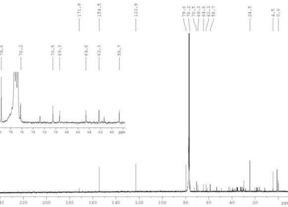
the presence of 12 carbons, comprising seven quaternary, being one at 171.9 ppm, attributed to a conjugated carbonyl group, and further six in the 76.2 to 58.7 range, as well as one methyl, one methyne, one methylene and two olefin (Table 1). Both olefin hydrogens at d 7.56 and 6.23 showed long-range
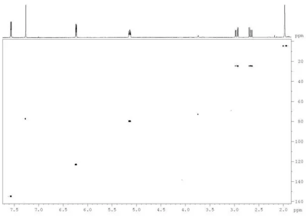
1H-13C correlations in the HMBC experiment with carbon
at d 171.9, confirming the α-β-unsaturated lactone system, as well as with methyne carbon at 79.6 ppm, evidencing a five-member lactone ring. Additionally, methyne hydrogen at 5.14 ppm showed correlations with methylene carbon at 24.5 ppm, which is attached to the lactone ring, as evidenced in the 1H NMR spectrum. Furthermore, 1H-13C long-range
correlation of methylene hydrogens H-1’ with carbons C-2’, C-3’, C-4’ and C-5’, as well as those of the methyl hydrogens H-8’ with carbons C-7’, C-6’, C-5’ and C-4’, enabled us to identify the polyacetylene system and the unequivocal
13C NMR chemical shift assignments of carbons C-2’ to
C-7’ (Table 1). The low 13C NMR chemical shift at 4.5 ppm
observed for methyl group C-8’ is due to high shielding effect of the triple bonds. Therefore, polyacetylene 1 is named as 5-octa-2,4,6-triynyl-furan-2(5H)-one, which was compared with previously published polyine signals.13-16
More than 750 naturally occurring polyacetylene derivatives belong predominantly to the family Asteraceae, subtribe Heliantheae. They are also present in several other plant families (Apiaceae, Araliaceae), as well as certain basidiomycete fungi. Straight chain polyacetylenes are the most common, while thiophenes and thiarubrines, sulphur heterocycles produced from polyacetylenes, are not widely distributed.17 However, the occurrence of a polyacetylene in the
genus Vernonia is described for the first time in this work. Polyacetylenes have been attracting attention due to their wide spectrum of biological activities, such as plant growth inhibition18,19 and their antifungal,20-22 antibacterial17,23 and
insecticidal activities,24 indicating an important role in the
protection and developmental physiology of the plant. Moreover, polyacetylenes have been proven to be cytotoxic against some cancer cell lines.25,26
The polyacetylene 1 cytotoxicity was investigated using MTT assay on two tumor cells (B16F10 and HeLa) and one non-tumor (L929) cell. Succinate-dehydrogenase, a mitochondrial enzyme which is present in living cells, cleaves the tetrazolium ring, converting the MTT to an insoluble purple formazan.27 Significant cytotoxicity
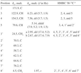
Table 1. NMR spectroscopy data for the polyacetylene 1a
Position dC, mult. dH, mult. (J in Hz) HMBC 1H-13C b
2 171.9, C
3 122.9, CH 6.23, dd (5.7; 1.9) 2, 4, and 5 4 154.5, CH 7.56, dd (5.7; 1.5) 2, 3, and 5 5 79.6, CH 5.14, dddd
(7.9; 5.2; 1.9; 1.5) 3, 4, 1’ and 2’ 1’ 24.5, CH2 a 2.95, dd (17.4; 5.2)
b 2.67, dd (17.4; 7.9)
4, 5, 2’, 3’, 4’, 5’ and 6’ 4, 5, 2’, 3’, 4’, 5’ and 6’ 2’ 70.5, C
3’ 69.3, C 4’ 58.7, C 5’ 62.3, C 6’ 64.6, C 7’ 76.2, C
8’ 4.5, CH3 1.97, s 2’, 3’, 4’, 5’, 6’ and 7’ aThe experiments were taken at 293 K at 400.13 and 100.61 MHz for 1H and 13C, respectively in CDCl
Buskuhl et al. 1329 Vol. 20, No. 7, 2009
was observed for the HeLa tumor cells with an IC50 of 158.1 µmol L-1 after 24 h treatment, showing a reduction
of 41% in growth of HeLa tumor cells with the application of 543 µmol L-1 (P < 0.001) of polyacetylene 1 (Figure 1C).
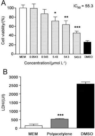
On the other hand, the L929 non-tumor cells were resistant to exposure to polyacetylene 1 at the tested concentrations (Figure 1A). However, no significant difference in cytotoxic effect was observed between the B16F10 tumor and L929 non-tumor cells, indicating specific cytotoxicity for HeLa tumor cells (Figure 1).
However, significant cytotoxicity was observed for the L929 non-tumor cells after just one-hour of treatment with polyacetylene 1 (IC50 55.3 µmol L-1) (Figure 2A).
Therefore, the necrotic cell death of the L929 non-tumor cells was evaluated after 1 hour, by lactate dehydrogenase
(LDH) assay, with lower LDH activity (525 ± 44 U/l) being observed in the presence of polyacetylene 1 compared with the positive control (2585 ± 114 U/l) (Figure 2B), indicating non-specific cell death, associated with necrosis, for L929 cells in short contact. After an initial period of stress, probably generated by the necrosis, the remaining cells began a new proliferative cycle, reducing their cytotoxicity levels (IC50 1130 µmol L-1 after 24 h).
In order, to evaluate other mechanisms related to the cytotoxicity of polyacetylene 1, apoptotic changes in the L929 non-tumor cells were verified. After 24 h in the presence of polyacetylene 1, only 22.7% of the cells showed ethidium bromide staining, while the remaining 77.3% cells showed staining only using Annexin V-FITC, indicating early apoptotic stages. On the other hand, the positive control (DMSO) showed extensive ethidium bromide staining, indicating necrotic action. The same approach was adopted for the B16F10 and HeLa tumor cells. In these cases, the necrotic cells were evidenced by the presence of an intact cell nucleus stained with ethidium bromide, as well as apoptotic cells, identified by nuclear fragmentation and apoptotic bodies and cells with cytoskeletal alterations,
Figure 1. Cytotoxicity effect of polyacetylene 1 in L929 (A) B16F10 (B)
and HeLa (C) cells for 24 hours. MEM and DMSO were used as negative and positive control, respectively. Bars represent mean values ± SD. *p < 0.05 **p < 0.01 ***p < 0.001 ANOVA followed by Tukey.
Figure 2. L929 cell viability (A) and LDH release (B) in L929 cells after
A New Polyacetylene from Vernonia scorpioides (Lam.) Pers. (Asteraceae) J. Braz. Chem. Soc.
1330
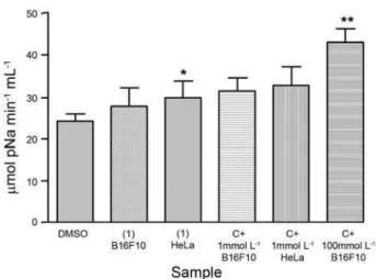
which were observed by using Annexin V-FITC. As a result, this analysis revealed an association of necrotic and apoptotic processes. However, the cells stained with ethidium bromide comprised less than 25% of the total, indicating a predominant apoptotic process. Therefore, caspase-3 activity in B16F10 and HeLa tumor cells, after 24 h of incubation, was also evaluated as an indicator of apoptosis induction, since different upstream pathways leading to apoptosis depend on caspase-3 induction for final apoptotic execution. Figure 3 shows the effect of polyacetylene 1, at the level of caspase-3 induction. There was no significant increase in caspase-3 activity in either group of cells at 1 µmol L-1, when compared with negative
control (Figure 3). A slight increase in caspase-3 activity was observed at 5 µmol L-1, and a twofold increase at
10 µmol L-1. These results are in agreement with those
obtained by Choi et al.28 in their studies of caspase-3 and
9 activities using the same approach and concentrations (5 and 10 µmol L-1) of test compound. Caspases belong
to the family of proteins that play a key role in the execution of apoptosis, and are responsible for many of the morphological features normally associated with this form of cell death.29 Isoforms 3 and 7 are the main caspases
family members involved in the induction and execution of the programmed cell death process.28,29 Therefore, caspase-3
activation is essential for DNA fragmentation to occur in apoptosis induced by a variety of stimuli. In line with these observations, our experiments clearly demonstrate that apoptotic cell death might account, at least in part, for the antitumor activity of polyacetylene 1. Notably, the data in the present study suggest that apoptotic cell death may be occurring, at least in part, via a caspase-dependent mechanism. To confirm the occurrence of apoptosis induced by polyacetylene 1, nuclear damage to DNA was assessed by comet assay using medulla cells. Negative control was performed with DMSO, which causes cell membrane disruption, with low DNA genotoxicity, while methyl methanesulfonate (MMS) at 40 µmol L-1 was used as
positive control, which is responsible for alkylation of DNA and extensive genotoxicity. The assay indicated a genotoxic distribution of polyacetylene at 54.3 µmol L-1 in levels 1-4,
mainly in level 3 (see Experimental section). By contrast, the negative control showed this distribution in levels 0-1, while the positive control showed a damage index in levels 3-4, particularly the latter. The damage index determination show a significant genotoxicity of polyacetylene 1, which was 49% higher than the negative control, and only 20% lower than the positive control (Figure 4). It is speculated that the extensive genotoxicity could explain the activation of apoptosis, due to extensive damage to the DNA, and the induction of apoptosis through p53 activation, or else
the apoptosis activated the caspase-3 apoptotic enzyme, followed by DNA fragmentation, or even both events occurring at the same time. In previous unpublished results, polyacetylene 1 induced DNA fragmentation, observed by DNA gel electrophoresis, with an association of unspecified genotoxicity and a ladder-like pattern, which is a typical character of DNA cleavage between nucleosomes, indicating DNA apoptosis profile damage for HeLa cells. Non-polar polyacetylene 1 showed genotoxic activity, and its hydrophobicity could be one reason for this behavior, due to its high capacity to cross the cell and nuclear membranes. In future studies, some chemical modification of polyacetylene 1 could be planned, generating a less toxic compound, reducing the unspecific die by necrosis or genotoxicity, and increasing the process of apoptosis.
Figure 3. Caspase-3 (µmol pNa min-1 mL-1) activity in HeLa and B16F10,
after 24 h of exposure to polyacetylene 1. DMSO was used as negative control, while paclitaxel (C+) at 1 and 100 mmol L-1 were used as positive control. Bars represent Mean ± SD. *p < 0.05 **p < 0.01 ANOVA followed by Tukey.
Figure 4. Damage index in the bone marrow cells after 1 hour of exposure
Buskuhl et al. 1331 Vol. 20, No. 7, 2009
Experimental
General procedures
The melting point was determined using a QUIMIS Q-340S23 micromelting point apparatus. The UV spectrum was obtained in MeOH using a Shimadzu UV-Vis 2401PC spectrophotometer. The IR spectrum was measured in KBr pellets using a Bomen Hartmann & Braun B-100 spectrophotometer. All the NMR data were recorded at 293 K in CDCl3 using a Bruker AVANCE 400 spectrometer operating at 9.4 T, observing 1H and 13C at 400.13 and
100.61 MHz, respectively.
1H-13C correlation (HSQC and HMBC) experiments
were optimized for an average coupling constant 1J(C,H)
of 140 Hz and LRJ(C,H) of 8 Hz, respectively. All the NMR
chemical shifts (d) were given ppm related to TMS as internal reference (0.00 ppm), and all the pulse programs were supplied by Bruker. Low-resolution ESI-MS and ESI-MS/MS data were acquired in the negative ion mode, using a Bruker Esquire 6000 ESI-ion trap instrument, while the high-resolution HR-TOF-MS measurement was carried out using a hybrid quadrupole reflector orthogonal time-of-flight high resolution Micromass Q-TOF mass spectrometer, equipped with an electrospray source.
Plant material
Flowers and leaves of Vernonia scorpioides were collected from wild specimens of “restinga” forest (a distinct type of coastal tropical and subtropical moist broadleaf forest) in Navegantes in November 2006 and identified by Dr. Ana Claudia Araújo, of the Universidade do Vale do Itajaí (UNIVALI). A voucher specimen (M. Biavatti 11) was deposited at the Barbosa Rodrigues Herbarium (HBR), Itajaí, Santa Catarina, Brazil.
Extraction and isolation
Fresh flowers and leaves of Vernonia scorpioides (3 kg) were extracted with ethanol (6 L) at room temperature, in absence of light, for seven days. After solvent reduction to 1/6 of initial volume under reduced pressure and the addiction of water (500 mL), the extract was submitted to liquid-liquid fractioning using solvents with increasing polarities, to give n-hexane (1.2 g), dichloromethane (2.3 g), and ethyl acetate (1.4 g) fractions. The dichloromethane fraction was initially subjected to silica gel column chromatography (CC) (60-230 mesh) eluted with n-hexane (500 mL), followed by dichloromethane (800 mL), ethyl acetate (700 mL) and methanol (400 mL), yielding one
subfraction for each of the four solvents. Additional chromatography separation of the dichloromethane subfraction (420 mg) by silica gel CC (230-400 mesh), using n-hexane with increasing concentrations of ethyl acetate (0-50%) as eluent, and collecting eluate portions of 30 to 50 mL each (47 flasks), resulted in polyacetylene 1 (6 mg, flasks 6, 7 and 8).
5-Octa-2,4,6-triynyl-furan-2(5H)-one (1)
Colorless crystals (CHCl3), mp 98-100 °C; UV (MeOH)
λmax/nm: (log ∈) 207 (5.58); IR (KBr) νmax/cm-1: 3081, 2515,
2438, 1786, 1289, 791, 679, 631, 551, 329; 1H and 13C
NMR data shown in Table 1; ESI-MS m/z 183.0 [M – H] −;
ESI-MS/MS (daughter ions, 25%) m/z 139 [M – H – CO2]−
(100), 87 (3); HR-TOF-MS (ESI positive) m/z 185.0597 [M + H]+ (calc. for C
12H8O2 + H
+, 185.0603).
Cells and cell culture conditions
The fibroblast L929 (Mus musculus C3H/NA) (BCRJ
CR020/ATCC CCL1), B16F10 melanoma (Mus musculus
C57BL/6J) (BCRJ CR010), and HeLa from cervix adenocarcinoma (Homo sapiens) (BCRJ CR050) were obtained from the Cell Bank of Rio de Janeiro (BCRJ), Brazil. The cells were maintained in minimal essential medium (MEM) supplemented with 0.2% sodium bicarbonate (Invitrogen/GIBCO), 10% heat inactivated fetal calf serum (FCS), 1% non-essential aminoacids (Invitrogen/GIBCO), two mmol L-1 L-glutamine, and antibiotic PCN 0.1%
(Invitrogen/GIBCO). The cells were maintained in a fully humidified atmosphere (95% air, 5% CO2) at 37 oC. Cell
viability was analyzed before each assay by trypan blue dye exclusion assay, which was always greater than 98%. For the damage index, (Comet assay) bone marrow cells were obtained from femur bones of Mus musculus, (Swiss). The ethical animal research committee of UNIVALI approved the protocol (# 236/07). The cells were obtained after vivisection using an O2/CO2 chamber, and the femur was obtained by surgery. Cell viability was analyzed before each assay, by trypan blue dye exclusion assay, which was always greater than 95%.
Cytotoxicity assay
Cell viability was determined using the cell proliferation reagent MTT (3-[4,5-dimethylthiazol-2-yl]-2,5-diphenyltetrazolium bromide) a tetrazolium salt that is cleaved by mitochondrial dehydrogenages in viable cells.27 100 µL Containing 2 × 10⁴ of cell suspension was
A New Polyacetylene from Vernonia scorpioides (Lam.) Pers. (Asteraceae) J. Braz. Chem. Soc.
1332
of 0.0543, 0.543, 5.43, 54.3, 543.0 µmol L-1. After one
and 24 h incubation, 20 µL of the proliferation reagent MTT at concentration of 5 mg mL-1 in phosphate-buffered
saline (PBS, pH 7.4) was added to each well and the cells were incubated at 37 oC for 4 h in humidified atmosphere
with 5% CO2. Formazan precipitates were solubilized by addition of 100 µL of DMSO. After 10 min at room temperature, spectrophotometric absorbance at 570 nm of each well was measured on a microculture plate reader. Cell viability percentage was calculated as [1-(absorbance of experimental wells/absorbance of control wells) × 100].
Lactate dehydrogenase assay
Lactate dehydrogenase (LDH) is a stable cytosolic enzyme that is released upon membrane damage and was used as an index of cytotoxicity. The LDH activity was measured using a commercial cytotoxicity assay kit (DiaSys). L929 cells containing 5 × 10⁵ of cell suspension
were placed in each of 6-well plates and incubated at 37 ºC for 1 hour in a humidified atmosphere with 5% CO2, in the presence of polyacetylene 1.30 Maximal LDH release of the
cells was determined by addition of lysis solution (0.8% Triton X-100), followed by incubation at 37 oC for 45 min.
Absorbance at 360 nm was determined photometrically and activity was represented as DA/min × 2000 (U/I). LDH activity released by cells cultured in absence or presence of polyacetylene 1, was compared with the negative control.
Detection of apoptosis
Apoptotic changes in L929, B16F10, and HeLa cells were evaluated using the Annexin V-FITC/ethidium bromide apoptosis detection kit. Cells culture containing 2 × 10⁴ of cell suspension in each well plate was cultured
in the presence of polyacetylene 1 for 24 h. Determination of phosphatidylserine externalization was followed by in situ detection protocols. Briefly, after 24 h treatment, the cells were washed with cold PBS and incubated in 100 µL of MEM, containing 1 µL of annexin V-FITC (12 mg mL-1) and 50 µL of ethidium bromide (2 µg mL-1)
at room temperature for 15 min in the dark. The cells were evaluated using an Olympus CKX41SF2 microscope, a CKX41-RFA fluorescence system, and an Olympus Q-color3 system equipped with 480 and 520 nm filters.30
Caspase-3 activity assay
Caspase-3 activity was measured in cell lysates using a SIGMA caspase-3 assay Kit, according to the manufacturer’s protocols. Briefly, HeLa and B16F10 cells were cultured for
24 h (37 oC, 5% CO
2) in the presence of polyacetylene. The
cells were then collected and washed with PBS (twice) and the assay plate incubated for 30 min at room temperature. The cleavage of substrate Z-DEVD-pNa for caspase-3 activity was quantified by measurement of absorbance at 405 nm on a spectrophotometric microplate reader.
Damage index
Nuclear damage to the DNA in the bone marrow cells was assessed by means of the comet assay method, according to Singh et al.31 with some modifications.32 The
slides were first coated with a 1% normal agarose layer. Next, approximately 2 × 10⁴ cells were suspended in 50 µL
of 1.0% low melting point agarose and layered onto the slides, which were immediately covered with cover slips. After agarose solidification at 4 °C for 5 min, the cover slips were removed and the slides were treated with polyacetylene 1 at 54.3 µmol L-1for 1 hour. Afterwards, the slides were
washed with cold PBS (three times), and immersed for 1 h at 4 °C in fresh lysis solution (2.5 mol L-1 NaCl,
100 mmol L-1Na
2 EDTA, 10 mmol L
-1Tris, 1% sodium
lauryl sarcosinate, pH 10) containing 1% Triton X-100 and 10% of DMSO. The slides were equilibrated in alkaline solution (1 mmol L-1 Na
2 EDTA, 300 mmol L
-1NaOH,
pH 10) for 30 min. Electrophoresis was carried out for 30 min at 25 V, 300 mmol L-1. Afterwards, the slides were
then neutralized by washing three times with 0.4 mol L-1
Tris buffer (pH 7.5) at 5 min intervals. The slides were then stained with ethidium bromide (20 µg mL-1). The images
were scored using a fluorescent microscope (Olympus CKX41SF2/CKX41-RFA equipped with a 510-569 nm excitation filter). Based on the extent of strand breakage, the cells were classified, according to their tail length, in five levels, ranging from 0 (no visible tail) to 4 (head of comet still detectable, but most of the DNA in the tail).33
Comparative damage was performed by measurement of the damage index using the equation: Σ (Level 0 x 0) + (Level 1 x 1) + (Level 2 x 2) + (Level 3 x 3) + (Level 4 x 4), with a maximum score of 100.34
Statistical analysis
Buskuhl et al. 1333 Vol. 20, No. 7, 2009
Tukeytest. The differences were considered statistically significant from the controls at p < 0.05.
Acknowledgments
The authors are grateful to Prof. Dr. Q. B. Cass and Dr. J. C. Barreiro of the Federal University of São Carlos, Brazil, for the ESIMS and ESI-MS/MS analysis and Dr. A. C. Araújo of the University of Vale do Itajaí for the botanical identification of plant material, as well as to CAPES, CNPq and FINEP for its financial support and fellowships.
Supplementary Information
Supplementary information containing 1D and 2D NMR, MS, IR and UV data is available free of charge at http://jbcs.org.br, as a PFF file.
References
1. Bohlmann, F.; Jakupovic, J.; Gupta, R. K.; King, R. M.; Robinson, H.; Phytochemistry1981,20,473.
2. Bohlmann, F.; Zdero, C.; King, R. M.; Robinson, H.;
Phytochemistry1982,21,695.
3. Kuo, Y. H.; Kuo, Y. J.; Yu, A. S.; Wu, M. D.; Ong, C. W.; Yang, L. M.; Huang, J. T.; Chen, C. F.; Li, S. Y.; Chem. Pharm. Bull.
2003, 51, 425.
4. Koul, J. L.; Koul, S.; Singh, C.; Taneja, S. C.; Shanmugavel, M.; Kampasi, H.; Saxena, A. K.; Qazi, G. N.; Planta Med.2003,
69, 164.
5. Jisaka, M.; Ohigashi, H.; Takegawa, K.; Huffman, M. A.; Koshimizu, K.; Biosci. Biotechnol. Biochem. 1993, 57, 833. 6. Izevbigie, E. B.; Bryant, J. L.; Walker, A.; Exp. Biol. Med. 2004,
229, 163.
7. Nergard, C. S.; Diallo, D.; Michaelsen, T. E.; Malterud, K. E.; Kiyohara, H.; Matsumoto, T.; Yamada, H.; Paulsen, B. S.;
J. Ethnopharmacol. 2004, 91, 141.
8. Lambertini, E.; Piva, R.; Khan, M. T.; Lampronti, I.; Bianchi, N.; Borgatti, M.; Gambari, R.; Int. J. Oncol.2004, 24, 419. 9. Cabrera, A. L.; Klein, R. M.; Fl. Ilustr. Catarin.1980, 354. 10. Freire, M. F. I.; Abreu, H. S.; Cruz, L. C. H.; Freire, R. B.;
Braz. J. Microbiol. 1996, 27, 1.
11. Leite, S. N.; Palhano, G.; Almeida, S.; Biavatti, M. W.;
Fitoterapia 2002, 73, 496.
12. Pagno, T; Blind, L. Z; Biavatti, M. W; Kreuger, M. R. O.; Braz. J. Med. Biol. Res. 2006, 39, 1483.
13. Hearn, M. T. W.; Turner, J. L.; J. Chem. Soc., Perkin Trans. 2
1976, 1027.
14. Muller, N.; Pritchard, D. E.; J. Chem. Phys. 1959, 31, 768. 15. Muller, N.; Pritchard, D. E.; J. Chem. Phys. 1959, 31, 1471. 16. Höbold, W.; Radeglia, R.; Klose, D.; J. Prakt. Chem. 1976, 318,
519.
17. Guillet, G.; Philogene, B. J. R.; O’Meara, J.; Durst, T.; Arnason, J. T.; Phytochemistry 1997, 46, 495.
18. Satoh, A.; Narita, E.; Nishimura, H.; Biosci., Biotechnol., Biochem. 1996, 60, 152.
19. Yamazoe, S.; Hasegawa, K.; Shigemori, H.; Phytochemistry
2007, 68, 1706.
20. Chobot, V.; Buchta,V.; Jahodarova, H.; Pour, M.; Opletal, L.; Jahodar L.; Harant, P.; Fitoterapia2003, 74, 288.
21. Hadacek, F.; Greger, H.; Phytochem. Anal. 2000, 11, 137. 22. Constabel, C.P.; Towers, G.H.N.; Planta Med.1989, 55, 35. 23. Towers, G.H.N.; Abramowski, Z.; Finlayson, A.J.; Zucconi, A.;
Planta Med.1985, 51, 225.
24. Jahodář, L.; Klečáková, J.; Chem. Listy 1999, 93, 320. 25. Ebermann, R.; Alth, G.; Kreitner, M.; Kubin, A.; J. Photochem.
Photobiol. B1996, 36, 95.
26. Pellati, F.; Calo, S.; Benvenuti, S.; Adinolfi B.; Nieri P.; Melegari, M.; Phytochemistry2006, 67, 1359.
27. Mosmann, T.; J. Immunol. Methods1983, 65, 55.
28. Choi, H. J.; Yee, S. B.; Park, S. E.; Im, E.; Jung, J. H.; Chung, H. Y.; Choi, Y. H.; Kim, N. D.; Cancer Lett. 2006, 232, 214. 29. Salvesen, G. S.; Dixit, V. M.; Cell1997, 91, 443.
30. Cruz-Chamorro, L.; Puertollano, M. A.; Puertollano, E.; Cienfuegos, G. A.; Pablo, M. A.; Peptides2006,27, 1201. 31. Singh, S.; Lowe, D. G.; Thorpe, D. S.; Rodriguez, H.; Kuang,
W. J.; Dangott, L. J.; Chinkers, M.; Goeddel, D. V.; Garbers, D. L.; Nature1988, 334, 708.
32. Collins, A. R.; Dusinska, M. In Methods in Molecular Biology; Armstrong, D., ed.; Humana Press: NJ, 2002, vol. 186. 33. Cavalcanti, B. C.; Costa-Lotufo, L. V.; Moraes, M. O.; Burbano,
R. R.; Silveira, E. R.; Cunha, K. M.; Rao, V. S.; Moura, D. J.; Rosa, R. M.; Henriques, J. A.; Pessoa, C.; Food Chem. Toxicol.
2006,44, 388.
34. Liu, W.; Christenson, S.; Standage, S.; Shen, B.; Science2002,
297, 1170.
Received: July 3, 2008 Web Release Date: July 6, 2009
S
u
p
p
le
m
e
n
ta
ry
In
fo
rm
a
ti
o
n
J. Braz. Chem. Soc., Vol. 20, No. 7, S1-S10, 2009. Printed in Brazil - ©2009 Sociedade Brasileira de Química 0103 - 5053 $6.00+0.00
*e-mail: [email protected]
# Present address: Laboratório de Farmacognosia, Departamento de Ciências Farmacêuticas, Centro de Ciências da Saúde, Universidade Federal de Santa Catarina, Trindade, 88040-900 Florianopolis-SC, Brazil
A New Polyacetylene from Vernonia scorpioides (Lam.) Pers. (Asteraceae)
and its in vitro Antitumoral Activity
Humberto Buskuhl,a Rilton A. Freitas,a Franco Delle Monache,b Andersson Barison,c Francinete R. Campos,c Yuri E. Corilo,d Marcos N. Eberlind and Maique W. Biavatti*,a,#
aCentro de Ciências da Saúde, Universidade do Vale do Itajaí, 88302-202 Itajaí-SC, Brazil
bDipartimento di Chimica e Tecnologia delle Sostanze Biologicamente Attive, Università “La Sapienza”, Roma, Italy
cDepartamento de Química, Centro Politécnico, Universidade Federal do Paraná, 81530-900 Curitiba-PR, Brazil
dInstituto de Química, Universidade Estadual de Campinas, 13083-970 Campinas-SP, Brazil
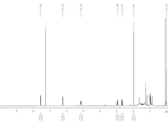
Figure S1. 1H NMR spectrum of the polyacetylene (1) in CDCl
A New Polyacetylene from Vernonia scorpioides (Lam.) Pers. (Asteraceae) J. Braz. Chem. Soc.
S2
Figure S2. Expansions of the 1H NMR spectrum of the polyacetylene (1) in CDCl
Buskuhl et al. S3 Vol. 20, No. 7, 2009
Figure S3. Simulated (Top) and experimental (Bottom, CDCl3 at 400 MHz) 1H NMR signal in order to understand the multiplicity of H-5 at 5.14 ppm
A New Polyacetylene from Vernonia scorpioides (Lam.) Pers. (Asteraceae) J. Braz. Chem. Soc.
S4
Figure S4. 13C{1H} NMR spectrum of the polyacetylene (1) in CDCl
3 at 100 MHz. Insert is an expansion showing the signal from C-2’ to C-7’.
Figure S5. 1H-1H correlation map from COSY NMR experiment of the polyacetylene (1) in CDCl
Buskuhl et al. S5 Vol. 20, No. 7, 2009
Figure S6. 1H-13C one-bond correlation map from HSQC NMR experiment of the polyacetylene (1) in CDCl
3 at 400 and 100 MHz.
Figure S7. 1H-13C long-range correlation map from HMBC NMR experiment of the polyacetylene (1) in CDCl
A New Polyacetylene from Vernonia scorpioides (Lam.) Pers. (Asteraceae) J. Braz. Chem. Soc.
S6
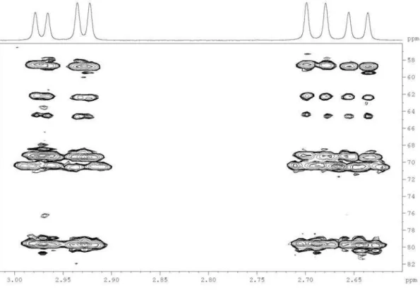
Figure S8. Expansion of the 1H-13C long-range correlation map from HMBC NMR experiment of the polyacetylene (1) in CDCl
3 at 400 and 100 MHz, showing the correlations for both H-1’ at 2.67 and 2.95 ppm.
Figure S9. Expansion of the 1H-13C long-range correlation map from HMBC NMR experiment of the polyacetylene (1) in CDCl
Buskuhl et al. S7 Vol. 20, No. 7, 2009
Figure S10. Expansion of the 1H-13C long-range correlation map from HMBC NMR experiment of the polyacetylene (1) in CDCl
3 at 400 and 100 MHz, showing the correlations with the olefinic C-2 and C-3 and the carbonyl C-4.
A New Polyacetylene from Vernonia scorpioides(Lam.) Pers. (Asteraceae) J. Braz. Chem. Soc.
S8
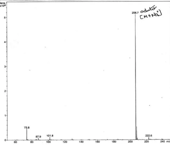
Figure S12. ESI-MS of the polyacetylene (1) in MeOH, negative mode.
Buskuhl et al. S9 Vol. 20, No. 7, 2009
Figure S14. HR-ESI-TOF-MS of the polyacetylene (1), positive mode.
A New Polyacetylene from Vernonia scorpioides(Lam.) Pers. (Asteraceae) J. Braz. Chem. Soc.
S10