Received: September 6, 2009 Accepted: May 23, 2010
Conflict of Interest Statement: The authors state that there are no financial and personal conflicts of interest that could have inappropriately influenced their work.
Copyright: © 2010 Santos et al.; licensee EDIPUCRS. This is an Open Access article distributed under the terms of the Creative Commons Attribution-Noncommercial-No Derivative Works 3.0 Unported License.
Cytotoxicity of carbamide peroxide bleaching
gel on L929 cells
Citotoxicidade do gel clareador peróxido de carbamida
em células L929
Rogério Lacerda dos Santos a
Matheus Melo Pithon a
Fernanda Otaviano Martins a
Maria Teresa Villela Romanos a
a Federal University of Rio de Janeiro, Rio de
Janeiro, RJ, Brazil
Correspondence: Rogério Lacerda dos Santos Rua Ipatinga, 170, Planalto Divinópolis, MG – Brazil 35501-191
E-mail: [email protected] or [email protected]
Abstract
Purpose: To test the hypothesis that the higher the concentration of carbamide peroxide, the greater is its cytotoxicity to fibroblast cells.
Methods: Three concentrations of carbamide peroxide (10%, 16%, and 22%) used in home bleaching techniques were evaluated regarding their cytotoxic effect on gingival tissues. The materials were divided into three groups as follows: Group C10 (White Gold Home 10%, Dentsply), Group C16 (White Gold Home 16%, Dentsply, and Group C22 (Nite White 22%, ACP Discus Dental). The cytotoxicity essay was carried out using cell cultures (mouse fibroblast L929 cell line) in which the viable cells were determined by means of the dye-uptake method performed at 2, 4, and 8 hours. Data were analyzed by analysis of variance (ANOVA) with multiple comparisons and Tukey’s test (P<0.05).
Results: The results showed statistically significant differences between Groups C10, C16, C22, and the cell control at 2, 4, and 8 hours (P<0.05). The amount of cell lysis increased proportionally to the exposure time to the materials studied.
Conclusion: The 22% carbamide peroxide group was more toxic than the other two groups (16% and 10% concentration) regardless of the exposure time.
Key words: Toxicity; hydrogen peroxide; dental bleaching
Resumo
Objetivo: Testar a hipótese que quanto maior a concentração do peróxido de carbamida, maior a citotoxicidade provocada em células fibroblásticas.
Metodologia: Foram avaliadas 3 concentrações de peróxidos de carbamida (10%, 16% e 22%) usados na técnica de clareamento caseiro, divididos em 3 grupos: grupo C10 (White Gold Home 10%, Dentsply), grupo C16 (White Gold Home 16%, Dentsply) e grupo C22 (Nite White 22%, ACP Discus Dental) quanto ao efeito citotóxico nos tecidos gengivais. O ensaio de citotoxicidade foi realizado utilizando cultura de células (linhagem L929, fibroblastos de camundongos) e submetidos ao teste para células viáveis em vermelho neutro (“dye-uptake”) no tempo de 2, 4 e 8 h. Os dados foram analisados estatisticamente por análise de variância e teste de Tukey (P<0,05).
Resultados: Houve diferença estatisticamente significante entre os grupos C10, C16 e C22 com o grupo CC (controle de células) nos tempos de 2, 4 e 8 h (P<0,05). A quantidade de lise celular aumentou diretamente proporcional ao tempo de exposição dos materiais com as culturas de células.
Conclusão: O peróxido de carbamida 22% foi mais citotóxico que os peróxidos de carbamida nas concentrações de 16 e 10% independentemente do tempo avaliado.
Introduction
Dental bleaching has been used for more than one hundred years (1), and since then several techniques and chemicals have been employed to whiten the teeth. The use of hydrogen peroxide to bleach teeth with pulp vitality began in the early 1900’s (2). Nowadays, two main bleaching techniques exist:
the in-ofice bleaching and the supervised at-home bleaching
techniques (3).
The at-home bleaching technique, which was introduced by Haywood and Heymann (3) in 1989, consists of using carbamide peroxide-based bleaching agents at concentrations
lower than those used in the ofice (16% and 22%) or hydrogen peroxide (from 1.5% to 7.5%). The patients
themselves use these chemical agents daily (1-8 hours) by means of a personal tray during periods ranging from 2 to 4 weeks (4). In general, bleaching agents comprise hydrogen peroxide or products releasing hydrogen peroxide, such as
10% carbamide peroxide, which breaks down into 6.4% urea and 3.6% hydrogen peroxide. The hydrogen peroxide
dissociate into water and oxygen, whereas urea breaks down into carbon dioxide and ammonia (5).
All dental bleaching agents, from carbamide peroxide to hydrogen peroxide, ionize and decompose to produce free radicals, which are highly unstable and react very
easily with other organic substances (6). The bleaching
principle is based on the fact that these agents react with highly conjugated organic molecules by breaking down their electronic conjugation and altering their molecular energy absorption, thus resulting in changes in their optical
structure (7). This can affect the absorption spectrum of the
composite, transforming its long wavelength (dark) into a shorter wavelength, that is, resulting in a lighter shade (8).
However, these free-radical reactions are not speciic to the
pigment molecules of the tooth only, as they can react with
other organic structures as well (6). The reactive
oxygen-derived species are known to cause damage to living cells due to the oxidative stress they provoke (9). This oxidative stress can cause apoptosis, DNA damage (genotoxicity), and cell cytotoxicity (9) observed in bacteria and cultured cells (10).
Hydrogen peroxide was shown to have a weak local carcinogenic-inducing potential (10). The mechanism is unclear (10), but it appears unlikely that oral health products which hydrogen peroxide release will enhance cancer risk in individuals except in those who have an
increased risk of oral cancer due to tobacco use, alcohol abuse, or genetic predisposition (11). Therefore, the possible alterations caused by the use of bleaching agents indiscriminately can potentially cause damage to dental tissues (12).
By definition, cytotoxicity of a given agent means destructive effect on cells (13). So far, there are very few studies reporting cytotoxicity effects of bleaching agents
on pulp cells (14), ibroblasts (9), and odontoblastic cells.
Because the application of such bleaching agents occurs close to gingival tissue, the objective of the present study was to test the hypothesis that the higher the concentration of carbamide peroxide, the greater the cytotoxic effect on
ibroblast cells.
Methods
Three concentrations of carbamide peroxide (10%, 16%, 22%) commonly used in the at-home bleaching technique
were assessed in order to determine their possible cytotoxic effect on gingival tissues. The materials were divided into
three groups: Group C10 (White Gold Home 10%, Dentsply, Petrópolis, Rio de Janeiro, Brasil), Group C16 (White Gold Home 16%, Dentsply, Petrópolis, Rio de Janeiro, Brasil), and Group C22 (Nite White 22%, ACP Discus Dental, Culver,
CA, USA) (Table 1). To verify the cell response to extreme situations, other three groups were included in the study: Group CC (cell control), consisting of cells not exposed to any material; Group C+ (positive control), consisting of Tween 80 (Polyoxyethylene-20-sorbitan); and Group C (negative control), consisting of PBS solution (phosphate-buffered saline) in contact with the cells.
Culture of L929 cells (mouse ibroblasts), obtained from
the American Type Culture Collection (ATCC, Rockville, MD, USA), were used in the present study and maintained in Eagle’s minimum essential medium (Cultilab, Campinas, Brazil). To the cell culture were added 0.03 mg/mL glutamine (Sigma, St. Louis, Missouri, USA), 50 mg/mL of garamicine (Schering Plough, Kenilworth, New Jersey, USA), 2.5 mg/ mL fungizone (Bristol-Myers-Squibb, New York, USA),
0.25% sodium bicarbonate solution (Merck, Darmstadt,
Germany), 10 mM HEPES (Sigma, St. Louis, Missouri, USA),
and 10% bovine foetal serum (Cultilab, Campinas, Brazil) as
growth medium or no bovine foetal serum for maintenance
medium only. The cell culture was incubated at 37o C for
48 hours.
Bleaching agents Basic Composition Manufacturer
White Gold Home 10% Carbamide peroxide, thickening agent, flavour, deionised water
Dentsply White Gold Home 16% Carbamide peroxide, thickening agent, flavour,
deionised water
Dentsply Nite White 22% Propylene glycol, C3H8O3, carbamide peroxide,
sílica, hydrogen peroxide, KNO3, emulsifying
wax, NF, hydroxypropyl cellulose, flavour, deionised water, KOH, dimethicone
ACP Discus Dental
Dye-Uptake
The so-called “dye-uptake” technique, which is based on the incorporation of neutral red by the living cells, was employed
to determine the cytotoxicity of the materials (15,16). The
bleaching agents were evaluated at 2, 4, and 8 hours because according to their manufacturers they are commonly used during periods ranging from 2 to 8 hours a day. This period of time represents the contact of carbamide peroxide with the cells for 2, 4, and 8 hours for analysis of cytotoxicity. Volumes of 100 µL of L929 cell suspension were distributed
into 96-well microplates. After 48 hours, the growth medium
was replaced with 100 µL of the culture medium (Eagle’s MEM) obtained from the mixture (1 mL of culture medium) with 0.5 mL of carbamide peroxide. The positive and negative control cells comprised culture mediums obtained from their mixture (1 mL) with, respectively, 0.5 mL Tween 80 and 0.5 mL PBS solution. The experiment was performed 4 times, with a total of 15 samples for each group.
After 2, 4, and 8 hours of incubation, 100 mL of 0.01%
neutral red solution (Sigma, St. Louis, Missouri, USA) were added to each well of the microplaque that was then
incubated at 37oC for 3 hours for allowing the living cells
to absorb the dye. Next, after discarding the dye, 100 µL of
4% formaldehyde solution (Vetec, Rio de Janeiro, Brazil) were added to PBS (130 mM NaCl; 2 mM KCl; 6 mM
Na2HPO4 2H2O; 1 mM K2HPO4; pH=7.2) for 5 minutes
to promote cell ixation. Next, 100 mL of 1% acetic acid solution (Vetec, Rio de Janeiro, Brazil) with 50% methanol
were added in order to remove the dye. After 20 minutes, a spectrophotometer (BioTek, Winnoski, Vermont, USA) was used for data reading at wavelength of 492 nm
(λ=492 nm).
Data were compared by using analysis of variance (ANOVA) and Tukey’s test to assess any difference between groups at
a signiicance level of 5%.
Results
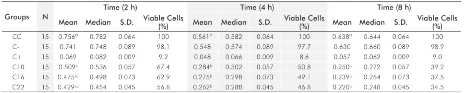
The results showed statistically signiicant differences between the Groups C10 (10% carbamide peroxide), C16 (16% carbamide peroxide), and C22 (22% carbamide
peroxide) in relation to the Group CC (cell control) at 2, 4, and 8 hours (P<0.05). No statistically signiicant differences
were observed between Groups C10 and C16 as well as between Groups C16 and C22 at 2 hours, and the same occurred between Groups C10, C16, and C22 at 4 and 8 hours
(Table 2). The amount of cell lysis increased in direct pro- portion to the exposure time of the materials to cell cultures. Group C10 had the lowest cytotoxicity values for all periods of time studied. Group C22 was notoriously more
cytotoxicity compared to Groups C10 and C16 (Table 2),
thus suggesting that the level of cytotoxicity is directly related to the carbamide peroxide concentration in the bleaching agents.
Discussion
All materials or chemical agents developed for application to dental or medical areas have to be submitted to previous tests for cytotoxicity and biocompatibility in order to evaluate their biological properties and risk factors before being introduced to the market. In Dentistry, a comprehensive evaluation of the dental material needs to follow a series of tests established by ANSI/ADA, ISO, and FDI, which are divided into three categories: initial tests, secondary tests, and pre-clinical tests. The initial tests are aimed at determining the toxic effects of a given material by using several methods, such as the cell cultures.
Tooth whitening has been increasingly sought by the patients
in the dental ofice, however, the effect of these bleaching
agents on gingival and dental tissues has been questioned. Because these bleaching agents act close to the gingival tissue, the objective of the present study was to test the hypothesis that the concentration of carbamide peroxide
affects its cytotoxicity to ibroblast cells.
Benetti et al. (17) have shown that the higher the concentration
of bleaching agent applied to the dental enamel, the greater is the penetration of hydrogen peroxide into the pulp chamber. According to Robertson and Melf (18), heat seems to have a direct relation to the increased diffusion of bleaching agents through the dental structures, including the pulp. Both hydrogen peroxide and heat are important components of the technique used for dental bleaching. Heat is applied in order to accelerate the chemical reaction between hydrogen peroxide and dental structures, that is, the cytotoxic effect of the bleaching agent will be enhanced if the material is in contact with the oral tissues.
Table 2. Descriptive statistics* for the optical density of the tested bleaching agents.
Groups N
Time (2 h) Time (4 h) Time (8 h)
Mean Median S.D. Viable Cells
(%) Mean Median S.D.
Viable Cells
(%) Mean Median S.D.
Viable Cells (%)
CC 15 0.756ª 0.782 0.064 100 0.561ª 0.582 0.064 100 0.638ª 0.644 0.064 100
C- 15 0.741 0.748 0.089 98.1 0.548 0.574 0.089 97.7 0.630 0.660 0.089 98.9
C+ 15 0.069 0.082 0.009 9.2 0.048 0.066 0.009 8.6 0.057 0.062 0.009 9.0
C10 15 0.509b 0.536 0.057 67.4 0.284b 0.302 0.057 50.8 0.250b 0.272 0.057 39.2
C16 15 0.475bc 0.498 0.073 62.9 0.275b 0.298 0.073 49.1 0.239b 0.254 0.073 37.5
C22 15 0.429cd 0.454 0.045 56.8 0.262b 0.288 0.045 46.8 0.220b 0.248 0.045 34.5
Some in vitro studies have evaluated the cytotoxic effects of the components of bleaching products by means of cell cultures (9,19). Cytotoxicity tests in cell cultures can
help evaluate the effects resulting from a speciic agent
concentration through the damage caused to either dental structures or biochemical pathways within the cells (20). Koulaouzidou et al. (21), who assessed the cytotoxicity of
hydrogen peroxide in ibroblastic cells cultivated in vitro,
reported that all products caused deinite cytotoxic effects
on the cells.
In the present study, one can observe the cytotoxic aspect of the materials after their exposure to cell culture.
Carbamide peroxide concentration of 22% caused more
cell death in comparison to other concentrations during the three experimental periods, thus suggesting that the level of cytotoxicity is directly related to the carbamide peroxide concentration existing in the bleaching agent as all the materials studied have thickening agent that, in
turn, may inluence the cytotoxicity. Woovelton et al. (22)
determined that adding a thickening agent (ex. Carbopol – carboxypolyethylene polymer) to the bleaching agent reduces the potential cytotoxic effect of this material on the cells, possibly due to its capacity to increase the viscosity and delay the release of hydrogen peroxide. This supports the idea that carbamide peroxide without thickening agent may have a more cytotoxic effect compared to that found in the present study.
The amount of cell lysis observed in the present study increased in direct proportion to exposure time of the
materials to cell cultures, a inding also corroborated by
Koulaouzidou et al. (21) and González-Ochoa (23), who
performed in vitro studies on the cytotoxicity of bleaching agents and reported that the cytotoxic effects on viable cells are both dose-dependent and time-dependent.
In clinical trials that used 10% carbamide peroxide in custom made trays, 25 to 40% of the patients reported
gingival irritation during treatment (24). The damage to the gingival tissue caused by the carbamide peroxide in the oral cavity can manifest itself in the form of mild hyperemia to ulceration. It is therefore advisable that the tray be designed to prevent gingival exposure by the use of a tray that has
irm contact with the teeth.
The metabolic processes in animal systems are more complex and dynamic than in cell cultures (13). In addition, when an agent is added to the cell culture it becomes readily available to the cells, which does not occur in the living systems. It is important to understand that highly cytotoxic dental materials, when applied directly to cell cultures, may not
cause signiicant risks to the pulp-dentine complex as the
dentine acts as a biological barrier that reduces or dilutes the soluble components of the materials (25). However, in contact with gingival tissues, cytotoxic dental materials may cause more severe damages.
Conclusions
This study suggests that the use of low concentrations of carbamide peroxide is less cytotoxic than the bleaching agents containing higher concentrations of such agent, and the risk of damage to gingival tissues depends on carbamide peroxide concentration and bleaching agent composition, besides exposure time to gingival tissues.
References
1. Haywood VB. History, safety, and effectiveness of current bleaching techniques and applications of the nightguard vital bleaching technique. Quintessence Int 1992;23:471-88.
2. Ames JW. Removing stains from motlled enamel. J Am Dent Assoc 1937;24:1674-7.
3. Haywood VB, Heymann HO. Nightguard vital bleaching. Quintessence Int 1989;20:173-6.
4. Haywood VB. Nightguard vital bleaching: current concepts and research. J Am Dent Assoc 1997;128 Suppl:19S-25S.
5. Feinman RA, Goldstein RE, Garber DA. Bleaching teeth. Chicago: Quintessence Books; 1987.
6. Kawamoto K, Tsujimoto Y. Effect of the hydroxyl radical and hydrogen peroxide on tooth bleaching. J. Endod 2004;30:45-50.
7. Sun G. The role of lasers in cosmetic dentistry. Dent Clin North Am 1998;44:83-9.
8. Seghi RR, Denry I. Effects of external bleaching on indentation and abrasion characteristics of human enamel in vitro. J Dent Res 1992;71:1340-1344.
9. Kanno S, Shouji A, Asou K, Ishikawa M. Effects of naringin on hydrogen peroxide-induced cytotoxicity and apoptosis in P388 cells. J Pharmacol Sci 2003;92:166-70.
10. Dahl JE, Pallesen U. Tooth bleaching--a critical review of the biological aspects. Crit Rev Oral Biol Med 2003;14:292-304. 11. SCCNFP (1999). Scientific Committee on Cosmetic Products
and Non-Food Products intended for Consumers. Hydrogen
peroxide and hydrogen peroxide releasing substances in oral health products. SCCNFP/0058/98. Summary on http://europa. eu.int/comm/food/fs/sc/sccp/out83_en.html and http://europa. eu.int/comm/food/fs/sc/sccp/out89_en.html [Accessed on 2002 October 31].
12. Potocnik I, Kosec L, Gaspersic D. Effect of 10% carbamide peroxide bleaching gel on enamel microhardness, microstructure, and mineral content. J Endod 2000;26:203-6.
13. Li Y. Biological properties of peroxide-containing tooth whiteners. Food Chem Toxicol 1996;34:887-904.
14. Anderson DG, Chiego DJ, Jr., Glickman GN, McCauley LK. A clinical assessment of the effects of 10% carbamide peroxide gel on human pulp tissue. J Endod 1999;25:247-50.
15. Pithon MM, dos Santos RL, Martins FO, Romanos MTV. Avaliação in vitro da citotoxicidade de parafusos expansores palatinos. Rev odonto ciênc 2009;24:168-72.
16. Pithon MM, dos Santos RL, Martins FO, Romanos MTV. Cytotoxicity of dental alginates Rev odonto ciênc 2009;24:270-3.
17. Benetti AR, Valera MC, Mancini MN, Miranda CB, Balducci I. In vitro penetration of bleaching agents into the pulp chamber. Int Endod J 2004;37:120-4.
18. Robertson WD, Melfi RC. Pulpal response to vital bleaching procedures. J Endod 1980;6:645-9.
20. Wataha JC, Hanks CT, Strawn SE, Fat JC. Cytotoxicity of components of resins and other dental restorative materials. J Oral Rehabil 1994;21:453-62.
21. Koulaouzidou E, Lambrianidis T, Konstantinidis A, Kortsaris AH. In vitro evaluation of the cytotoxicity of a bleaching agent. Endod Dent Traumatol 1998;14:21-5.
22. Woolverton CJ, Haywood VB, Heymann HO. Toxicity of two carbamide peroxide products used in nightguard vital bleaching. Am J Dent 1993;6:310-4.
23. González-Ochoa JG. Histological assessment of dental pulpar after vital bleaching with 10% carbamide peroxide. [Thesis]. Indiana: University School of Dentistry; 2002.
24. Tam L. Clinical trial of three 10% carbamide peroxide bleaching products. J Can Dent Assoc 1999;65:201-5.