NEUROSCIENCE
Auditory evoked potential in handled and non-handled
iron-deicient rats
Luciene de Fatima Rocinholi
1,2, João-José Lachat
3and José Eduardo Dutra de Oliveira
31 Universidade Estácio de Sá - Resende, Rio de Janeiro, Brazil
2 Instituto Brasileiro de Medicina de Reabilitação, Rio de Janeiro, Brazil 3 Universidade de São Paulo, Ribeirão Preto, Brazil
Abstract
Iron deiciency alters metabolism, neurotransmission, glial integrity and the cortical myelin layer, besides increasing myelinization time. Environmental stimulation (handling) improves morphological, biochemical, electrophysiological and behavioral aspects of both well-nourished and malnourished animals. The objective of the present study was to determine the effects of an iron-deicient diet and of handling on the brainstem auditory evoked potential (BAEP) of rats during development. Ninety-six male rats were divided since birth into Well-nourished (W, 35 mg iron/kg) and Anemic (A, 4 mg iron/kg) groups, and subdivided into Handling (H) and No Handling (NH). Body weight, hemoglobin (Hb), hematocrit (Ht), latencies of waves I, II, III IV, I-IV interpeak interval, and response threshold to auditory stimuli were evaluated at 18, 22, and 32 days. W animals presented higher Hb and Ht levels than A animals at 18, 22 and 32 days. The animals presented longer latencies of waves I, II, III and IV and I-IV interpeak interval of BAEP at 18 than at 22 and 32 days, and AH18 rats presented longer latencies of waves I and II than AH22 and AH32 rats, and longer wave I latency than WH18 animals. Iron deiciency increased the latencies of BAEP waves, suggesting damage to the myelin layer, especially during the early development, and the effects of handling were more evident along time in anemic animals. Keywords: iron deiciency, brainstem auditory evoked potential, handling, wave latency.
Received 5 September 2008; received in revised form 7 November 2008; accepted 10 November 2008. Available online 22 December 2008
Luciene F. Rocinholi - Departamento de Psicologia da Universidade Estácio de Sá – Campus Resende e Instituto Brasileiro de Medicina de Reabilitação – Uni-IBMR – Praia de Botafogo – Rio de Janeiro, Brazil. J-J. Lachat - Departamento de Cirurgia e Anatomia da Faculdade de Medicina de Ribeirão Preto – Universidade de São Paulo – Campus de Ribeirão Preto – FMRP-USP, Brazil. J.E. Dutra de Oliveira – Departamento de Clínica Médica da Faculdade de Medicina de Ribeirão Preto – Universidade de São Paulo – Campus de Ribeirão Preto – FMRP-USP, Brazil. Correspondence regarding this article should be directed to: José Eduardo Dutra de Oliveira Rua Lafaiete, 1222 / 71 – Centro - Ribeirão Preto – SP, Brazil, 145015-080. E-mail: [email protected]
Introduction
The idea of investigating the auditory evoked potentials of iron-deicient animals was based on an initial study that showed the effects of malnutrition (reduced amount of protein in the diet) on the brainstem auditory evoked potentials (BAEPs) of rats during development. The study was conducted in the laboratory of Nutrition and Behavior at the Faculty of Philosophy, Sciences and Letters of Ribeirão Preto, USP, under the direction of Prof. Luiz Marcellino de Oliveira (Rocinholi, De Oliveira & Colafêmina, 2001a, 2001b).
Both malnutrition and iron deiciency can produce changes in the myelin layer of different cortical brain regions and in the entire telencephalon (Felt et al., 2006; Wu et al., 2008). Although iron is detected throughout the rat brain, it is mainly located in the lateral and medial globus pallidus, in the substantia nigra, dentate nucleus, as well as in the putamen, nucleus ruber, thalamus, caudate nucleus (Morris, Candy, Oakley, Bloxham, & Edwardson, 1992), hippocampus (Wu et al., 2008) and, to a lesser extent, in the striatum (François, Nguyen-Legros & Percheron, 1981). This microelement is mainly located in oligodendrocytes (Connor & Benkovic, 1992), which are the cells responsible for myelination of the central nervous system, and may also be present in the interstitial spaces and be associated with nerve cell processes (Hill & Switzer, 1984).
Iron deiciency may alter metabolism, neurotransmission and glial integrity and may also increase the myelination time of the nervous system (Schmidt, Waldow, Grove, Salinas, & Georgieff, 2007) and reduce the magnitude of the acoustic startle response (Unger et al., 2006). However, the effects of protein- or iron-deicient diets on the myelin layer that envelops the speciic neurons of the brainstem have not been reported in the literature.
and some studies have shown that malnutrition can promote alterations in this area, increasing the latency of BAEP waves (Rocinholi et al., 2001b). We assumed that this effect could be due to changes produced by malnutrition in the myelin layer, and since iron deiciency can also produce this type of alteration (Wu et al., 2008) we felt that it was reasonable to investigate the effects of an iron-deicient diet on the BAEP and on the response threshold to auditory stimuli.
On the other hand, environmental stimulation (handling and enriched environment) can improve the morphological (Lemaire, Lamarque, Le Moal, Piazza& Abrous, 2006; Lucion, Pereira, Winkelman,Sanvitto & Anselmo-Franci, 2003; Carughi, Carpenter, & Diamond, 1989), biochemical (Sullivan & Dufresne, 2006; Wiggins, Fuller & Enna, 1984), electrophysiological (Rocinholi et al., 2001a, 2001b) and behavioral (Chapillon, Patin, Roy, Vincent,& Caston, 2002; Rocinholi, Almeida & De Oliveira, 1997) aspects of adequately fed animals, of animals receiving protein-deicient diets and also of stressed animals (a situation also occurring in malnourished animals). In addition, it can also reduce the differences between control and experimental animals (malnourished and stressed) (Lemaire et al., 2006; Rocinholi et al., 2001a, 2001b).
Thus, the objective of the present study was to investigate the effects of an iron-deicient diet and of handling on the BAEP of rats during development, a highly vulnerable period during which the myelin layer is still forming in the nervous system (Connor & Benkovic, 1992).
Methods
Subjects
On the day of birth, 33 litters consisting of 6 male neonates from the animal facility of the University of São Paulo in Ribeirão Preto, were obtained from a larger pool of pups and placed with a lactating female (n=198 pups). However, only 96 animals (n=8 per group) were used in this study, the remaining being used in another study of our laboratory. Lactating females were maintained on isocaloric diets until the end of lactation (21 days). The isocaloric diet, prepared according to AOAC - Association of Official Agricultural Chemists (Cunniff, 1995) recommendations, consisted of 20% casein, 0.15% choline, 0.27% salt mixture, 10% vitamin mixture, 20% cornstarch (Maizena®), 5% corn oil, 0.5% DL methionine and 49.38% glucose H2O. Dams of the Well-nourished (W) group received the diet enriched with 35 mg iron/kg, while dams of the Anemic (A) group received diets containing 4 mg iron/kg (Felt & Lozof, 1996). After weaning, the pups were maintained on the same diet of the lactation period until 32 days of age.
The animals (n=96) were weighed on the 18th, 22nd and 32nd days of age and maintained on a 12L:12D cycle at 23-25º C. These conditions meet the standards for the care of laboratory animals as outlined in the
Guide for the Care and Use of Laboratory Animals
of the National Research Council (National Research Council, 1996).
Apparatus
During the lactation period the animals were housed in polyethylene cages (41 x 33 x 16 cm) and after weaning they were housed in the plastic cages with different measure (30 x 19 x 11.5) with stainless steel lids, food troughs and glass drinking spouts. All animals were weighed with a Filizola® scale.
BAEPs were recorded using Bio-Logic-Traveller Unit 2 TDH-39 insertion earphones and platinum contact electrodes. The rectal temperature at 36.5-37.0º C was monitored with a digital clinical thermometer.
Procedure
All material used for animal housing and care was previously washed with a 30% (v/v) nitric acid solution and rinsed with deionized water in order to prevent contamination with iron present in the environment.
Handling
Half the pups in both diet groups were exposed to individual handling ive days per week from birth to the test day (18th, 22nd or 32nd). Handling consisted of holding the animal in one hand and stroking its dorsal region for 3 minutes with the thumb of the other hand. The handling procedure during the lactation and post-weaning period has been described by Rocinholi et al. (2001b). The following groups were studied (n=8 per group): Well-nourished No handling (WN), Well-nourished Handling (WH), Anemic No Handling (AN), and Anemic Handling (AH). Independent rat groups were tested on the 18th (WN18, WH18, AN18 and AH18), 22nd (WN22, WH22, AN22 and AH22) and 32nd (WN32, WH32, AN32 and AH32) days of age.
Hematology
The determinations were performed in the Laboratory of Clinical Analyses of the Faculty of Pharmaceutical Sciences of Ribeirão Preto, USP. A 1 ml blood sample was collected from each pup by cardiac puncture using a 10% aqueous solution of potassium ethylene diaminotetraacetate as anticoagulant. Hematologic determinations were performed on the same day of collection in order to assess the following parameters: hemoglobin concentration by the cyanomethemoglobin method and hematocrit by the microhematocrit method (Simmons, Ng, Harker & Hockaday, 1989).
Brainstem Auditory Evoked Potentials (BAEPs)
in an acoustically and electrically isolated room. BAEPs were evoked by clicks of the rarefaction polarity and intensities of 80, 70, 60, 50, 40, 30 DB n HL or until the threshold, delivered at a rate of 21.1 clicks/second with 100 µs of pulse duration each. Stimuli were presented through earphones (TDH-39) inserted into the auditory conduit of the animal’s left ear. The wave threshold was determined as the lowest intensity able to evoke this wave. Recordings with more than 200 artifacts (noise) were excluded.
The evoked signals were then ampliied and iltered with a bandpass of 150-3000 Hz, and fed into the signal processor for averaging. Each BAEP recording consisted of the average response to 1000 clicks and was taken in duplicate. The different wave peaks were identiied by two independent observers taking into account the morphology of each record. Data were collected for the latencies of waves I, II, III and IV, and for the I-IV interpeak interval, as described elsewhere (Rocinholi et al., 2001a, 2001b).
Statistics
Body weight, hemoglobin (Hb), hematocrit (Ht) and auditory threshold were compared by three-way ANOVA for independent measures: diet (well-nourished, malnourished), stimulation (Handling, No Handling), and age (18, 22 and 32 days).The latencies of waves I, II, III and IV and the I-IV interpeak interval were log transformed and compared by four-way ANOVA: diet (well-nourished, malnourished), stimulation (Handling, No Handling), age (18, 22 and 32 days), and intensity (80, 70, 60, 50, 40, 30 dB – waves I, II, III and IV and I-IV interpeak interval), with intensity as a repeated measure. Post hoc analysis of all measures was
conducted by the Newman-Keuls test (p < .05).
Results
Pup Body Weight
The ANOVA revealed an effect of diet (F(1,81) =126.4414), stimulation (F(1,81) = 5.3823), age (F(2,81) = 468.42), diet x stimulation (F(1,81) =7.27), diet x age (F(2,81) = 20.26), and diet x stimulation x age (F(2,81) = 9.13) on pup body weight. The post hoc Newman-Keuls
test showed that W rats weighed more than A rats, H rats weighed more than N rats, and young animals weighed less than older animals (18 < 22 < 32 days) (p < .05).
Regarding the diet x stimulation interaction, the post hoc analysis revealed that WN and WH animals weighed
more than AN and AH animals, respectively. In addition, the WH group weighed more than the WN group (p < .05).
The diet x stimulation x age interaction showed that groups WN and WH weighed less at 18 days than at 22 and 32 days, and also less at 22 days than at 32 days. The WN group weighed less than the WH group at 18 (37.54 x 41.77 g) and 32 (108.94 x 116.5 g) days. WN (108.94 g) and WH (116.5 g) animals weighed more than AN (72 g) and AH (73.9 g) animals at 32 days of age. WH animals weighed more than AH animals at 32 days of age. WH animals weighed more than AN animals at 18 (41.77 x 36.44 g) and 22 (60 x 48.4 g) days. AN animals weighed less at 18 (41.27 g) and 22 (46.5 g) days than at 32 (72 g) days. The weight of AH animals at 18 days (36.44 g) was lower than the weight of AN animals at 18 days (41.28 g) and than the weight of AH animals at 22 (48.4 g) and 32 (73.9 g) days; AH animals weighed more at 32 than at 22 days (p < .05).
Hemoglobin (Hb) and Hematocrit (Ht)
ANOVA revealed an effect of diet (F (1,75) = 469.5394), age (F(2,75 ) = 6.3157), and diet x age interaction (F(2,75) = 56.4509) on Hb levels in the pups. The post hoc analysis
showed that W rats had a higher concentration of Hb than A rats (p < .05). The same analysis also showed that the animals had a smaller amount of Hb at 18 and 22 days than at 32 days of age. The post hoc analysis of the diet x age
interaction showed that W animals had a greater amount of Hb than A animals at 18, 22 and 32 days of age. Regarding the percentage of Ht, the ANOVA indicated an effect of diet (F (1,75) = 403.62), age (F(2,75) = 21.54) and diet x age interaction (F(2,75) = 70.69). The post hoc analysis showed
that W rats had a higher %Ht than A rats and that the %Ht was lower at 18 and 22 days than at 32 days. Also, W rats had a lower %Ht at 18 and 22 days compared to 32 days, and a higher %Ht than A rats at 18, 22and 32 days of age. In addition, A rats had a higher %Ht at 18 and 22 days than at 32 days of age. As the statistical analyses did not show stimulation effects on Hb (F(1,75) = 0,2488; p>0,05) and Ht (F(1,75) = 0,9712; p>0,05), the NH and H rats were gathered in the same diet group (W or A) (Table 1).
Auditory Evaluation
ANOVA showed effects of age on waves I (F(2,32) = 8.23), II (F(2,36) = 7.17), III (F(2,54) = 20.29) and IV
Table 1. Hemoglobin (Hb) and Hematocrit (Ht) of pups tested
at 18, 22 and 32 days of age (Mean + SEM). Groups: W
(Well-nourished, Handling plus No Handling, n=16) and A (Anemic, Handling plus No Handling, n=16) on the 18th, 22nd and 32nd
days of age. ANOVA and the Newman-Keuls test were used. Hb and Ht: (*) W > A on the 18th, 22nd and 32nd days of age (p
< .05) and (#) 18 and 22 < 32 d (p < .05); Ht: (**) A 18th and A
22nd > A 32nd (p < .05).
Hb Group W Group A
18 Days 7,73 + 0.25 (*) 5.47 + 0.15
22 Days 7.70 + 0.27 (*) 5.09 + 0.16
32 Days(#) 11.57 + 0.37 (*) 4.32 + 0.14
Ht Group W Group A
18 Days 25.13 + 0.69 (*) 18.13 + 0.42
22 Days 25.00 + 0.94 (*) 17.00 + 0.60
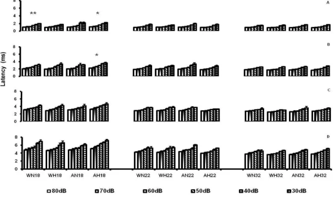
(F(2,26) = 28.32); and of intensity on waves I (F(5,190) = 420.84), II (F(5,180) = 339.21), III (F(4,21) = 278.34) and IV (F(5,130) = 186.35), as well as an effect of the diet x intensity interaction for waves I (F(5,190) =2.79), II (F(5,180) = 2.57) and IV (F(5,13) = 3.19), and of diet x stimulation x age interaction for waves I (F(2,38) = 3.90) and II (F(2,36) = 3.90).
Post hoc comparisons showed that the animals
presented longer latencies of waves I, II, III and IV of the BAEP at 18 than at 22 and 32 days. However, the latencies were longer at 22 than at 32 days only for waves I and IV (p < .05). Regarding the effect of intensity, the multiple comparison test revealed that all animals presented lower latencies for waves I, III and IV at higher intensities compared to lower ones (80 < 70 < 60 < 50 < 40 < 30dB). For wave II, the effect of intensity was demonstrated as starting only at 70 dB (80 and 70 < 60 < 50 < 40 < 30dB) (p < .05) and the diet x intensity interaction indicated the same effect in both the W and A groups (p <.05). In this same direction, the diet x intensity interaction revealed that W and A animals had lower latencies for waves I and IV at higher intensities compared to lower ones (W80 < W70 < W60 < W50 < W40 < W30dB and A80 < A70 < A60 < A50 < A40 < A30dB). The post hoc analysis for
the diet x stimulation x age interaction showed that AH18 rats had longer latencies of waves I and II than AH22 and AH32 rats, as well as a longer latency of wave I than WH18 rats (p < .05) (Figure 1).
ANOVA indicated effects of age (F(2,26) = 21.43) and intensity (F(5,130) = 30.90) on the I-IV interpeak interval. The post hoc test indicated that the interpeak
intervals were greater at 18 than at 22 days, and that these two groups had greater I-IV intervals than rats tested at 32 days. The post hoc analysis of the intensity
variable showed that all groups presented a greater I-IV interpeak interval when submitted to higher intensities (80dB) compared to lower ones (30dB) (80 < 70 < 60 < 50 < 40 < 30dB) (p < .05).
ANOVA did not show effects of diet (F(1,83) = 0.01; p > .05), stimulation (F(1,83) = 0.01; p > .05) or age (F(2,83) = 2.08; p > .05) on the threshold of the responses to auditory stimuli.
Discussion
Iron is a necessary component for red blood cell formation and for cell metabolism(Schmidt et al., 2007). Its deiciency may cause a reduction in the amount of serum iron (Cunha, van Ravenzwaay, Mellert & Kaufmann,2008) and in hematocrit and liver iron stores (Bourque, Iqbal, Reynolds, Adams & Nakatsu, 2008), components whose deiciency may impair tissue growth and consequently reduce the body weight of individuals receiving iron-poor diets (Eseh & Zimmerberg, 2005). In the present study, the iron-deicient diet imposed during the lactation period produced a lower body weight
Figure 1. Latency of waves I (A), II (B), III (C) and IV (D) of the Brainstem Auditory Evoked Potential (BAEP) in response to a click intensity of 80, 70, 60, 50, 40 and 30dB. The vertical bars represent the mean ± SEM for the groups. Groups: WN18, WN22, WN32 (Well-nourished, No Handling, n=8), WH18, WH22, WH32 (Well-nourished, Handling, n=8), AN18, AN22, AN32 (Anemic, No Handling, n=8) and AH18, AH22, AH32 (Anemic, Handling, n=8) on the 18th, 22nd and 32nd days of age.
compared to animals that received a diet with adequate amounts of iron according to the AOAC (Cunniff, 1995). These data demonstrate the importance of dietary iron for the maintenance of the quantity of cells in tissues and also of the animals’ weight, in agreement with previous studies that reported a reduction of body weight in rats with iron deiciency (Eseh & Zimmerberg, 2005). In the present study, AH rats had lower body weights than AN animals at 18 days, with the effects of handling being similar to those observed in malnourished rats of the same ages (Rocinholi et al., 2001b) in which malnutrition is known to cause a reduction in the rate of cell division (Morgane, Austin-LaFrance, Bronzino, Tonkiss & Galler, 1992). However, in contrast to what was demonstrated in previous studies, WH animals had higher weights than WN animals, showing an inverse effect of handling on anemic and control animals at this age. Although the iron-deicient diet reduced the body weight of anemic rats compared to controls, revealing inadequate development, all groups presented weight gain along time.
The iron-deicient diet, in addition to reducing the body weight of group A animals, also caused lower Hb and %Ht compared to W rats of the same age (18, 22 and 32 days), demonstrating that this diet promoted a deicient nutritional condition in anemic rats, as also reported in the study by Cunha et al. (2008). Thus, it permitted the investigation of the effects of anemia on the BAEP of the rats. Although the body weight of all animals increased until the end of the experiment, only W animals showed an increase in Hb and Ht at 32 days compared to rats of 18 and 22 days of age. Anemic animals, although they gained body weight, presented lower quantities of Hb and Ht at 32 days than at 18 and 22 days of age. These results reveal that the imposition of the deicient diet up to 32 days continued to reduce the iron stores in the organism along the period evaluated, reducing Ht and Hb. However, the other components of the diet were adequate, a fact that may have provided the body weight gain, although the animals require iron for cell metabolism and growth and for the adequate development of body tissues (Schmidt et al., 2007).
BAEP recordings of the animals studied here revealed that age is an important factor that determines the maturation of the auditory pathway of the brainstem, since waves I, II, III and IV, formed by the arrival of nervous impulses to the nuclei that compose this pathway, present longer latencies in young animals (18 days) than in older ones (22 and 32 days). Since the conduction velocity of nervous impulses is directly associated with the thickness of the myelin layer and with the caliber of the axons of the neurons that compose the pathways of the nervous system, we may suggest that in the present study the younger animals were probably going through the process of myelinization, which starts at about 19 days of life and reaches the post-lactation phase (Connor & Benkovic, 1992). Thus, we may suggest that the waves have longer latencies because the nervous
impulse occurs at a lower velocity due to the fact that the myelin layer is still forming.
The analysis of the diet x stimulation x age interaction revealed that waves I and II had longer latencies in group AH at 18 days of age than at 22 and 32 days. This effect was not observed in the WH group, showing that handling reduced the effect of age in W animals and was unable to produce a similar effect in group A. Iron-deiciency can alter metabolism, neurotransmission and glial integrity and can increase the time of nervous system myelinization (Schmidt et al., 2007), while on the other hand handling can promote increased thickness of the myelin layer in the corpus callosum (Lima, 1992) and hippocampus (Iuvone, Geloso & Dell’Anna, 1996), and reduce the latency of BAEP waves in rats (Rocinholi et al., 2001b). The data suggest that the effects of handling over time became evident only in W animals since these animals have a substrate for an earlier formation of the myelin layer, a fact that does not occur in A rats due to the low amount of iron in their diet.
Since AH animals showed longer latencies of waves I and II and since these waves depend on the integrity of myelin in the system, we may assume that there was an alteration in the myelin layer of auditory nerve neurons (responsible for the formation of wave I) and of the cochlear nuclei (responsible for the formation of wave II). However, there are no reports of morphological analyses of the structures of brainstem auditory pathways of stimulated animals.
Although an increase in the latency of waves I and II was not observed in WH animals, the AH group presented greater wave I latency than the WH group at 18 days of age, showing that diet is the one factor responsible for this increase, since the myelin layer is being formed during this period of development (Connor & Benkovic, 1992), and also revealing a greater vulnerability of peripheral structures (auditory nerve) to the effects of iron deficiency, since no increase occurred in the latency of waves III and IV.
Similarly, BAEP recordings showed a shorter latency of the I-IV interpeak interval only in young animals compared to older animals, indicating the effect of age. However, no diet effect was observed, demonstrating that the brainstem was not intensely affected by the iron-deicient diet, as was the case for malnourished animals (Rocinholi et al., 2001a, 2001b).
Acknowledgment
This work was supported by grant nº 98/02989-8 from the Fundação de Amparo à Pesquisa do Estado
de São Paulo. We would like to thank Mr. Domingos
Edmundo Pitta for technical assistance and Prof. Dr. Ana Maria de Souza, of the Faculdade de Ciências Farmacêuticas de Ribeirão Preto – USP, for checking Ht and Hb analyses. L.F.R. was recipient of a FAPESP fellowship Proc. nº 98/02987-5.
References
Bourque, S.L., Iqbal U., Reynolds J.N., Adams, M.A., & Nakatsu, K.
(2008). Perinatal iron deiciency affects locomotor behavior and water maze performance in adult male and female rats. Journal of Nutrition, 138(5), 931-937.
Carughi, A., Carpenter, K.J., & Diamond, M.C. (1989). Effect of environmental enrichment during nutritional rehabilitation on body growth, blood parameters and cerebral cortical development of rats. Journal of Nutrition, 119, 2005-2016.
Chapillon, P., Patin, V., Roy, V., Vincent, A., & Caston, J. (2002). Effects
of pre and postnatal stimulation on developmental, emotional, and cognitive aspects in rodents: a review. Developmental Psychobiology, 41(4), 373-387.
Connor, J.R., & Benkovic, S.A. (1992). Iron regulation in the brain: Histochemical, biochemical and molecular considerations. Ann. Neurology, 32, S51-S61.
Darwin, C. (1877/2000). A expressão das emoções no homem e nos animais. São Paulo: Companhia das Letras.
Cunha, G.C., van Ravenzwaay, B., Mellert, W., & Kaufmann, W. (2008). Iron deiciency causes duodenum mucosal hyperplasia in male Wistar rats. Toxicology Letters, 177(3), 156-167.
Cunniff, P. (1995). Oficial methods of analysis of AOAC International. Maryland: Association of Oficial Agricultural Chemists. Eseh, R., & Zimmerberg, B. (2005). Age-dependent effects of
gestational and lactational iron deiciency on anxiety behavior in rats. Behavioral Brain Research, 164(2), 214-221.
Felt, B.T., Beard, J.L., Schallert, T., Shao, J., Aldridge, J.W.,
Connor, J.R., Georgieff, M.K., & Lozoff, B. (2006). Persistent neurochemical and behavioral abnormalities in adulthood despite early iron supplementation for perinatal iron deiciency anemia in rats. Behavioral Brain Research, 171(2), 261-270.
Felt, B.T., & Lozoff, B. (1996). Brain iron and behavior of rats are not normalized by treatment of iron deiciency anemia during early development. Journal of Nutrition, 126(3), 693-701.
François, C., Nguyen-Legros, J., & Percheron, G. (1981). Topographical and cytological localization of iron in rat and monkey brains. Brain Research,215(1-2), 317-322.
Hill, J.M., & Switzer, R. C. (1984). The regional distribution and cellular localization of iron in the rat brain. Neuroscience, 11(3), 595-603.
Iuvone, L., Geloso, M.C., & Dell’Anna, E. (1996). Changes in open ield behavior, spatial memory, and hippocampal parvalbumin immunoreactivity following enrichment in rats exposed to
neonatal anoxia. Experimental Neurology, 139, 25-33.
Jewett, D.L., & Romano, M.N. (1972) Neonatal development of auditory system potentials averaged from the scalp of rat and cat. Brain Research, 36, 101-115.
Lemaire, V., Lamarque, S., Le Moal, M., Piazza, P.V., & Abrous, D.N. (2006). Postnatal stimulation of the pups counteracts prenatal stress-induced deicits in hippocampal neurogenesis. Biology Psychiatry, 59(9), 786-792.
Lima, J.G. (1992). Estudo morfológico e morfométrico do corpo caloso de ratos submetidos a diferentes tipos de dieta e à estimulação ambiental. Unpublished Doctoral Thesis, University of São Paulo.
Lucion, A.B., Pereira, F.M., Winkelman, E.C., Sanvitto, G.L., &
Anselmo-Franci, J.A. (2003). Neonatal handling reduces
the number of cells in the locus coeruleus of rats. Behavioral Neuroscience, 117(5), 894-903.
Morgane, P., Austin-LaFrance, R.J., Bronzino, J., Tonkiss, J., & Galler, J. R. (1992). Malnutrition and the developing central nervous system. In R.L. Isaacson & K.F. Jensen (Eds.), The vulnerable brain and environmental risks, v.1 (pp. 3-43). New York: Plenum Press.
Morris, C. M., Candy, J. M., Oakley, A. E., Bloxham, C. A., & Edwardson, J. A. (1992). Histochemical distribution of non-haem iron in the human brain. Acta Anatomica, 144, 235-257.
National Research Council. (1996). Guide for the Care and Use of Laboratory Animals.
Rocinholi, L.F., Almeida, S.S., & De Oliveira, L.M. (1997). Response threshold to aversive stimuli in stimulated early protein-malnourished rats. Brazilian Journal of Medical Biological Research, 30, 407-413.
Rocinholi, L.F., De Oliveira, L.M., & Colafêmina J.F. (2001a). Malnutrition and environmental stimulation in rats: Interpeak intervals of the brainstem auditory evoked potentials. Nutritional Neuroscience, 4, 189-198.
Rocinholi, L.F., De Oliveira, L.M., & Colafêmina, J.F. (2001b). Malnutrition and environmental stimulation in rats: Wave latencies of the brainstem auditory. Nutritional Neuroscience, 4, 199-212.
Simmons, D., Ng, L.L., Harker, M., & Hockaday, T.D. (1989). Glycemic control affects cellular sodium metabolism. Diabetes Research, 10(1), 13-15.
Sullivan, R.M., & Dufresne, M.M. (2006). Mesocortical dopamine and HPA axis regulation: Role of laterality and early environment.
Brain Research, 1076(1), 49-59.
Schmidt, A.T., Waldow, K.J., Grove, W.M., Salinas, J.A., & Georgieff,
M.K. (2007). Dissociating the long-term effects of fetal/neonatal iron deiciency on three types of learning in the rat. Behavioral Neuroscience, 121(3), 475-82.
Unger, E.L., Paul, T., Murray-Kolb, L.E., Felt, B., Jones, B.C., &
Beard, J.L. (2007). Early iron deiciency alters sensorimotor
development and brain monoamines in rats. Journal of Nutrition, 137(1), 118-124.
Wiggins, R. C., Fuller, G., & Enna, S.J. (1984). Undernutrition and the development of brain neurotransmitter systems. Life Sciences, 35(21), 2085-2094.
Wu, L.L., Zhang, L., Shao, J., Qin, Y.F., Yang, R.W., & Zhao, Z.Y.