Effects of malnutrition and sensory-motor stimulation on
auditory evoked potentials
Juraci Gonçalves de Lima, Carolina Araújo Rodrigues Funayama, Luiz Marcellino de Oliveira
†,
Maria Rossato and José Fernando Colafêmina
Universidade de São Paulo, Ribeirão Preto, Brazil.
† in memoriam
Abstract
There is evidence that the auditory evoked potential (AEP) is altered by malnutrition both in laboratory animals and in humans. The objective of the present study was to determine whether changes in the AEP caused by malnutrition could be reversed by nutritional rehabilitation and sensorymotor and environmental stimulation during hospitalization. Six children aged 5-33 months with severe malnutrition (kwashiorkor, marasmus and marasmic-kwashiorkor) were admitted to the Pediatric Ward of a University Hospital. Normal age and sex-matched children from the hospital day-care center were enrolled as a control group. The AEP was tested in an electrically and acoustically isolated room using a Nicolet CA 2000 microcomputer. Clicks of 90; 80; 70 and 60 dBn HL were presented through earphones.The results suggest that malnutrition leads to an increase in wave I latencies in patients with marasmus, and in waves I, III and V in those with kwashiorkor or marasmic-kwashiorkor type at 90 dB HL. At discharge, all but one patient with kwashiorkor showed reduced latencies of waves I, III and V compared to the values on admission. Despite the small sample, these preliminary results pointed out that the process of sensory stimulation used in our study in a properly directed, systematic and individualized manner showed encouraging results in terms of AEP recovery in these children. Keywords: malnutrition, infants, nutritional rehabilitation, auditory evoked potential, stimulation.
Received 30 September 2008; received in revised form 9 December 2008; accepted 16 December 2008. Available online 30 December 2008
Juraci Gonçalves de Lima, Carolina Araújo Rodrigues Funayama, Luiz Marcellino de Oliveira, Maria Rossato and José Fernando Colafêmina – Universidade de São Paulo de Ribeirão Preto. Correspondence regarding this article should be directed to: Dr. Carolina A.R. Funayama – Universidade de São Paulo, Hospital das Clínicas, Ribeirão Preto, SP, Brazil, 14048-900. E-mail [email protected]
Introduction
Many investigators have shown that nutrition is one of the basic factors for the development of the central nervous system (CNS) and that nutritional deiciency impairs CNS functioning (Bedi, 1987; Bedi, Thomas, Davies & Dobbing, 1980; Cragg, 1972; Cravioto, Delicardie, & Birch, 1966; Dobbing, 1968; Dobbing & Sands, 1971; Finger & Stein 1982; Lima, 1992; Mourek, Himwich, Myslivecet, & Callison, 1967; Quirk, Mejya, Hesse, & Su, 1995; Smart, Dobbing, Adlard, Linch, & Sands, 1973; Sobotka, Cook, & Brodie, 1974; Stein, Finger, & Hart, 1983; Wang & Xu, 2007).
There is evidence since the 1950’s and 1960’s that protein-calorie malnutrition is associated with retarded brain growth (Stock & Smythe, 1963), reduced cerebral cellularity (Winick & Rosso, 1969),
reduced myelination (Fishman, Prensky, & Dodge, 1969) and, in the neurophysiological ield, changes in electroencephalogram (Engel, 1956; Nelson & Dean, 1959) and evoked potential tracings (Kawai, Nakamura, & Matsuo, 1989; Flinn, Barnet, Lydick, & Lackner, 1993; Hernández et al., in press).
Auditory Evoked Potentials (AEP) are very sensitive measures related to brain functions and have been used by many researchers with diverse objectives in humans, as in the pioneering studies by Hecox and Galambos (1974); and in experimental studies in animals (Buchwald & Huang, 1975; Shipley, Buchwald, Norman, & Guthrie, 1980; Plantz, Williston, & Jewett, 1981).
the period of rehabilitation. The above data suggested that these abnormalities could be due to deiciencies in the myelination process that would result in decreased synaptic eficiency in the auditory system.
Some studies have shown CNS recoveryin laboratory animals (Bedi & Bhide, 1988; Renner & Rosenzweig, 1987; Rocinholi, de Oliveira, & Colafêmina, 2001), and it is well known that relatively simple programs of sensorymotor stimulation of malnourished children are effective in promoting a better development of these children (Carrazza et al., 1993; Grantham-McGregor, Powell, & Walker, 1991; Grantham-McGregor, Schoield, & Harris, 1983; Grantham-McGregor, Schoield, & Powell, 1987; Granthan-McGregor, Stewart, & Schoield, 1980; Nahar et al., in press; Walker, Chang, Powell, & Grantham-McGregor, 2005).
Studies evaluating electrophysiological parameters have pointed to diverse nervous system compromise of auditory (Durmaz, Karagol, Deda, & Onal, 1999; Vandana & Tandon, 2006), visual (Durmaz et al., 1999; McDonald, Joffe, Barnet, & Flinn, 2007), corticospinal (Karak, Misra, Garg, & Katiyar, 1999), and somatosensory paths (Hesse, Rivera, de Díaz, & Quirk, 1998), as well as inter-hemispheric modulation (Pinto & Guedes, 2008), but no reference to the stimulation effects on evoked potentials in malnourished infants was found in the literature. Thus, the objective of the present study was to determine the effects of malnutrition (kwashiorkor, marasmus and marasmic-kwashiorkor) on AEPs and to establish whether changes could be reversed by nutritional rehabilitation associated with daily and individual sensorymotor and environmental stimulation.
Methods
Participants
After approval by the local Committee of Ethics and provision of signed informed consents by the adults responsible for the infants, the study was conducted on six children admitted to the Pediatric Unit of the University Hospital (HC) of Ribeirão Preto, São Paulo University,
presenting one of the following types of malnutrition: kwashiorkor (cases N1 and N5), marasmus (N2 and N3) and marasmic kwashiorkor (N4 and N6) according to the classiication of McLaren, Pleet and Read (1967). Data concerning the identiication of the children at birth and during hospitalization are presented in Table 1. All patients but one (N4 - 2.300g - low weight at birth) were born at term with adequate weight for the gestational age, according to Sala (1977). The children were submitted to two AEP evaluations, one at admission and the other just before discharge from the hospital after nutritional rehabilitation. During hospitalization the children were submitted to sensory and environmental stimulation. Six healthy children matched by age and sex from the hospital day-care center were enrolled as controls. Control children were submitted to only one AEP evaluation after investigation of the middle ear by impedance measurement and by the stapes relex. Subjects with other pathologies interfering with the air or sensorial ear conduction and presenting growth or neurodevelopmental problems were excluded from the study.
Sensory-motor Stimulation
Stimulating activities were performed as described by Celedon (1983), taking into account the children’s age and development, evaluated by the Bayley Infant Development Scale (1993), soon after admission to the hospital. Based on the Bayley Scale psychomotor score, each child was assigned to an age group and the stimulation process was started with the activities listed in Table 2.
The children were submitted to daily stimulation sessions of one hour, between 1:30 and 2:30 pm, in a separate room prepared for the stimulation process. Three well-trained professionals were in charge of the stimulation process and received previous training in order to obtain agreement regarding the application of the technique proposed at the beginning of the study. During the irst days of hospitalization, before the stimulation program, the three professionals took turns caring for each child, so that he/she would adapt to the new environment (the hospital) and to establish a process of child-stimulator interaction important for the fulillment of the objectives proposed.
Table 1. Signs and symptoms of malnourished children Mo- months d- days *For the same age and sex of socioeconomic group IV of the table of Marques et al. (1974) ** MacLaren et al. (1967) + Present - Absent Kw-kwashiorkor MIII-marasmus degree III Mix - Marasmic-kwashiorkor.
Patient Age Weight (g) Expected Weight(g)*
Weight(g)
deicit Weight(g) discharge Edema Skin lesions
Hair
changes Hepatomegaly Albumin (g/dl) Points** Class
N1 F 20mo 9d 9190 11862 2253 10180
+ - + - 3,4 10 Kw
N2 M 6mo27d 4840 8278 4153 6740
- + - - 6 5 M III
N3 M 7mo8d 3700 8278 5531 5760
- - + - 4 5 MIII
N4 M 5mo26d 4200 7387 4314 5190
+ + - + 6,7 8 Mix
N5 F 34mo5d 7820 14869 4741 9400
+ - + + 4 10 Kw
N6 F 4mo26d 2760 6810 5983 4570
Table 2. Objectives proposed and activities in the process of sensorymotor stimulation of children at different age ranges. DD dorsal decubitus VD – ventral decubitus.
OBJECTIVES AND ACTIVITIES
0-3 months
To acquire prehension, visual, auditory integration and cervical control: - hold objects of different shapes, sizes, colors, sounds. DD - follow a light source (large lashlight) and objects in the horizontal, vertical and circular directions. VD - follow light sources and objects in the vertical direction (up and down).
4-5 months
To facilitate object manipulation: - Exploration of different objects; VD: grasp objects at different heights; dorsal decubitus, grasp objects. To acquire schemes of trunk dislocation and balance: - placed now on the right, now the left; DD, with support under the axillae, bring to the sitting position in a movement cadenced on music. To acquisition and reinforcement of sound:- DD, rhythmic exercises in limb lexion and extension with the help of the sound, repeating casually emitted sounds.
6-8 months
To reinforce the sitting position and change in decubitus:-.sitting up, to manipulate and explore different and new objects. To stimulate crawling:- Sitting up, to turn to a ball thrown now to the left, now to the right. DD, try to reach objects placed now on the left, now on the right. DV, try to reach objects initially near and after more far.
9-12 months
To reinforce hand dexterity:- it, stack up or group chips locks, rings, beating a drum; hit the keys of a piano with hands and ingers rhythmically imitating the examiner and orders. To place in the standing position and help to walk:- walk with support with two hands, then with one and then holding on to furniture. Acquisition of irst words, attitudes:- point and name igures on cards (daddy, mommy, meow, bow wow).To recognize body parts: - Indicate parts of the body on him-herself or on a doll.
13-18 months
To reinforce activities of the previous group, increase coordination, improve ine movements and language: - grasp and arrange balls, stack, it and group smaller objects, model sand, clay, hug, blow a kiss, say please, say goodbye, dance and mimic.
19-24 months
To reinforce ine motor coordination and balance.:- object manipulation as in the previous stage, walk along a straight line and in a circle; walking, construct a line and a circle with an object on the head, jump, run, squat and rise to the sound of music , listen to stories and to construct sentences looking at illustrative igures.
Any activity to be performed with the child needed to be accepted by him/her and the sequence of activities was not rigid but depended on the interest and development of each child.
AEP Evaluation
Following regular otoscopic examination, the infants were evaluated in an electrically and acoustically isolated room. All of them received 16% chloral hydrate at the dose of 1.2 ml/kg body weight 30 minutes before the AEP test to reduce artifacts of muscular origin to a minimum and thus obtain satisfactory recordings before each evaluation. Brain stem electrical activities were recorded
and consisted of monaural rarefaction clicks generated by pulses of 100 µs duration. White noise was presented in the contralateral ear to mask with a differential of 40 dBn HL between the two stimuli. The clicks were presented at stimulation rates of 11.4/sec at intensities of 90, 80, 70 and 60 dBn HL, with a low ilter (high - pass) of 150 Hz and high ilter (low - pass) of 3000 HZ (12 dB/ octave). The scanning speed was 100ms. Responses to 2000 clicks with an accepted rejection level of up to 20% were averaged and stored in a loppy disk, while wave latencies were simultaneously evaluated with a mobile cursor on the screen with digital reading. The data were plotted using a CITO HM 8510 printer/plotter.
Statistics
A paired t-test was used for the comparison of waves and interpeak latencies at different intensities of dBn HL, and qualitative analysis was performed taking into consideration the small sample.
Results
The mean latencies of the wave I, III and V and the interpeak intervals I-III, III-V and I-V are shown in Table 3 for 70 dBn HL and in Table 4 for 90 dBn HL. Only both levels, 70 and 90 dBn HL, were presented, since the results for 60 and 70 dBn HL were similar, as well as for 80 and 90 dBn HL (t-test p > .05).
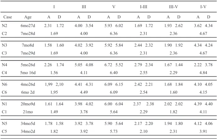
Before the stimulation program and the nutritional treatment, patient N3 had a good registry for analysis only at 90 dBn HL, and the latencies of waves I, III and V were lower than those obtained in the control infant. Patients N1 (kwashiorkor) and N4 (marasmic-kwashiorkor, the only one who was small according to the gestational age at birth in the sample) showed increases in latencies of waves I, III and V in both 90 and 70 dBn HL. Patient N2 (marasmus degree III) showed increased latencies of wave I at 90 dBn HL, and of waves I, III, V at 70 dBn HL. Patient N6 (marasmic-kwashiorkor) showed increase in the latency of waves I (90 dBn HL) and V (70 dBn HL); the other latencies were equivalent to those of controls in both levels. Patient N5 (kwashiorkor) showed increased latencies of wave V, and waves I and III similar to control at both 90 and 70 dBn HL. At admission, the interpeak I-III was larger for N4 (both levels) and N1 (90 dBn HL); III-V for N5 (70 dBn HL), N1 and N6 (both levels); and I-V was larger for N6 (70 dBn HL) and N5 (both levels). At discharge, all but one infant (N1 - kwashiorkor) showed decrease in the latencies of waves I, III and V, compared to the values at admission. The latencies of wave I remained above the respective controls for patients N1, N2, N3, N4, and N6 even at discharge (70 dBn HL). At discharge, ive patients (N2, N3, N4, N5, N6) at 90 dBn HL and three (N4, N5 and N6) at 70 dBn HL had latencies of waves III or V lower than their respective controls.
Discussion
Evidence that malnutrition causes changes in AEP measurements in children has been provided sparsely in the literature (Barnet et al., 1978; Bartel et al., 1986; Durmaz et al., 1999; Odabas et al., 2005).
Taking into account that the most used AEP reaches the level of the brainstem, studies involving higher paths and cortical areas are necessary, focusing on their vulnerability, damage, and persistence of the effects of malnutrition on higher cortical activity with present and future consequences for development. Flinn et al. (1993) investigated potentials recorded in cortical areas, with the child’s name as a stimulus. They observed that malnourished infants had a lower number of wave peaks and amplitudes than controls, after a nutritional recovery period of about four months.
Although it is not our scope to address speciic aspects of the stimulation procedure in this report, the effects of different types of stimuli on behavior (Cabral & Almeida, 2008), and the best results on visual learning when multisensory stimuli are presented in a congruent way (Kim, Seitz, & Shams, 2008) are important issues for stimulation strategies in the clinical ield.
As in the present work, in human clinical settings we can not attribute the results exclusively to the environmental and sensory stimulation, since nutritional treatment and medicines were often prescribed. To sort out such variables, methods in animal experiments have been developed. AEP analysis of handling stimulation without food supplementation in rats (Rocinholi et al., 2001) showed that stimulation alone reduced wave latencies, suggesting the possibility of reversion of some of the damage caused by malnutrition.
Table 3. Latencies of waves I, III, V and interpeak values I-III, III-V and I-V recorded at 90 dB in six malnourished children and controls (C). A – admission D - discharge mo – months d - days.
I III V I-III III-V I-V
Case Age A D A D A D A D A D A D
N2 6mo27d 2.31 1.72 4.00 3.54 5.93 6.02 1.69 1.72 1.93 2.62 3.62 4.34
C2 7mo28d 1.69 4.00 6.36 2.31 2.36 4.67
N3 7mo8d 1.58 1.60 4.02 3.92 5.92 5.84 2.44 2.32 1.90 1.92 4.34 4.24
C3 7mo29d 1.69 4.00 6.36 2.31 2.36 4.67
N4 5mo26d 2.26 1.74 5.05 4.08 6.72 5.52 2.79 2.34 1.67 1.44 2.22 3.78
C4 5mo 16d 1.56 4.11 6.40 2.55 2.29 4.84
N6 4mo26d 1,99 2,10 4.41 4.31 6.09 6.15 2.42 2.21 1.68 1.84 4.10 4.05
C6 6mo 2d 1,95 4.49 6.09 2.54 1.60 4.15
N1 20mo9d 1.61 1.64 3.98 4.02 6.00 6.04 2.37 2.38 2.02 2.02 4.39 4.40
C1 21mo 1.49 3.78 5.64 2.29 1.82 4.11
N5 34mo5d 1.78 1.58 3.92 3.78 5.90 5.64 2.17 2.20 1.94 1.80 4.12 4.06
C5 34mo2d 1.82 3.92 5.73 2.10 2.31 3.91
not fully developed in newborns, although it is not clear whether this initial delay in the latency of wave I is the result of immaturity of the middle and inner ear or of neuronal immaturity.
Concerning the interpeak latencies, our indings of kwashiorkor infants with III-V and I-V latencies longer than those of children with marasmus are in accordance with the indings of Durmaz et al. (1999). Bartel et al. (1986) found I-III, III-V and I-V intervals uniformly distributed among the degrees of malnutrition, studying 22 children, also showing differences between the two ears, which was further supported by Odabas et al. (2005). In the present sample we did not analyze the data for each ear.
The analysis of covariates in malnutrition and AEP studies has been rarely mentioned in the literature. The children’s height was mentioned by Barnet et al. (1978), inding higher AEP waves amplitude in children with shorter stature. Other covariates, such as iron deiciency and albuminaemia, were analyzed by Odabas et al. (2005), showing in malnourished infants with iron deiciency longer than controls the mean latency of wave I on the left side and the mean III-V interpeak on the right side; in malnourished infants without iron deiciency, the mean I-III interpeak on the right ear was longer than controls, and no difference was found between low and normal serum albumin within the malnourished groups.
The SGA (small for gestational age) condition has not been studied among the possible confounding variables in the ield of malnutrition. Odabas et al.
(2005) mention the exclusion of SGA infants in their criteria for subject selection. In the present study, the ive-month-old patient who was born SGA, was the only one presenting increased I-III interpeak interval and above the control values at 70 dBn HL, in both the admission and discharge AEP tracings, suggesting the need to examine the cochlear station since neonatal period for the SGA patients.
According to Bartel et al. (1986), AEP abnormalities were still evident after one year of nutritional follow-up of marasmic children, probably relecting long-term effects of malnutrition on cerebral function. Durmaz et al. (1999) found different results comparing malnourished infants to controls on admission in a nutritional program and discharge. These authors reported differences at the time of admission, but not at discharge, in line with our results. In spite of the limitations of the present analysis, an interesting inding was the fact that the malnourished group showed lower latencies of waves I, III and V at discharge compared with the latencies at admission, and in three patients (one kwashiorkor) the latencies of waves III or V at discharge were lower than those of their respective controls. Thereby, we can suggest that the process of sensory and environmental stimulation may have been effective in bringing about a recovery fromor prevention of the effects of malnutrition.
results in terms of AEP recovery in these children. Even if they attend day-care centers using some type of stimulation, these children are exposed to a more collective and not individually directed process.
Acknowledgements
The authors owe debts of gratitude to Psychologist Dorothy Bono, Dr Naul Motta de Souza, and the staff of the Pediatrics Ward at the University Hospital of Ribeirão Preto, São Paulo University.
References
Barnet, A.B., Weiss, I.P., Sotillo, M.V., Ohbrich, E.S., Srkuvich, M.Z., & Cravioto, J. (1978). Abnormal auditory evoked potentials in early infancy malnutrition. Science, 201(4354), 450-452. Bartel, P.R., Robinson, E., Conradie, J.M., & Prinsloo, J.G. (1986).
Brainstem auditory evoked potentials in severely malnourished children with kwashiorkor. Neuropediatrics, 17(4), 178-182. Bayley, N. (1993). Bayley scales of infant development. Texas: The
Psychological Corporation.
Bedi, K.S. (1987). Lasting neuroanatomical changes following undernutrition during early life. In: J. Dobbing (Ed.), Early nutrition and later achievement (pp. 1-49). New York: Academic Press.
Bedi, K.S., & Bhide, P.G. (1988). Effects of environmental diversity on brain morphology. Early Human Development, 17(2-3), 107-143.
Bedi, K.S., Thomas, Y.M., Davies, A.A., & Dobbing, J. (1980). Synapse-to-neuron ratios of the frontal and cerebellar cortex of 30-day-old and adult rats undernourished during early post natal life. Journal of Comparative Neurology, 193(1), 49-56.
Buchwald, J.S., & Huang, C. (1975). Far-ield acoustic response:
origins in the cat. Science, 189(4200), 382-384.
Cabral, A., & Almeida, S.S. (2008). Effects of tactile stimulation and underwater trauma on the behavior of protein-malnourished rats in the elevated plus-maze test. Psychology & Neuroscience, 1(1), 63-66.
Carrazza, F.R., Patah, D., Marco, S., Godoy, C.M., Pieri, S., & Issler, H. (1993) Normalização do Quociente de Desenvolvimento de lactantes desnutridos graves estimulados pelas suas mães. Revista Paulista de Pediatria, 11(2), 174-177.
Celedon, J.M. (1983). Nutrición e inteligencia en el niño. Santiago: Ediciones de la Universidad de Chile.
Cragg, B.G. (1972). The development of cortical synapses during starvation in the rat. Brain, 95(1), 143-150.
Cravioto, J., Delicardie, E.R., & Birch, H.G. (1966). Nutrition, growth and neurointegrative development: an experimental and ecologic study. Pediatrics, 38(2), 319-367.
Dobbing, J. (1968). Effects of experimental undernutrition on development of the nervous system. In: N.S. Scrimshaw & J.E. Gordon (Eds.), Malnutrition learning and behavior (pp. 181-202). Cambridge: MIT Press.
Dobbing, J., & Sands, J. (1971). Vulnerability of developing brain. IX. The effect of nutrition growth-retardation on the timing of the brain growth spurt. Biology of the Neonate, 19(4), 363-378. Durmaz, S., Karagöl, U., Deda, G., & Onal, M.Z. (1999). Brainstem
auditory and visual evoked potentials in children with protein-energy malnutrition. Pediatrics International, 41(6), 615-619. Engel, R. (1956). Abnormal brain wave patterns in kwashiorkor.
Electroencephalography and Clinical Neurophysiology, 8(3), 489-500.
Finger, S., & Stein, D.G. (1982). Brain Damage and recovery: research and clinical perspectives. New York: Academic Press. Fishman, M.A., Prensky, A.L., & Dodge, P.R. (1969). Low content of
cerebral lipids in infants suffering from malnutrition. Nature, 221(5180), 552-553.
Flinn, J.M., Barnet, A.B., Lydick, S., & Lackner, J. (1993). Infant malnutrition affects cortical auditory evoked potentials. Perceptual and Motor Skills, 76(3 Pt2), 1359-1362.
Grantham-McGregor, S., Powell, C.A., & Walker, S.P. (1991). Nutritional supplementation, psychosocial stimulation and Table 4. Wave latencies I, III, V and interpeak values I-III, III -V and I-V recorded at 70 dB in six malnourished children (N) and controls (C). A – admission D - discharge mo – months d - days.
I III V I-III III –V I-V
Case Age A D A D A D A D A D A D
N2 6mo27d 2.52 2.28 4.43 3.90 6.46 6.31 1.91 1.25 2.02 2.87 3.94 4.12
C2 7mo28d 1.82 4.02 6.30 2.20 2.28 4.48
N3 7mo8d --- 1.91 --- 4.37 --- 6.30 2.46 1.89 4.35
C3 7mo29d 1.82 4.02 6.30 2.20 2.28 4.48
N4 5mo26d 2.87 2.00 5,68 4,42 7.12 6.16 2.81 2.42 1.44 1.74 4.25 4.16
C4 5mo 16d 1.92 4,36 6.69 1.96 2.33 4.29
N6 4mo26d 2,59 2,44 4.78 4.80 6.93 6.81 2.19 2.36 2.15 2.00 4.34 4.36
C6 6mo 2d 2,06 5.23 6.79 2.37 1.56 4.03
N1 20mo9d 2.25 2.36 4.26 4.53 6.43 6.49 1.90 2.17 2.28 1.96 4.18 4.14
C1 21mo 1.76 3.96 5.97 2.20 2.01 4.21
N5 34mo5d 2.39 2.07 4.63 4.08 6.40 6.07 2.20 2.01 2.31 1.09 4.01 4.00
mental development of stunted children: the Jamaican study. Lancet, 338(8758), 1-5.
Grantham-McGregor, S., Schoield, W., & Harris, L. (1983). Effect
of phychosocial stimulation on mental development of severely malnourished children: an interim report. Pediatrics, 72(2), 239-243.
Grantham-McGregor, S., Schoield, W., & Powell, C.A. (1987).
Development of severely malnourished children who received psychosocial stimulation: six-year follow-up. Pediatrics, 79(2), 247-254.
Grantham-McGregor, S., Stewart, M.E., & Schoield, W. (1980). Effect
of long-term psychosocial stimulation on mental development of severely malnourished children. Lancet 2(8198), 785-789. Hecox, K., & Galambos, R. (1974). Brainstem auditory responses in
human infants and adults. Archives of Otolaryngology, 99(1), 30-33.
Hernández, A., Burgos, H., Mondaca, M., Barra, R., Núñez, H., Pérez, H., Soto-Moyano, R, Sierralta, W., Fernández, V., Olivares, R., & Valladares, L. (in press). Effect of prenatal protein malnutrition on long-term potentiation and BDNF protein expression in the rat entorrhinal cortex after neocortical and hippocampal tetanization. Neural Plasticity.
Hesse, H., Rivera, M.F., de Díaz, I., & Quirk, G.J. (1998). Central somatosensory conduction time in severely growth-stunted children. American Journal of Clinical Nutrition, 67(1), 93-96. Isaac, M.L. (1999) Estudo da maturação das vias auditivas por
meio dos potenciais auditivos evocados de tronco cerebral em crianças pré-termo e a termo até 18 meses de idade. Unpublished Doctoral Thesis. Universidade de São Paulo.
Jiang, Z.O., Zheng, M.S., Sun, D.K., & Liu, X.Y. (1991). Brainstem auditory evoked responses from birth to adulthood: normative date of latency and interval. Hear Research, 54(1), 67-74. Karak, B., Misra, S., Garg, R.K., & Katiyar, G.P. (1999). A study of
transcranial magnetic stimulation in older (>3 years) patients of malnutrition. Neurology India, 47(3), 229-233.
Kawai, S., Nakamura, H., & Matsuo, T. (1989). Effects of early postnatal undernutrition on brainstem auditory evoked potentials in weanling rats. Biology of the Neonate, 55(4-5), 268-274. Kim, R.S., Seitz, A.R., & Shams, L. (2008). Beneits of Stimulus
Congruency for Multisensory Facilitation of Visual Learning.
PLoS ONE, 3(1), e1532. doi: 10.1371/ journal.pone.0001532.
Lima, J.G. (1992). Estudo morfológico e morfométrico do corpo caloso de ratos submetidos a diferentes tipos de dietas e à estimulação
sensorial e ambiental. Unpublished Doctoral Thesis. Universidade
de São Paulo.
Marques, R.M., Berquó, E., Yunes, J., & Marcondes, E. (1974). Crescimento de crianças brasileiras: peso e altura segundo idade e sexo. Anais Nestlé, Suppl. 2.,
McDonald, C.G., Joffe, C.L., Barnet, A.B., & Flinn, J.M. (2007). Abnormal lash visual evoked potentials in malnourished infants: an evaluation using principal component analysis. Clinical Neurophysiology, 118(4), 896-900.
McLaren, D.S., Pleet, P.L., & Read, W.W.C. (1967). A simple scoring system for classifying the severe forms of protein-calorie malnutrition of early childhood. Lancet, 1(7489), 533-535. Mourek, J., Himwich, W.A., Myslivecet, J., & Callison, D.A. (1967).
The role of nutrition in the development of evoked cortical responses in rat. Brain Research, 6(2), 241-251.
Nahar, B., Hamadani, J.D., Ahmed, T., Tofail, F., Rahman, A., Huda, S.N., & Grantham-McGregor S.M. (in press). Effects of psychosocial stimulation on growth and development of severely malnourished children in a nutrition unit in Bangladesh. European Journal of Clinical Nutrition.
Nelson, G.K., & Dean, R.F.A. (1959). The electroencephalogram in African children: Effects of kwashiorkor and a note on the
newborn. Bulletin of the World Health Organization, 21, 779-82.
Odabas, D., Caksen, H., Sar, S., Tombul, T., Kisli, M., Tuncer, O., Yuca, K., & Yilmaz C. (2005). Auditory brainstem potentials in children with protein energy malnutrition. International Journal ofPediatricOtorhinolaryngology, 69(7), 923-8.
Pinto, A.V., & Guedes, R.C. (2008). Direct evidence of
inter-hemispheric modulation by callosal ibers: a cortical spreading
depression study in wellnourished and early-malnourished adult rats. Experimental Brain Research, 186(1), 39-46.
Plantz, R.G., Williston, J.S., & Jewett, D.L. (1981). Effects of
undernutrition on development of far-ield auditory brainstem
responses in rat pups. Brain Research, 213(2), 326-329. Quirk, G.L., Mejya, W.R., Hesse, H., & Su, H. (1995). Early malnutrition
followed by nutritional restoration lowers the conduction velocity and excitability of the corticospinal tract. Brain Research, 670(2), 277-282.
Renner, J.M., & Rosenzweig, M.R. (1987). Enriched and impoverished environment: effects on brain and behavior. New York: Spring-Verlag.
Rocinholi, L.F., de Oliveira, L.M., & Colafêmina, J.F. (2001). Malnutrition and environmental stimulation in rats: wave latencies of the brainstem auditory evoked potentials. Nutritional Neuroscience, 4(3), 199-212.
Sala, M.M. de. (1977). Estudo do crescimento intrauterino na segunda metade da gestação. Determinação dos percentis 10º, 25º, 50º, 75º e 90º, do peso placentário, índice placentário, peso e estatura fetal. Unpublished Thesis. Universidade de São Paulo. Salamy, A., & Mckean, C.M. (1976). Postnatal development of
human brainstem potentials during the irst year of life.
Electroencephalography and Clinical Neurophysiology, 40(4), 418-426.
Shipley, C., Buchwald, J.S., Norman, R., & Guthrie, D. (1980). Brain stem auditory evoked response development in the kitten. Brain Research, 182(2), 313-26.
Smart, J.L., Dobbing, J., Adlard, B.P.F., Linch, A., & Sands, J. (1973). Vulnerability of developing brain: Relative effects of growth restriction during the fetal and suckling periods on behavior and brain composition in adult rats. The Journal of Nutrition, 103(9), 1327-1338.
Sobotka, J., Cook, M.P., & Brodie, R.E. (1974). Neonatal malnutrition: neurochemical hormonal and behavioral manifestations. Brain Research, 65(3), 443-457.
Stein, D.G., Finger, S., & Hart, T. (1983). Brain damage and recovery: problems and perspectives. Behavioral and Neural Biology, 37(2), 185-222.
Stock, M.B., & Smythe, P.M. (1963) Does undernutrition during infancy inhibit brain growth and subsequent intellectual development? Archives of Disease in Childhood, 38, 546-552. Vandana, Tandon, O.P. (2006). Auditory evoked potential responses in
chronic malnourished children. Indian Journal of Physiology & Pharmacology, 50(1), 48-52.
Walker, S.P., Chang, S.M., Powell, C.A., & Grantham-McGregor, S.M. (2005). Effects of early childhood psychosocial stimulation and nutritional supplementation on cognition and education in growth-stunted Jamaican children: prospective cohort study. Lancet, 366(9499), 1756-1758.
Wang, L., & Xu, R.J. (2007). The effects of perinatal protein malnutrition on spatial learning and memory behaviour and brain-derived neurotrophic factor concentration in the brain
tissue in young rats. Asia Paciic Journal of Clinical Nutrition, 16(1), 467-472.