Impact of Side Branch Predilation on
Percutaneous Coronary Intervention in
Complex Coronary Bifurcation Lesions
Ricardo A. Costa
1, Fausto Feres
2, Rodolfo Staico
3, J. Ribamar Costa Jr.
4, Dimytri Siqueira
5,
Luiz F. Tanajura
6, Alexandre Abizaid
7, Amanda G. M. R. Sousa
8, J. Eduardo Sousa
9, Antonio Colombo
10ABSTRACT
Background: The treatment of coronary bifurcation lesions with single stenting in the main vessel and provisional side branch stenting may be limited by the degree of anatomi-cal/morphological complexity. Side branch predilation, a procedural step that is typically avoided, may be required to maintain side branch patency. The impact of side branch predilation on the immediate results of percutaneous coro-nary intervention in complex corocoro-nary bifurcation lesions
was investigated. Methods: Between May, 2008 and August,
2009, 59 patients with single coronary bifurcation lesions and significant involvement of the main and side branches were included in the study. The main exclusion criteria were the involvement of the left main coronary artery, ST-elevation acute myocardial infarction (< 72 hours) and in-stent
re-stenosis. Results: The mean age of patients was 61.2 ± 11
years, 25.4% were female, and 30.1% had diabetes mellitus. Lesions were most prevalent in the left anterior descending artery/diagonal branch (86.4%). During the procedure, 8.5% (5/59) of lesions had unsuccessful side branch predilation, and four of these bifurcations were treated with two stents. In the multivariate model, side branch stenosis at baseline was the only significant predictor of unsuccessful side branch predilation (odds ratio 1.15, 95%CI: 1.01–1.30; P = 0.04), and side branch stenosis > 87.6% was identified as the most accurate cut-off value to predict failure in the
receiver operating characteristic (ROC) curve. Conclusions:
Side branch predilation was associated with immediate side
1 PhD. Physician, Interventionist Cardiologist of the Invasive Car-diology Service at the Instituto Dante Pazzanese de Cardiologia. São Paulo, SP, Brazil.
2 PhD. Physician, Interventionist Cardiologist of the Invasive Car-diology Service at the Instituto Dante Pazzanese de Cardiologia. São Paulo, SP, Brazil.
3 PhD. Physician, Interventionist Cardiologist of the Invasive Car-diology Service at the Instituto Dante Pazzanese de Cardiologia. São Paulo, SP, Brazil.
4 PhD. Physician, Interventionist Cardiologist of the Invasive Car-diology Service at the Instituto Dante Pazzanese de Cardiologia. São Paulo, SP, Brazil.
5 PhD. Physician, Interventionist Cardiologist at the Instituto Dante Paz-zanese de Cardiologia and Hospital do Coração. São Paulo, SP, Brazil. 6 PhD. Head of the Medical Section of Coronary Angioplasty at the Instituto Dante Pazzanese de Cardiologia. São Paulo, SP, Brazil.
Original Article
RESUMO
Impacto da Pré-Dilatação do Ramo Lateral no Procedimento de Intervenção Coronária Percutânea
em Lesões de Bifurcação Coronária Complexas
Introdução: O tratamento de lesões de bifurcação com implante de stent único no vaso principal e stent provisional no ramo lateral pode ser limitado pelo grau de complexidade anatômica/ morfológica da lesão. A pré-dilatação do ramo lateral, um passo do procedimento geralmente evitado, pode ser necessária para manter a patência do ramo lateral. Investigamos o impacto da pré-dilatação do ramo lateral nos resultados imediatos de inter-venção coronária percutânea em lesões de bifurcação coronária
complexas. Métodos: Entre maio de 2008 e agosto de 2009, 59
pacientes com lesão de bifurcação coronária única e compro-metimento signiicativo de vaso principal e ramo lateral foram incluídos no estudo. Os principais critérios de exclusão foram: envolvimento do tronco de coronária esquerda, infarto agudo do miocárdio com elevação do segmento ST (< 72 horas) e reestenose intrastent. Resultados: A média de idade foi de 61,2 ± 11 anos, 25,4% eram do sexo feminino e 30,1% tinham diabetes. As lesões mais frequentemente se localizaram em artéria descendente anterior/ramo diagonal (86,4%). Durante o procedimento, 8,5% (5/59) das lesões tiveram pré-dilatação do ramo lateral sem sucesso, e 4 dessas bifurcações foram tratadas com 2 stents. No modelo multivariado, a estenose do ramo lateral no pré-procedimento foi o único preditor signiicativo de pré-dilatação sem sucesso do ramo lateral (odds ratio 1,15, intervalo de coniança de 95% 1,01-1,30; P = 0,04), e estenose > 87,6% no ramo lateral foi identiicada na curva ROC como
© 2012 Elsevier Editora Ltda. and Sociedade Brasileira de Hemodinâmica e Cardiologia Intervencionista. All rights reserved.
7 Tenured Professor. Director of the Invasive Cardiology Service at the Instituto Dante Pazzanese de Cardiologia. São Paulo, SP, Brazil. 8 Tenured Professor. Head Director of the Instituto Dante Pazzanese de Cardiologia. São Paulo, SP, Brazil.
9 Tenured Professor. Head of Structural Disease Intervention Center of the Instituto Dante Pazzanese de Cardiologia. São Paulo, SP, Brazil. 10 Guest Professor in Medicine of Columbia University. Director of the Invasive Cardiology Department of the Centro Columbus and Istituto Scientiico Universitario San Raffaele. Milan, Italy.
Correspondence to: Ricardo A. Costa. Serviço de Cardiologia Invasiva – Instituto Dante Pazzanese de Cardiologia. Av. Dr. Dante Pazzanese, 500 – Ibirapuera – São Paulo, SP, Brazil – CEP 04012909
E-mail: [email protected]
branch failure in < 10% of cases, and the only significant predictor in the multivariate model was side branch stenosis severity (> 85%) at baseline.
DESCRIPTORS: Coronary artery disease. Coronary stenosis. Coronary angiography. Angioplasty. Stents. Drug-eluting stents.
valor de corte com maior acurácia para predizer o insucesso. Conclusões: A pré-dilatação do ramo lateral esteve associada a falência imediata do ramo lateral em < 10% dos casos e o único preditor signiicativo na análise multivariada foi a gravidade da estenose (> 85%) no ramo lateral no pré-procedimento.
DESCRITORES: Doença da artéria coronariana. Estenose coronária. Angiograia coronária. Angioplastia. Stents. Stents farmacológicos.
C
oronary bifurcation lesions represent up to 20% of lesions submitted to percutaneous coronary interven-tion (PCI).1 Notably, the use of PCI in bifurcation lesions has been associated with complex procedures and relatively low success rates (< 90%), primarily due to the occurrence of side branch complications.2–7 In recent years, several percutaneous techniques have been developed for the treatment of bifurcation lesions; however, the risk of side branch impairment (failure) through dissection, low disturbance, occlusion, and/or signiicant residual stenosis remains of great concern to interventionists.8,9 Among the currently used technical approaches is treatment with a single stent, that is, a planned stent implantation in the main vessel and stent implantation in the side branch only if necessary. This simple approach is known as a provisional strategy.10 Alternatives include more complex treatments, such as stenting in both branches of the bifurcation (the main vessel and the side branch).11 Speciically, side branch predilation as an initial step in the PCI procedure in bifurcations (regardless of the technique employed) remains controversial, as it can cause impairment of the branch and dificulty of access; however, its per-formance, mainly in lesions with complex morphology, including severe disease at the side branch ostium, may be critical for maintaining branch patency during the procedure.12This study aimed to evaluate the impact of side branch predilation on the immediate outcomes in a cohort of patients with complex coronary bifurcation lesions undergoing PCI with drug-eluting stent (DES) implantation, in a comparative study between a pro-visional strategy and the elective implant of 2 stents.
METHODS
Study design and population
The present study represents a subanalysis of a prospective clinical, randomised study conducted in a single centre (Instituto de Cardiologia Dante Pazza-nese, São Paulo, SP, Brazil) between May of 2008 and August of 2009.13 The primary inclusion criteria were age ≥ 18 years; clinical presentation of stable or unstable angina or presence of silent ischemia; and presence of a single de novo lesion involving
coronary bifurcation with significant impairment of the main vessel and side branch (‘true’ bifurcation lesion), with a branch lesion extending beyond the ostium (> 5 mm). In contrast, the main exclusion criteria were myocardial infarction within 72 hours of the index procedure; renal failure, with a basal serum creatinine level ≥ 2 mg/dL; allergy, hypersen-sitivity, and/or contraindication to antithrombotic therapy with aspirin, thienopyridines (clopidogrel) and heparin; lesion located in the left main coronary artery; stent restenosis; and left ventricular ejection fraction < 30%.
The investigation protocol was in accordance with the Declaration of Helsinki related to research in humans and was approved by the ethics committee of the institution. All patients signed an informed consent prior to the index PCI procedure.
Procedure
The technical approach proposed by the protocol included side branch predilation with subsequent randomisation, depending on the predilation out-come. The side branch predilation procedure was performed with a semi-compliant balloon-catheter with a diameter approximately 0.5 mm smaller than that of the reference vessel. A successful side branch predilation was defined when the result of the control angiography showed a) residual stenosis < 50% (by visual estimation), b) a thrombolysis in myocardial infarction (TIMI) 3 flow, and c) the absence of any dissection. In cases of stenosis ≥ 50%, TIMI flow < 3 and/or the presence of dissection, side branch predilation was considered unsuccessful (side branch failure). Patients who had unsuccessful side branch predilation were treated according to the surgeon’s discretion. Those with successful side branch predi-lation were subsequently randomised to PCI with a single-stent strategy (provisional technique) vs. an elective implant of 2 stents (any technique) (Figure 1). All patients were treated exclusively with second-generation, everolimus-releasing XienceTM V DES (Abbott Vascular – Santa Clara, CA, USA), which has been previously described in detail.14
antiplatelet agents: acetylsalicylic acid (aspirin) and thienopyridine (clopidogrel). The pretreatment includes aspirin at a dose of 100–200 mg/day in cases of chronic use (> seven days) or a single dose of 500 mg adminis-tered > 24 hours prior to PCI; for clopidogrel, a loading dose of 300 mg was administered either > 24 hours before the intervention in elective cases or 600 mg were administered > 2 hours before the procedure in cases of acute coronary syndrome. After the PCI, the use of aspirin (100–200 mg/day) was recommended indei-nitely, and clopidogrel (75 mg/day) was administered for a minimum of 12 months. Regarding antithrombin therapy during the procedure, intravenous heparin was administered at a dose of 70–100 U/kg to maintain an activated coagulation time > 250 seconds (> 200 seconds in cases of concomitant administration of glycoprotein IIb/IIIa according to the surgeon’s discretion).
Definitions
Major adverse cardiac events (MACEs) were de-ined by the composite outcome of cardiac death, myocardial infarction, and target lesion revascularisa-tion. All deaths were considered to be cardiac unless a noncardiac cause could be clearly established by clinical or pathological analysis. Severe bleeding was deined according to the TIMI major bleeding criteria,16 which include intracranial haemorrhage, a decrease in haemo globin levels ≥ 5 g/dL, or an absolute decrease in hematocrit ≥ 15%. Angiographic success was deined by the presence of residual stenosis < 30% in the main
vessel and < 50% in the side branch, according to the quantitative coronary angiography (QCA) analysis, in addition to the presence of anterograde TIMI 3 low and the absence of dissection in the two bifurcation branches. Procedural success included angiographic success combined with the absence of MACE/severe bleeding during hospitalisation at the index procedure.
Angiographic analysis
The QCA and qualitative angiographic analyses were performed before and after the procedure. In general, these assessments followed the recommen-dations made by the Angiographic Subcommittee of the European Bifurcation Club.17 Image acquisition occurred after the intracoronary administration of nitrate (100–200 µg), unless clinically contraindicated, and included two to three orthogonal projections (separated > 30 degrees), which aimed to optimise the visualisation of the target lesion. Bifurcation le-sions were classified according to the Medina clas-sification.18 QCA analyses were performed off-line by experienced operators that were blinded to the procedure data through a validated and commercially available computer program (QAngio XA ® version 7.2, Medis – Leiden, the Netherlands) that incorpo-rates a tool for performing dedicated segmental QCA analysis for coronary bifurcations. Lesion extension was delimited by the distance between the points immediately before and after the target stenosis con-sidered to be free of the atheromatous process, i.e., the transition between the stenotic segment and the normal references (5–10 mm) of the proximal and distal main vessel. In the side branch, lesion exten-sion was considered from the ostium to the transition from the stenotic segment to the normal distal refer-ence (5–10 mm). The minimal lumen diameter and reference vessel diameter were used to calculate the stenosis diameter with the following formula:
DS (%) = [1 − (MLD/RD)] x 100,
in which DS = diameter stenosis, MLD = minimal lu-men diameter, and RD = reference diameter.
Statistical analysis
Quantitative variables were expressed as the mean and standard deviation and compared using Student’s t-test. Qualitative variables were expressed as absolute frequencies and percentages, and were compared by either the chi-squared test or Fisher’s exact test, as appropriate. A logistic regression model was used to evaluate the independent variables predictive of side branch predilation failure. The receiver opera-ting characteristic (ROC) curve was constructed to evaluate cut-offs of quantitative variables identified by logistic regression with greater accuracy. The level of significance was set at 5% (α).
Figure 1 – Flow chart and study protocol. PCI = percutaneous coronary intervention; SB = side branch; LMCA = left main coronary artery; TIMI = thrombolysis in myocardial infarction; MV = main vessel.
Patients with ‘true’* coronary bifurcation lesions (without LMCA involvement)
SB pre-dilation
No Yes
SUCCESS
– Stenosis < 50% – TIMI flow 3 – No dissection
PCI technique at the surgeon’s discretion
PCI with 1 stent vs. 2 stents (randomized arms)
RESULTS
The study included 59 patients (59 coronary bifur-cation lesions) who were submitted to the side branch predilation protocol. The mean age of the patients was 61.2 ± 11 years, 30.1% were diabetics, 39% had a previous acute myocardial infarction (AMI) (19% with AMI < 30 days) and 11.9% had renal failure (Table 1). Regarding the clinical presentation, stable angina showed the highest prevalence (42.4%, n = 25), followed by acute coronary syndrome in 25.4% (n = 15), asymptomatic after recent AMI in 20.3% (n = 12) and silent ischemia in 11.9% (n = 7). Table 2 shows the angiographic data. Most bifurcation lesions (86.4%) involved the left anterior descending artery and the diagonal branch; additionally, most were located in the proximal segment of the coronary artery. All considered lesions showed signiicant impairment of the two bifurcation branches, and lesions of type 1,1,1, according to the classiication of Medina,18 were found in 61% of cases. Regarding the left ventricular function, 36% of patients had some degree of dysfunction (< 50%).
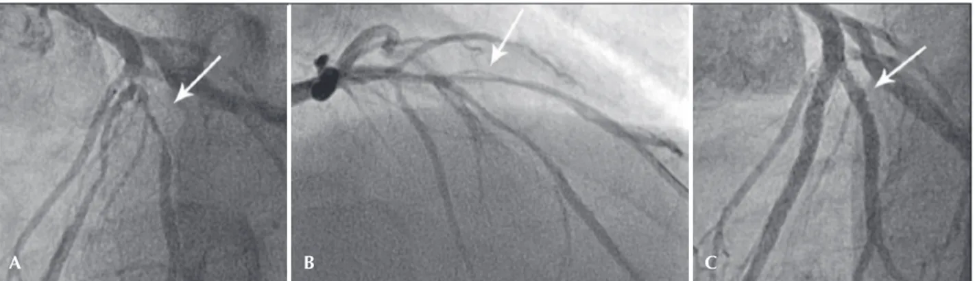
During the procedure, 8.5% of the lesions (5/59) showed side branch predilation failure due to dissection (4/5) or signiicant residual stenosis (1/5). Figure 2 shows
a case involving the left anterior descending artery/ diagonal branch with a lateral branch with severe and diffuse disease, which showed dissection after predilation and was treated with a mini-crush technique. Of the ive patients with side branch predilation failure, four were treated with a 2-stent strategy (T-stenting technique, two; mini-crush, two). The other patient with side branch predilation failure had severe lesion calciication, which proved to be non-responsive to successive dilation attempts with a balloon catheter; thus, he was ultimately treated with the single-stent technique, as it was not possible to deliver the stent to that branch. The pa-tients who achieved successful side branch predilation (54/59) were treated with a 2-stent technique in 59.3% (n = 32) and a single stent in 40.7% of cases (n = 22). Simultaneous post-dilation with a kissing-balloon was per-formed in 100% of the lesions that showed unsuccessful side branch predilation vs. 96.3% in successfully dilated lesions. At the end of the procedure, the angiographic success rate was 80% (4/5) in the group without side branch predilation success (one lesion showed signiicant residual stenosis of the side branch) vs. 98.2% (53/54) in the group with successful side branch predilation, in which one lesion showed side branch occlusion after
TABLE 1
Baseline Clinical Characteristics of Patients
Variable n = 59
Mean age, years Female gender, n (%) Diabetes mellitus, n (%) Using insulin
Arterial hypertension, n (%) Dyslipidemia, n (%) Smoking, n (%) Previous Current
Previous AMI (> 30 days), n (%) Previous PCI, n (%)
Previous CABG, n (%) Family history of CAD, n (%) History of CHF, n (%) Previous CVA, n (%)
Renal failure (serum basal creatinine ≥ 1.5 mg/dL), n (%)
61.2 ± 11 15 (25.4) 18 (30.1) 5 (8.5) 51 (86.4) 42 (71.2)
39 (66.1) 11 (18.6) 23 (39) 15 (25.4)
1 (1.7) 34 (57.6)
6 (10.2) 4 (6.8) 7 (11.9)
CVA = cerebrovascular accident; CABG = coronary artery bypass graft; CAD = coronary artery disease; AMI = acute myocardial infarction; CHF = congestive heart failure; PCI = percutaneous coronary intervention; n = number of patients.
TABLE 2
Angiographic Characteristics of Patients
Variable n = 59
Target-vessel, n (%) LAD
LCx RCA
Lesion location
(coronary segment), n (%) Proximal
Middle Distal
Medina18 classiication, n (%) 1,1,1
1,0,1 0,1,1
Eccentricity (MV/SB), n (%) Moderate to severe calciication (MV/SB), n (%)
Pre-procedural TIMI low 3 (MV/SB), n (%)
51 (86.4) 7 (11.9)
1 (1.7)
40 (67.8) 15 (25.4) 4 (6.8)
36 (61) 4 (6.8) 19 (32.2) 40 (67.8)/27 (45.8) 33 (55.9)/26 (44.1)
56 (94.9)/58 (98.3)
treatment with a single stent (P = 0.09). The procedural success was similar for both groups (80% vs. 87%). In the successful predilation group, seven patients had MACE/bleeding, including periprocedural AMI (n = 6), and major bleeding (n = 1) associated with the use of a glycoprotein IIb / IIIa inhibitor.
The comparisons of the QCA data of the main vessels and side branches are shown in Tables 3 and 4, respectively. In general, the group with unsuccessful
side branch predilations had more extensive side branch lesions, a smaller minimal lumen diameter, and more severe obstruction compared with the side branches of the group with successful predilations. The multivariate analysis evaluated the presence of calciication, distal and proximal angles, eccentricity, lesion extension, reference vessel diameter, minimal lumen diameter, and stenosis diameter percentages. The only signiicant predictor of side branch predilation failure was the percentage of diameter stenosis at the ostium of the
A B C
Figure 2 – Image of a ‘true’ coronary bifurcation lesion located in the proximal segment of the anterior descending artery (A), which showed dissection of the diagonal branch after predilation with a balloon-catheter (B), treated with a 2-stent (mini-crush) technique with good angiographic results (C).
TABLE 3
Quantitative Coronary Angiography of the Main Vessel Comparing Lesions with Successful vs. Unsuccessful Predilation
Variable
SB Predilation
Unsuccessful (n = 5)
Successful
(n = 54) P
Pre-procedural Lesion lenght, mm RD, mm
MLD, mm DS, %
Post-procedural RD, mm Intra-segment MLD, mm DS, %
Acute gain, mm Intrastent MLD, mm DS, % Acute gain, mm
20.18 ± 10.17 2.94 ± 0.4 0.58 ± 0.37
80 ± 11.7
2.92 ± 0.2
2.39 ± 0.19 21.2 ± 0.04 1.82 ± 0.43
2.67 ± 0.46 17.2 ± 5.2 2.09 ± 0.38
21.2 ± 7.91 3.01 ± 0.39 0.75 ± 0.36 75 ± 11.7
3.04 ± 0.38
2.47 ± 0.37 18.7 ± 0.09 1.75 ± 0.49
2.7 ± 0.36 16.4 ± 0.07 1.96 ± 0.49
0.85 0.75 0.42 0.45
0.29
0.42 < 0.001
0.75
0.9 0.75 0.48
Values expressed as the means ± SD.
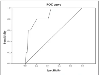
side branch by QCA analysis (odds ratio 1.15 for each percentage unit; 95% conidence interval: 1.01–1.30; P = 0.04). The analysis of the ROC curve (Figure 3) with a cut-off value of the side branch percent diameter stenosis > 87.6% before the procedure was identiied as a predictor of side branch predilation failure, with an accuracy of 86.5%.
DISCUSSION
In the current analysis, a technical approach was proposed with side branch predilation prior to stenting implant in bifurcation lesions. However, the systematic performance of such a procedure has been the subject of controversy, as lumen enlargement at the side branch ostium location can lead to dissections, which can hinder or even prevent access to the side branch through the stent struts in cases of treatment with the provisional strategy.10 Another issue has been the possibility that side branch predilation promotes guidewire recrossing in the side branch through the proximal struts, which
could also compromise, at least in theory, the structural stent conformation in the main vessel in relation to the side branch origin.19 Conversely, side branch predilation has been indicated in cases where access to the side branch is dificult or when the stenosis is severe and calciied.10,19 In these situations, such a procedure may facilitate access to the side branch, while providing an increased vascular lumen. In general, the dificulty or impossibility of access (or recrossing) to the side branch remains a limiting factor for PCI in coronary bifurcation lesions. Previous studies have identiied several predic-tors of poor access to the side branch, which include signiicant calciication involving the proximal segment of the main vessel or the side branch, severe stenosis with massive plaque build-up in the proximal segment of the main vessel, excessive tortuosity in the proximal segment of the bifurcation lesion, distal angle > 90 degrees, and compromised side branch low.20,21
The present study included only coronary bifurcation lesions with signiicant side branch impairments, with TABLE 4
Quantitative Coronary Angiography of the Side Branch Comparing Lesions with Successful vs. Unsuccessful Predilation
Variable
SB Predilation
Unsuccessful (n = 5)
Successful
(n = 54) P
Pre-procedural Lesion extension, mm RD, mm
MLD, mm SD, %
Post-procedural RD, mm Intra-segment MLD, mm DS, %
Acute gain, mm Intrastent* MLD, mm DS, %
Acute gain, mm Ostium 5 mm MLD, mm DS, %
Acute gain, mm
12.62 ± 8.77 2.53 ± 0.66
0.5 ± 0.58 82.4 ± 18.5
2.52 ± 0.42
1.7 ± 0.75 33.2 ± 31.5
1.2 ± 0.97 (n = 4)* 2.09 ± 0.26
17 ± 17.9 1.53 ± 0.76
1.87 ± 0.82 32.5 ± 34.4 1.37 ± 0.89
9.92 ± 5.71 2.58 ± 0.58 0.73 ± 0.4 71.4 ± 15.1
2.63 ± 0.51
1.9 ± 0.48 29.1 ± 18.2 1.17 ± 0.61 (n = 32)* 2.31 ± 0.29 19.3 ± 11.4 1.65 ± 0.51
2.02 ± 0.56 27.1 ± 18.5 1.3 ± 0.67
0.55 0.89 0.45 0.29
0.61
0.64 0.82 0.95
0.13 0.8 0.76
0.72 0.75 0.88
Values expressed as the means ± SD.
* Number of lesions treated with side branch stenting, i.e., two-stent technique.
lesions extending beyond the ostium. The pre-procedural angiographic analysis of the side branch showed mean values of the lesion extension and degree of obstruction in patients in the randomised study arms (group with successful side branch predilation) of 9.9 mm and 71%, respectively. The group with unsuccessful side branch predilation had more complex lesions, including a mean lesion length and degree of obstruction of the side branch of 12.6 mm and 82%, respectively. Nonetheless, these differences were not signiicant, which was likely due to the small sample size. Additionally, 44% of lesions had moderate to severe calciication in the side branch. Previous studies observed that the risk of occlusion of the side branch is proportional to the degree of stenosis. An analysis by Chaudhry et al.22 demonstrated that for each increase of 10 percentage points in the degree of stenosis, the risk of side branch impairment increased by 23% during a percutaneous approach with single stenting. In the present study, the presence of calciication was also associated with a compromised side branch (P = 0.06). It is noteworthy that the side branch involvement was associated with the occurrence of periprocedural myocardial infarction.
Therefore, the use of a technical approach with systematic side branch predilation in the present study is justiied by the complexity of the included lesions. Furthermore, the side branch predilation failure in this situation could lead to early identiication of the morphologies that are unsuitable for treatment with a provisional strategy, as changing to the 2-stent strategy can be dificult, and there is no guarantee of success, especially due to side branch involvement.20 In such cases, techniques that allow for a primary stent implant in the side branch, such as crush or Culotte stenting, should be preferred.11 In the present study, side branch predilation failure occurred in 8.5% of cases, and the only independent predictor identiied in the multivariate analysis was the degree of stenosis of the side branch.
Speciically, a degree of stenosis of 87.6% was identiied as a poor response predictor cut-off of the side branch predilation. However, further studies that are properly designed and have adequate statistical power are needed to completely evaluate the impact of side branch predi-lation on complex coronary bifurcation lesions.
CONCLUSIONS
The side branch predilation procedure in complex coronary bifurcation lesions treated by PCI was as-sociated with relatively low immediate side branch failure rates (< 10%). Nonetheless, this procedure was able to identify at an early stage certain morpholo-gies that are most likely inadequate for the provi-sional strategy approach, which must be considered when selecting the PCI technique in this complex subgroup. In the multivariate analysis, the degree of side branch stenosis before the procedure was the only independent predictor of the predilation failure of the identified side branch.
CONFLICTS OF INTEREST
The authors declare no conlicts of interest.
REFERENCES
1. Tsuchida K, Colombo A, Lefèvre T, Oldroyd KG, Guetta V, Guagliumi G, et al. The clinical outcome of percutaneous treatment of bifurcation lesions in multivessel coronary artery disease with the sirolimus-eluting stent: insights from the Ar-terial Revascularization Therapies Study Part II (ARTS II). Eur Heart J. 2007;28(4):433-42.
2. Meier B, Gruentzig AR, King SB 3rd, Douglas JS Jr, Hollman J, Ischinger T, et al. Risk of side branch occlusion during coronary angioplasty. Am J Cardiol. 1984;53(1):10-4.
3. Weinstein JS, Baim DS, Sipperly ME, McCabe CH, Lorell BH. Salvage of branch vessels during bifurcation lesion angioplasty: acute and long-term follow-up. Cathet Cardiovasc Diagn. 1991;22(1):1-6.
4. Melikian N, Di Mario C. Treatment of bifurcation coronary lesions: a review of current techniques and outcome. J Interv Cardiol. 2003;16(6):507-13.
5. Yamashita T, Nishida T, Adamian MG, Briguori C, Vaghetti M, Corvaja N, et al. Bifurcation lesions: two stents versus one stent-immediate and follow-up results. J Am Coll Cardiol. 2000;35(5):1145-51.
6. Hildick-Smith D, de Belder AJ, Cooter N, Curzen NP, Clayton TC, Oldroyd KG, et al. Randomized trial of simple versus complex drug-eluting stenting for bifurcation lesions: the British Bifurcation Coronary Study: old, new, and evolving strategies. Circulation. 2010;121(10):1235-43.
7. Costa RA, Moussa ID. Percutaneous treatment of coronary bifurcation lesions in the era of drug-eluting stents. Minerva Cardioangiol. 2006;54(5):577-89.
8. Costa RA, Kyono H, Costa M, Russell M, Moussa ID. Coronary artery bifurcation lesions: anatomy. In: Moussa ID, Colombo A, editors. Tips and tricks in interventional therapy of coronary bifurcation lesions. London: Informa Healthcare; 2010. 9. Latib A, Colombo A, Sangiorgi GM. Bifurcation stenting:
current strategies and new devices. Heart. 2009;95(6):495-504. Figure 3 – Receiver operating characteristic (ROC) curve constructed
to evaluate the cut-off of the percentage of the side branch stenosis before the procedure to more accurately predict predilation failure.
Specificity ROC curve
1.0
1.0 0.8
0.8 0.6
0.6 0.4
0.4 0.2
0.2 0.0
0.0
Sensiti
10. Lefèvre T, Darremont O, Albiero R. Provisional side branch stenting for the treatment of bifurcation lesions. EuroInterven-tion. 2010;6 Suppl J:J65-71.
11. Latib A, Moussa I, Sheiban I, Colombo A. When are two stents needed? Which technique is the best? How to perform? EuroIntervention. 2010;6 Suppl J:J81-7.
12. Hildick-Smith D, Lassen JF, Albiero R, Lefèvre T, Darremont O, Pan M, et al. Consensus from the 5th European Bifurcation Club Meeting. EuroIntervention. 2010;6(1):34-8.
13. Costa RA. Achados ultrassonográicos em lesões de bifurcação coronária tratadas com stent único versus estratégia com dois stents [tese de doutorado]. São Paulo: Instituto Dante Pazzanese de Cardiologia, Universidade de São Paulo; 2011.
14. Serruys PW, Ong AT, Piek JJ, Neumann FJ, van der Giessen WJ, Wiemer M, et al. A randomized comparison of a durable polymer everolimus-eluting stent with a bare metal coronary stent: the spirit irst trial. EuroIntervention. 2005;1(1):58-65. 15. King SB 3rd, Smith SC Jr, Hirshfeld JW Jr, Jacobs AK, Morrison
DA, Williams DO, et al. 2007 focused update of the ACC/ AHA/SCAI 2005 guideline update for percutaneous coronary intervention: a report of the American College of Cardiology/ American Heart Association Task Force on Practice Guidelines: 2007 Writing Group to Review New Evidence and Update the ACC/AHA/SCAI 2005 Guideline Update for Percutaneous Coronary Intervention, Writing on Behalf of the 2005 Writing Committee. Circulation. 2008;117(2):261-95.
16. Chesebro JH, Knatterud G, Roberts R, Borer J, Cohen LS, Dalen J, et al. Thrombolysis in Myocardial Infarction (TIMI) Trial, Phase I: a comparison between intravenous tissue plasminogen acti-vator and intravenous streptokinase. Clinical indings through hospital discharge. Circulation. 1987;76(1):142-54.
17. Lansky A, Tuinenburg J, Costa M, Maeng M, Koning G, Popma J, et al. Quantitative angiographic methods for bifurcation lesions: a consensus statement from the European Bifurcation Group. Catheter Cardiovasc Interv. 2009;73(2):258-66.
18. Medina A, Suarez de Lezo J, Pan M. A new classiication of coronary bifurcation lesions. Rev Esp Cardiol. 2006; 59(2):183.
19. Albiero R, Boldi E. Provisional stenting technique for non-left main coronary bifurcation lesions: patient selection and technique. In: Moussa I, Colombo A, editors. Tips and tricks in interventional therapy of coronary bifurcation lesions. London: Informa Healthcare; 2010.
20. Burzotta F, De Vita M, Sgueglia G, Todaro D. How to solve dificult side branch access? EuroIntervention. 2010;6 Suppl J:J72-80. 21. Costa RA, Costa MA, Moussa ID. Bifurcation lesion
morpho-logy and intravascular ultrasound assessment. Int J Cardiovasc Imaging. 2011;27(2):189-96.