Cop
yright
© ABE&M t
odos os dir
eit
os r
eser
vados
.
149 Arq Bras Endocrinol Metab. 2012;56/2
case report
Does undetectable basal Tg
measured with a highly sensitive
assay in the absence of antibodies
and combined with normal
ultrasonography ensure the
absence of disease in patients
treated for thyroid carcinoma?
Tireoglobulina basal indetectável medida com ensaio ultrassensível na ausência de anticorpos e combinada com ultrassonograia normal assegura ausência de doença em pacientes tratados de câncer de tireoide?
Pedro Weslley Rosário1, Augusto Flávio Campos Mineiro Filho1
SUMMARY
It has been proposed that, in patients treated for well-differentiated thyroid carcinoma, unde-tectable basal thyroglobulin (Tg) levels measured with a highly sensitive assay in the absence of anti-thyroglobulin antibodies (TgAb) and combined with negative neck ultrasonography (US) ensured the absence of disease. We report a series of ive patients with well-differentiated (pa-pillary) carcinoma submitted to total thyroidectomy with apparently complete tumor resection, followed by remnant ablation with 131I (100-150 mCi), who had no distant metastases upon initial post-therapy whole-body scanning. When tumor recurrence or persistence was detected, these patients presented undetectable basal Tg (0.1 ng/mL) in the absence of TgAb, and US sho-wed no anomalies. Two patients had lymph node metastases, one had mediastinal metastases, bone involvement was observed in one patient, and local recurrence in one. We conclude that further studies are needed to deine in which patients undetectable basal Tg (negative TgAb) combined with negative US is suficient, and no additional tests are required. Arq Bras Endocrinol Metab. 2012;56(2):149-51
SUMÁRIO
Tem sido proposto que em pacientes tratados de carcinoma bem diferenciado da tireoide o encontro de valores basais indetectáveis de tireoglobulina (Tg), dosada por ensaios ultrassen-síveis, na ausência de anticorpos antitireoglobulina (TgAc), e combinado à ultrassonograia (US) cervical negativa, asseguraria ausência de doença. Reportamos aqui uma série de cinco pacientes com carcinoma bem diferenciado (papilífero), submetidos à tireoidectomia total, com ressecção tumoral aparentemente completa, seguida da ablação de remanescentes com 131I (100-150 mCi), sem metástases distantes na pesquisa de corpo inteiro pós-dose inicial, que, na ocasião em que a recorrência ou persistência tumoral foi detectada, apresentavam Tg basal indetectável (0.1 ng/ml), TgAc negativos e US sem anormalidades. Dois pacientes tinham me-tástases linfonodais, um tinha mediastinal, outro acometimento ósseo e um recorrência local. Concluímos que mais estudos são necessários para a deinição de que pacientes com Tg basal indetectável (sem TgAc) combinada à US sem anormalidades seria suiciente, dispensando tes-tes adicionais. Arq Bras Endocrinol Metab. 2012;56(2):149-51
Correspondence to: Pedro Weslley Rosário
Núcleo de Pós-Graduação da Santa Casa de Belo Horizonte Av. Francisco Sales, 1111, 8ºC 30150-221 – Belo Horizonte, MG, Brazil
Received on Jul/18/2011 Accepted on Feb/24/2012
1 Núcleo de Pós-Graduação da
Cop
yright
© ABE&M t
odos os dir
eit
os r
eser
vados
.
150 Arq Bras Endocrinol Metab. 2012;56/2
False-negative result of highly sensitive thyroglobulin
INTRODUCTION
M
easurement of serum thyroglobulin (Tg) is con-sidered to be the most sensitive method for the detection of persistent or recurrent disease in patients with differentiated thyroid carcinoma after initial thera-py. Obviously, the sensitivity of this marker is limited in patients with poorly differentiated tumors that do not secrete Tg, or in patients with circulating anti-thyro-globulin antibodies (TgAb) that interfere with serum Tg measurement (false reduction). In addition, positive serum Tg result depends on the secretion of suficient amounts, which can be detected by the assay. The use of highly sensitive assays enables the detection of even minimal amounts of Tg in the circulation, which would not be identiied by traditional assays. The exceptional cases of persistent/recurrent disease, in which basal (no TSH stimulation) Tg levels may be undetectable with these assays, would be due to small cervical lymph node metastases or local recurrence (1-7), which could be identiied by ultrasonography (US). Therefore, the observation of undetectable basal Tg levels measured by highly sensitive assays in patients with well-differen-tiated tumors, in the absence of circulating TgAb and combined with negative US, ensures the absence of disea se at that time, and as along as the results continue the same (1,2,4-6,8). We report here a series of ive patients who exhibited tumor persistence or recurrence despite the presence of this combination of indings.CASE REPORT
All patients had well-differentiated thyroid carcinoma (papillary type) and had undergone total
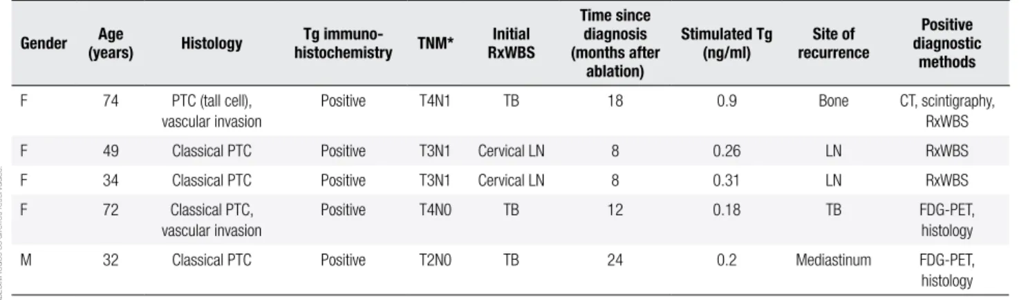
thyroidec-tomy with apparently complete tumor resection, follo-wed by remnant ablation with 131I (3.7-5.5 GBq). Initial post-therapy whole-body scanning (RxWBS) re-vealed no distant metastases. When tumor persistence or recurrence was detected, all patients presented unde-tectable basal Tg (0.1 ng/ml) in the absence of TgAb, and US showed no anomalies. The characteristics of each patient are shown in Table 1.
Tg was measured with a highly sensitive chemilumi-nescent assay (Tg Access, Beckman Coulter, Fullerton, CA), with functional sensitivity of 0.1 ng/mL. This as-say had been used in recent series (1,2,4,5,7). TgAb were determined by a chemiluminescent assay (Immu-lite 2000), with a detection limit of 20 IU/mL, and reference value of up to 40 IU/mL.
Ultrasonography was performed with a linear multi-frequency 10-MHz transducer. RxWBS was performed with a therapeutic dose of 131I (3.7-5.5 GBq), after stimulation with recombinant human TSH (rhTSH) and administration of a low-iodine diet for the 10 days before the examination. Anterior and posterior whole--body images were obtained 7 days after iodine admi-nistration. FDG-PET was also carried out after stimu-lation with rhTSH. Computed tomography (CT) was performed on 5-10 mm sequential sections.
DISCUSSION
Several studies have evaluated the performance of hi-ghly sensitive Tg assays in patients treated for differen-tiated thyroid carcinoma without circulating TgAb. All of these series included patients with persistent/recur-rent disease in the presence of undetectable basal Tg (1-7). Therefore, the observation of undetectable basal
Table 1. Characteristics and test results of the patients
Gender (years)Age Histology histochemistryTg immuno- TNM* RxWBSInitial
Time since diagnosis (months after
ablation)
Stimulated Tg (ng/ml)
Site of recurrence
Positive diagnostic
methods
F 74 PTC (tall cell), vascular invasion
Positive T4N1 TB 18 0.9 Bone CT, scintigraphy,
RxWBS
F 49 Classical PTC Positive T3N1 Cervical LN 8 0.26 LN RxWBS
F 34 Classical PTC Positive T3N1 Cervical LN 8 0.31 LN RxWBS
F 72 Classical PTC,
vascular invasion
Positive T4N0 TB 12 0.18 TB FDG-PET,
histology
M 32 Classical PTC Positive T2N0 TB 24 0.2 Mediastinum FDG-PET,
histology
* All M0.
Cop
yright
© ABE&M t
odos os dir
eit
os r
eser
vados
.
151 Arq Bras Endocrinol Metab. 2012;56/2
False-negative result of highly sensitive thyroglobulin
Tg even in the absence of TgAb, and measured with a highly sensitive assay does not rule out the need for imaging methods. Considering that these false-negative cases are cervical lymph node metastases or local re-currence (1-7), ultrasonography is the complementary method of choice. Even this combination (undetec-table basal Tg measured with a highly sensitive assay, negative TgAb, and normal ultrasonography) does not ensure the absence of long-term recurrence (6,7). Mo-reover, these cases of recurrence have been described even when Tg continued to be undetectable (6,7), su-ggesting that follow-up using exclusively basal Tg and TgAb is not suficient, even when the measurements are performed with new assays. Periodic ultrasonography is a valuable tool for the identiication of these cases (6,7).
More interestingly, Giovanella and cols. (3) reported the case of a patient with undetectable basal Tg, nega-tive TgAb and normal ultrasonography who had local recurrence detected by FDG-PET. Similarly, Brassard and cols. (7) reported the case of a patient who was diagnosed with distant (bone) metastases (not identi-iable by ultrasonography), although Tg continued to be undetectable and TgAb continued to be negative. These cases may be added to those reported here, in which the disease was detected even in patients with undetectable basal Tg measured with a highly sensitive assay and had negative TgAb, with ultrasonography showing no disease. It should be noted that the patients described by those authors (3,7), and the patients re-ported here had well-differentiated thyroid carcinoma, and immunohistochemistry for Tg was positive in the present series, a inding demonstrating the initial ability of the tumor to produce this protein.
It is recognized that these cases (metastases in pa-tients with undetectable basal Tg measured with a highly sensitive assay – without TgAb – combined with negative ultrasonography) are not common, and that the best approach to identify them (Tg stimulation, FDG-PET or others) is a challenge. Diagnostic whole-body scanning is clearly not an interesting option for these patients with negative basal Tg (9). These cases show that, in addition to the major concern related to their lower speciicity (10), the use of highly sensitive Tg assays also requires further studies investigating their sensitivity and the necessary complementary in-vestigation in cases of undetectable basal Tg. In fact,
the Latin American Thyroid Society (LATS) does not recommend the routine use of low functional sensitivity Tg assays until more evidence is published regarding their eficacy (10).
We conclude that further studies are needed to de-ine for which patients undetectable basal Tg (negative TgAb) combined with negative neck ultrasonography is suficient, and no additional tests are required.
Disclosure: no potential conlict of interest relevant to this article was reported.
REFERENCES
1. Rosario PW, Purisch S. Does a highly sensitive thyroglobulin (Tg) assay change the clinical management of low-risk patients with thyroid cancer with Tg on T4 < 1 ng/ml determined by traditional assays? Clin Endocrinol. 2008;68:338-42.
2. Iervasi A, Iervasi G, Ferdeghini M, Solimeo C, Bottoni A, Rossi L, et al. Clinical relevance of highly sensitive Tg assay in monitoring patients treated for differentiated thyroid cancer. Clin Endocrinol. 2007;67:434-41.
3. Giovanella L, Ceriani L, Ghelfo A, Mafioli M, Keller F, Spriano G. Thyroglobulin assay during thyroxine treatment in low-risk dif-ferentiated thyroid cancer management: comparison with recom-binant thyrotropin stimulated assay and inaging procedures. Clin Chem Lab Med. 2006;44:648-52.
4. Castagna MG, Tala Jury HP, Cipri C, Belardini V, Fioravanti C, Pasqui L, et al. The use of ultrasensitive thyroglobulin assays reduces but not abolishes the need for TSH stimulation in pa-tients with differentiated thyroid carcinoma. J Endocrinol Invest. 2011;34(8):e219-23. Epub 2011 Mar 7.
5. Smallridge RC, Meek S, Morgan MA, Gates GS, Fox TP, Grebe S, et al. Monitoring thyroglobulin in a sensitive immunoassay has comparable sensitivity to recombinant human TSH-stimulated thyroglobulin in follow-up of thyroid cancer patients. J Clin Endo-crinol Metab. 2007;92:82-7.
6. Giovanella L, Mafioli M, Ceriani L, De Palma D, Spriano G. Unsti-mulated high sensitive thyroglobulin measurement predicts out-come of differentiated thyroid carcinoma. Clin Chem Lab Med. 2009;47:1001-4.
7. Brassard M, Borget I, Edet-Sanson A, Giraudet AL, Mundler O, Toubeau M, et al. Long-term follow-up of patients with papillary and follicular thyroid cancer: a prospective study on 715 patients. J Clin Endocrinol Metab. 2011;96:1352-9.
8. Zöphel K, Wunderlich G, Smith BR. Serum thyroglobulin mea-surements with a high sensitivity enzyme-linked immunosorbent assay: is there a clinical beneit in patients with differentiated thy-roid carcinoma? Thythy-roid. 2003;13:861-5.
9. Rosario PW. Câncer de tireóide: seguimento com radioiodo. In: Volpi E, Friguglietti C, Kulcsar MA. Câncer da tireóide.
Aborda-gem multidisciplinar. 1th ed. São Paulo: AC Farmacêutica; 2010.
p. 280-3.