Cop
yright
© ABE&M t
odos os dir
eit
os r
eser
vados
.
Correspondence to: Ana R. Vlainich
Av. Paes de Barros, 1899, ap. 52 03115001 − São Paulo, SP, Brazil [email protected] Received on Feb/24/2010 Accepted on Mar/9/2011
Ultrasonography compared
to magnetic resonance
imaging in thyroid-associated
Graves’ ophthalmopathy
Comparação de ultrassonograia à ressonância magnética nuclear na oftalmopatia de Graves associada à tiroide
Ana R. Vlainich1, João H. Romaldini2, Ana B. Pedro2,
Chady S. Farah2, Cicero A. Sinisgalli Jr.3
ABSTRACT
Objective: To compare ultrasonography (US) to magnetic resonance imaging (MRI) and the clinical activity score (CAS) in Graves’ ophthalmopathy. Subjects and methods: Nineteen pa-tients underwent extraocular muscle thickness measurements by US and MRI, reflectivity by US and signal-intensity ratio by MRI. There were also twelve US control subjects. Results: US median thicknesses were greater than in controls. Correlation was found between US and MRI in the median thickness of the left eye rectus medial muscle as well as between signal-intensity ratio (SIR) and thickness by US. An inverse correlation was found between reflectivity and SIR in the inferior and lateral rectus. On associating the tests for detecting activity the best results were obtained with CAS plus MRI (sensitivity 75%), and US and MRI (positive predictive value 77% and specificity 80%). Conclusion: CAS and US results showed poor correlation with MRI results suggesting that they cannot replace each other but when combined these methods can improve the evaluation of thyroid-associated ophthalmopathy. Arq Bras Endocrinol Metab. 2011;55(3):184-8
Keywords
Graves’ disease; thyroid-associated ophthalmopathy; orbital ultrasonography; magnetic resonance imaging
RESUMO
Objetivo: Comparar a ultrassonografia (US) à ressonância magnética nuclear (RMN) e o índice de atividade clínica (IAC) na oftalmopatia de Graves. Sujeitos e métodos: Dezenove pacientes submetidos à medida da espessura dos músculos extraoculares por US e RMN, refletividade ao US e razão da intensidade de sinal (RIS) à RMN. Grupo controle para US de 12 indivíduos.
Resultados: Espessura mediana ao US foi maior que dos controles. Houve correlação entre US e RMN na espessura mediana dos músculos retos mediais dos olhos esquerdos e entre a RIS e a espessura ao US e correlação inversa entre refletividade e SIR nos retos inferior e lateral. Detectando atividade, os melhores resultados foram associando IAC e RMN (sensitividade de 75%) e US e RMN (valor preditivo positivo de 77% e especificidade de 80%). Conclusão: Re-sultados do IAC e US mostraram pouca correlação com a RMN, sugerindo que não podem ser substituídos, mas, quando combinados, esses métodos podem melhorar a avaliação da oftal-mopatia associada à tiroide. Arq Bras Endocrinol Metab. 2011;55(3):184-8
Descritores
Doença de Graves; oftalmopatia associada à tiroide; ultrassonograia orbitária; ressonância magnética nuclear 1 Ophthalmology, Services
at Hospital Servidor Publico Estadual de Sao Paulo (IAMSPE), São Paulo, SP, Brazil
2 Endocrinology, IAMSPE, São Paulo, SP, Brazil 3 Radiology, IAMSPE, São Paulo, SP, Brazil
INTRODUCTION
T
hyroid-associated ophthalmopathy (TAO) is an autoimmune disorder of the orbit associated with Graves’ disease (GD) which presents signs andCop
yright
© ABE&M t
odos os dir
eit
os r
eser
vados
.
clinical activity are associated with lymphocytic iniltra-tion and edema, whereas inactivity is associated with ibrosis (6,7), both producing proptosis (2,6,7). Treat-ments are more effective in the active phase, thus its determination is essential (1,4,7,8) but management fails in 50% of the cases probably due to the dificulty (4,6,7) in identifying the disease phase (8). The clinical activity score (CAS) is not enough to help diagnosis for this reason different imaging techniques that en-able visualization of the orbit contents and evaluation of extraocular muscles (EOM) have been tested (9-11). US enables differential diagnosis in proptosis and seems to reveal the existing inlammation (12-17). Computed tomography scan (CT) shows muscle thickness, diagno-ses compressive optical neuropathy and deines the de-gree of proptosis but does not detect activity (1,10,18). Therefore, MRI seems to be the most suitable method because it reaches the retraction of the upper eyelid (18), the activity of the muscles and the increase in orbital fat (1,4,19-23). To present, few studies have compared US with MRI in assessing TAO (20,22,24). In this study we compared TAO orbits within different activity levels using US, MRI, and CAS.
SUBJECTS AND METHODS
Nineteen patients were examined (15 women - 4 men) with uni- or binocular TAO, mean age and standard deviation were 39.3 ± 13.2 years (range 16 - 59 ye-ars), there were 6 (31%) smokers and the duration of the disease was 21.7 ± 16.4 months (3 - 60 months). CAS was assessed by the physicians (ABP and CSF) and ranged from 0 to 7, any score higher than 3 was considered to be active TAO disease. GD was diagno-sed badiagno-sed on characteristic signals and symptoms plus diffuse goiter, conirmed by suppressed serum TSH, elevated free total T4 levels, thyroid peroxidase (TPO-Ab), thyroglobulin (TgAb) and thyroid receptor an-tibodies (TRAb), and increase in thyroid radioactive iodine uptake. Hypothyroidism was diagnosed by ele-vated serum TSH and low free T4 levels. Proptosis was measured by Hertel exophthalmometer. The US con-trol group consisted of 12 age- and gender-matched subjects with normal clinical, laboratorial data, and ophthalmologic examinations. All patients underwent US and MRI, done no later than three weeks apart, with the control group submitted only to the US. Ex-clusion criteria were previous ocular surgery, problems in performing MRI (claustrophobia), or anything that
could hinder US performance (e.g. high myopia). The study was approved by the Medical Ethics Committee and written informed consent terms were obtained from all subjects.
Serum free T4 and TSH concentrations were me-asured by luoroimmunoassay (Delphia; Pharmacia), normal ranges were 0.8 to 2.0 ng/mL and 0.3 to 4.0 mU/L, respectively. TRAb was determined by TSH--receptor antibody kit supplied by RSR Ltd. (Cardiff, Wales, UK) and was expressed as the percentage of inhibition of 125-I-labelled bovine TSH binding to the TSH receptor, normal values were considered less than 11 IU/L. To determine TPOAb and TgAb, direct RIA system supplied by RSR Ltd was used and normal range was less than 1.0 IU/L.
The EOM assessed were the inferior rectus (IR),
rectus medial (RM), rectus lateral (RL) and rectus supe-rior (RS). The US was performed by the same examiner (ARV) with A&B USA Humphrey Instruments Inc. 10Mh model 837, in primary eye position, low gain, lo-garithmic curve, muscle diagonal section (2,12,25,26). Thickness (7,16) and relectivity were assessed using the maximum thickness inside the muscle capsules (12,16,17). Using printed images, the average of the height of all peaks from baseline to the top, between the external borders of the muscles, was calculated as a percentage of the sclerotic peak (12,16,17).
Orbital MRIs were analyzed by the same radiologist using a 1.5 Tesla MR unit head coil (General Electric Co., Milwaukee, USA). The pulse sequences were T1--weighted, spin-echo, axial (TR 366, TE12, 3 mm sec-tion thickness (ST)) and coronal plane (TR 500, TE 17, 4 mm ST) used for muscle thickness measurements, and T2- weighted, fast-spin-echo, axial (TR 3000, TE 103, 3 mm ST) and coronal plane (TR 300; TE98.7, 4 mm ST). Coronal plane was used to examine RS and IR and the axial plane to examine RM, RL, and ce-rebral substantia alba (SA). Furthermore, the T2 sig-nal intensity of the muscles was compared to the SA signal and expressed as a signal-intensity ratio (SIR) in order to reduce individual differences and to com-pensate for the slight variation in machine sensitivity when used on subsequent occasions. This ratio also eli-minates the need to submit the control group to MRI (10,21,24,27-29). An increase in SIR might indicate activity due to edema presence (11,18,27,28,30).
Rank-Cop
yright
© ABE&M t
odos os dir
eit
os r
eser
vados
.
-Sum, Fischer’s Exact, ANOVA and the Spearman coe-ficient. Statistical signiicance was set at 5% (P < 0.05).
RESULTS
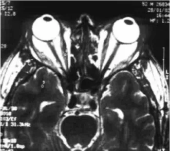
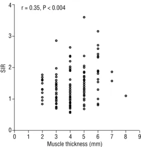
The mean CAS value of TAO patients was 2.4 ± 1.3 (range 1 to 6) and 63.1% of the patients were diagnosed as in active phase. However, using MRI 15 (78.9%) pa-tients showed activity (at least 1 muscle with SIR higher than 1.5) (21), while only 8 (42.1%) showed activity by US (relectivity smaller than 40% in at least one mus-cle). Proptosis (above 20 mm) was observed in 13 patients (68.4%) with a mean of 24.2 ± 2.2 mm (22 - 30 mm) that was signiicantly higher (P > 0.001) than in the control group (14.7 ± 2.1 mm; 12-18 mm). Figure 1 shows US image of one patient and igure 2 MRI-T2 in coronal section. Based on US measurements, there was signiicant difference using ANOVA analysis (P = 0.0006) of IR mean thickness compared to controls (Figure 3), whereas no differences were found in their respective relectivity. Correlation was found between median thicknesses RM in the left eye as measured by US and by MRI (r = 0.51; P < 0.02) (Figure 4), as well as between SIR and thickness by US (r = 0.35; P < 0.004) (Figure 5). However, there was no correla-tion between CAS or proptosis with any muscle value measured by US, MRI or smoking. Furthermore, an inverse correlation was noted between relectivity and SIR in IR (r = -0.63; P < 0.003) and RL (r = -0.39; P < 0.01) (Figure 6). An interesting analysis was obtained by associating the tests for detecting activity and the best results were achieved for CAS plus MRI reaching a sensitivity of 75%, and US and MRI with positive pre-dictive value of 77% and speciicity of 80% (Table 1).
Figure 1. Shows US B-mode image of right rectus muscle in a TAO patient.
Figure 2. Shows axial MRI image at T2 to evaluate lateral and medial rectus besides substantia alba.
MR: right medial rectus, ML: left medial rectus, LR: right lateral rectus, LL: left lateral rectus, SR: right superior rectus, SL: left superior rectus, IR: right inferior rectus, IL: left inferior rectus.
Figure 3. Significant Anova (P = 0.0006) of rectus muscle thickness measured by ultrasound in TAO patients and controls.
Figure 4. Spearman correlation between medial rectus in the left eye by ultrasound and magnetic resonance imaging in mm.
10.0
Controls TAO patients 7.5
5.0
2.5
0.0
MR ML LR LL SR SL IR IL
Muscle thickness (mm)
8
r = 0.51, P < 0.02 6
4
2
0
0.0 2.5 5.0 7.5 10.0
Muscle thickness of left medial
rectus MRI (mm)
Cop
yright
© ABE&M t
odos os dir
eit
os r
eser
vados
.
DISCUSSION
In the present study, no relationship was observed be-tween GD status (hyperthyroidism, euthyroidism, or hypothyroidism) and CAS of patients with TAO, con-irming data from other studies (3,6,7). Additionally, some authors found that ultrasound results are more sensitive than clinical exam (16,17). Prummel and cols. (12) showed that EOM in patients with TAO and active inlammation presented different relectivity, deducing that US relectivity is a reliable method to determine TAO disease activity.
Several explanations can be given for the poor cor-relation between US and MRI in EOM thickness mea-surement: external eye muscles are oval-shaped and US examinations are not made at a perfect perpendicular angle (14); low US accuracy could be due to the pre-sence of some artifacts or ibrosis often found in TAO EOM, which makes muscle delimitation very dificult (10,14,24), and the fact that MRI seems to be more accurate to visualize the back portion of the orbit (24).
Several methods have been tested to determine di-sease activity (1,25) and better response to immunosu-ppressive treatment (10). CAS helps predict response to treatment (4,8,16,27) but works better if combined with US relectivity (8-10,16,26,31). On the other hand, the orbital octreoctide scan method (10,30) and MRI are more expensive and not as readily available in healthcare facilities; furthermore, as stated by Hoh and cols. (21), MRI may fail to differentiate inactive fatty degeneration.
Similar to other studies (8,32,33), no correla-tion was found between CAS and relectivity or SIR, perhaps because CAS has wide a variation among ob-servers (1,4) or because the great majority of imaging methods assesses only the EOM and not orbital fat inlammation (4,8). We noted that combining the US and MRI resulted in signiicant increase in the positive predictive value and in speciicity. However, US does not provide the comprehensive amount of information on the EOM that is available with the use of MRI. SIR determination is considered to relect free water con-tent, thus an increase usually indicates edematous chan-ges during the active phase of TAO (23,29).
Although our data is limited by the absence of hi-ghly active orbits, CAS and US results were somehow poorly correlated to MRI results what suggests that they cannot replace each other but from a clinical point of view when used in combination these methods can
Figure 5. Spearman correlation between extraocular muscles signal-intensity ratio (SIR) by MRI and thickness measured by US.
Figure 6. Spearman inverse correlation found between US reflectivity and MRI SIR of the inferior (A) and lateral (B) rectus muscles.
Table 1. Calculating predictive positive value PpV, predictive negative value PnV, sensitivity and specificity expressed in % when associating the tests for detecting TAO activity. CAS considered > 3, reflectivity considered > 40% and SIR considered > 1.5
PpV PnV Sensitivity Speciicity
CAS plus reflexivity 44 58 25 77
CAS plus SIR 42 60 75 27
SIR plus reflexivity 77 27 25 80
r = 0.35, P < 0.004
3 4
SIR 2
1
0
0 1 2 3 4 5 6 7 8 9 Muscle thickness (mm)
100 A
B
r = -0.63, P < 0.003
r = -0.39, P < 0.01
75
50
25
0.5 1.0 1.5 2.0 2.5 3.0
1.0 1.5 2.0 SIR
Reflectivity
2.5 3.0 100
75
50
Cop
yright
© ABE&M t
odos os dir
eit
os r
eser
vados
.
help in the evaluation of this multifaceted and challen-ging disease.
Disclosure: no potential conlict of interest relevant to this article was reported.
REFERENCES
1. Bartalena L, Tanda ML. Graves’ ophthalmopathy. N Engl J Med. 2009;360:944-1001.
2. Villadolid MC, Yokoyama N, Izumi M, Nishikawa T, Kimura H, Ashi-zawa K, et al. Untreated Graves’ disease patients without clinical ophthalmopathy demonstrate a high frequency of extraocular muscle enlargement by magnetic resonance. J Clin Endocrinol Metab. 1995;80:2830-3.
3. Asman P. Ophthalmological evaluation in thyroid-associated ophthalmopathy. Acta Ophthalmol Scand. 2003;81:437-48. 4. Bartalena L, Pinchera A, Marcocci C. Management of Graves’
ophthal-mopathy: reality and perspectives. Endocr Rev. 2000;21:168-99. 5. Franzco DS, Cehn C, King G. Late reactivation of thyroid
orbitopa-thy. Clin Experiment Ophthalmol. 2004;32:46-50.
6. Wiersinga WM, Prummel MF. An evidence-based approach to the treatment of Graves’ ophthalmopathy. Endocrinol Metabol Clin North Am. 2000;29:297-319.
7. Wiersinga WM. Management of Graves’ ophthalmopathy. Nature Endocrinol Metab. 2007;3:396-404.
8. Mourits M, Koornneef I, Wiersinga WM, Prummel MF, Berghout A, Van der Gaag R. Clinical criteria for the assessment of disease ac-tivity in Graves’ ophthalmopathy: a novel approach. Br J Ophthal-mol. 1989;73:639-44.
9. Prummel MF. Graves’ ophthalmopathy: diagnosis and manage-ment. Eur J Nucl Med. 2000;27:373-6.
10. Kahaly GI. Imaging in thyroid-associated orbitopathy. Eur J Endo-crinol. 2001;145:107-18.
11. Bartley GB, Gorman CA. Diagnostic criteria for Graves’ ophthal-mopathy. Am J Ophthalmol. 1995;119:792-5.
12. Prummel MF, Suschulten MSA, Wersinga WM, Verbeek AM, Mourits MP, Koornneef L. A new ultrasonographic method to detect disease activity and predict response to immunosup-pressive treatment in Graves’ ophthalmopathy. Ophthalmology. 1993;199:556-61.
13. Char DH, Norman D. The use computed tomography and ultraso-nography in the evaluation of orbital masses. Surv Ophthalmol. 1982;27:49-63.
14. Byrne SF, Gendron EK, Glaser JS, Feuer W, Aha H. Diame-ter of normal extraocular in echography. Am J Ophthalmol. 1991;112:706-13.
15. Given-Wilson R, Pope RM, Mitchell MJ, Cannon R, McGregor A. The use of real-time orbital ultrasound in Graves’ ophthalmopa-thy: a comparison with computed tomography. Br J Radiology. 1989;62:705-9.
16. Gerding MM, Prummel MF, Wiersinga WM. Assessment of disea-se activity in Graves’ ophthalmopathy by orbital ultrasonography and clinical parameters. Clin Endocrinol. 2000;52:641-6. 17. Sabetti I, Toscano A, Specchia G, Balestrazz E. Alterations of the
internal reflectivity of extra-ocular muscles associated with
seve-ral clinical stages of Graves’ ophthalmopathy. Ophthalmologica. 1998;212(suppl I):107-9.
18. Goodall KL, Jackson A, Leatherbarrow B, Whitehouse RW. Enlar-gement of the tensor intermuscularis muscle in Graves’ ophthal-mopathy. A computed tomographic and magnetic resonance imaging study. Arch Ophthalmol. 1995;113:1286-9.
19. Hosten N, Sander B, Cordes M, Schubert CJ, Schorner W, Felix R. Graves’ ophthalmopathy: magnetic resonance imaging of the or-bits. Radiology. 1989;172:759-62.
20. Polito E, Leccisotti A. Magnetic resonance imaging in Graves’ orbitopathy: recognition of enlarged muscles and prediction of steroid response. Ophthalmologica. 1995;209:182-6.
21. Hoh HB, Laitt RD, Wakeley C, Kabala J, Goddard P. The STIR se-quence magnetic resonance imaging in the assessment of extra-ocular muscles in thyroid eye disease. Eye. 1994;8:506-10. 22. Lennerstrand G, Tiam S, Isberg B, Landau Högbeck I, Bolzani R,
Tallstedt L, et al. Magnetic resonance imaging and ultrasound measurements of extraocular muscles in thyroid-associated ophthalmopathy at different stages of the disease. Acta Ophthal-mol Scand. 2007;85:192-201.
23. Taoka T, Iwasaki S, Uchida H, Fukusumi A, Kichikawa K, Nakagawa H, et al. Enhancement pattern of normal extraocular muscles in dy-namic contrast-enhanced magnetic resonance imaging with fat suppression. Acta Radiologica. 2000;41:211-6.
24. Nagy VE, Toth J, Kaldi I, Damjanovich J, Mezosi E, Lenkey A, et al. Graves’ ophthalmopathy: eye muscle involvement in patients with diplopia. Eur J Endocrinol. 2000;142:591-7.
25. Prummel MF, Wiersinga WM. Immunomodulatory treatment of Graves’ ophthalmopathy. Thyroid. 1998;8:545-8.
26. Erickson BA, Harris G, Lewandowski MF, Murray KJ, Massaro BM. Echographic monitoring of response of extraocular muscles ir-radiation in Graves’ ophthalmopathy. Int J Radiation Oncology. 1995;31:651-60.
27. Stamato FJC, Maciel RMB, Manso PG, Wolosker AMB, Paiva ER, Lopes AC, et al. Colchicine in the treatment of the inflammatory phase of Graves’ ophthalmopathy: a prospective and randomized trial with prednisone. Arq Bras Oftalmol. 2006;69(6):811-6. 28. Troelstra A, Rijneveld WJ, Kooijman AC, Houtman WA.
Correla-tion between magnetic resonance scans of extraocular muscles and clinical symptoms in Graves’ ophthalmopathy. Doc Ophthal-mol. 1988;70:243-9.
29. Utech CI, Khatibnia U, Winter PF, Wulle KG. MR T2 relaxation time for the assessment of retrobulbar inflammation in Graves’ ophthalmopathy. Thyroid. 1995;5:185-93.
30. Gerding MM, Van der Zant FM, Van Royen EA, Koornneef L, Kren-ning EP, Wiersinga WM, et al. Octreotide-scintigraphy is a disease activity parameter in Graves’ ophthalmopathy. Clin Endocrinol. 1999;50:373-9.
31. Yanik B, Conkbayir I, Acaroglu G, Heckimoglou B. Graves’ ophthal-mopathy: comparison of the doppler sonography parameters with the clinical activity score. J Clin Ultrasound. 2005;33:375-80. 32. Delint PJ, Mouritis MPh, Kerlen CH, Scheenloop JJ, Wittebol-Post D. B-scan ultrasonography in Graves’ orbitopathy. Doc Ophthalmol. 1993;85:1-4.