ABSTRACT
ORIGINAL AR
Antonio Alberto Nogueira
Julio César Rosa e Silva
Ana Carolina Japur de Sá Rosa e Silva
Marcos Dias de Moura
CA-125 levels in women
with pelvic endometriosis
Gynecology and Obstetrics Department, Faculdade de Medicina de
Ribeirão Preto, Universidade de São Paulo, Ribeirão Preto, São Paulo,
Brazil
INTRODUCTION Endometriosis is characterized by the pres-ence of endometrial tissue, consisting of glands and/or stroma located outside the uterus. The incidence of the disorder in infertile patients ranges from 10 to 25%, with prevalence increasing to 60 to 70% in cases of chronic pelvic pain, and it may be present in 1 to 20% of asymptomatic women.1
One of the diagnostic markers already well established is CA-125, a high molecular weight glycoprotein (> 20,000 Da) of epi-thelial origin found in normal cells,2,3 and
which is produced in the celomic epithe-lium during embryonic development.4 The
OC-125 marker, a monoclonal antibody that recognizes an antigenic determinant in CA-125, was developed thereafter. In adult tissues, CA-125 has been detected in normal and neoplastic epithelium of celomic origin such as endometrium, endocervix, epithe-lial cells of Fallopian tubes and cancerous epithelial cells of the ovary. The fi rst study of this marker in patients with endometriosis detected elevated serum levels.5 The elevation
of this marker in women with endometriosis is due to its higher concentration in ectopic than in entopic endometrium. This increase is also due to infl ammatory reactions, which alter endothelial permeability, thereby allowing the marker to reach the circulation.2
Koninckx & Martin, in a study of CA-125 levels in patients with pelvic endometriosis6
concluded that superfi cial disease leads to a greater increase in this marker in peritoneal fl uid, whereas in deep disease CA-125 levels are higher in blood and lower in peritoneal fl uid. Abrão et al. evaluated serum CA-125, C-reactive protein, amyloid A protein and anticardiolipin antibodies during the men-strual phase and the middle follicular phase, and concluded that CA-125 was the marker found to be present at highest levels in patients
with advanced endometriosis when measured during the menstrual phase.1
Peritoneal fl uid CA-125 levels are higher than serum CA-125, but no signifi cant dif-ference is observed between the peritoneal fl uid levels of CA-125 in women with and without endometriosis7,8 Levels of CA-125
in peritoneal fl uid seem to be a more sensitive indicator of disease than the level in serum, and the measurement of CA-125 in peritoneal fl uid could be useful in the detection of early-stage endometriosis.9-11 The limited number
of studies combining peritoneal and serum samples and the small number of cases studied encouraged us to reevaluate the concentrations of CA-125 in peritoneal fl uid and serum in women with and without endometriosis.
OBJECTIVE The objective of the present study was to correlate CA-125 and 17β-estradiol (E2) levels in serum and peritoneal fl uid from women with and without pelvic endometriosis.
MATERIALS AND METHODS
Study design Study design
This was a prospective cross-sectional study.
Setting Setting
The study was conducted in the university hospital of Faculdade de Medicina de Ribeirão Preto, Universidade de São Paulo, which is a tertiary-level public hospital.
Subjects Subjects
Fifty-two consecutive women who un-derwent laparoscopy during the early fol-licular phase (because of clinical indication of infertility, pelvic pain or tubal ligation) were selected and divided into two groups. The endometriosis group consisted of 35 women
CONTEXT AND OBJECTIVE: One of the diag-nostic markers of endometriosis is CA-125, and elevated levels of this are caused by high concentrations in the ectopic endometrium. The objective of this study was to correlate CA-125 levels in serum and peritoneal fl uid from women with and without pelvic endometriosis.
DESIGN AND SETTING:This was a prospec tive, cross-sectional, controlled study of consecutive pa tients undergoing laparoscopy for infertility, pelvic pain or tubal ligation, during early follicu-lar phase, at the university hospital of Faculdade de Medicina de Ribeirão Preto.
METHODS: Fifty-two patients were divided into two groups: endometriosis group, consisting of 35 patients with biopsy-confi rmed pelvic endo-metriosis, and control group, consisting of 17 patients without endometriosis. CA-125 levels in serum samples and peritoneal fl uid were determined by chemiluminescence.
RESULTS: CA-125 levels in serum and peritoneal fl uid were higher in patients with advanced pel-vic endometriosis (means of 39.1 ± 45.8 U/ml versus 10.5 ± 5.9 U/ml in serum, p < 0.005; 1,469.4 ± 1,350.4 U/ml versus 888.7 ± 784.3 U/ml in peritoneal fl uid, p < 0.05), and showed a positive correlation between each other (correlation coeffi cient (r) = 0.4880). Women with more advanced degrees of endometriosis showed higher CA-125 levels in both serum and peritoneal fl uid (p = 0.0001).
CONCLUSION: There is a positive correlation between serum and peritoneal fl uid values of CA-125 in women with and without endome-triosis, and their levels are higher in peritoneal fl uid. Advanced endometriosis is related to higher levels in both serum and peritoneal fl uid.
with pelvic endometriosis confi rmed by biopsy and the control group consisted of 17 women without pelvic endometriosis. The exclusion criteria were the use of hormonal medications during the six months prior to the laparos copy, presentation of ovarian neoplasia or pelvic in-fl ammatory disease as an intraoperative fi nding, pregnancy, or refusal to participate.
The study was approved by the Research Ethics Committee of the university hospital, Faculdade de Medicina de Ribeirão Preto, Universidade de São Paulo, and all patients gave informed written consent to participate in the project.
Blood and peritoneal Blood and peritoneal fl uid collection fl uid collection
Blood samples were collected in the early follicular phase during the laparoscopy proce-dure, placed in dry test tubes, identifi ed and left to stand for 30 minutes to three hours, or
until blood clotting occurred. Peritoneal fl uid was aspirated from the anterior uterovesical space and from the posterior cul-de-sac during laparoscopy. The volume of peritoneal fl uid was noted. The peritoneal fl uid sample was placed in a sterile tube and centrifuged at 1,500 g for 10 min. The supernatant was col-lected and stored at -18º C until assayed. In women with pelvic endometriosis, the disease was staged according to the classifi cation of the American Society for Reproductive Medicine, as revised in 1996.
Laboratory analysis Laboratory analysis
CA-125 was measured using an OM-MA Immulite 2000 kit (DPC; Diagnostic Prod-ucts Corporation, Los Angeles, California, United States) by the chemiluminescence method in duplicate. All samples underwent analysis using the same kit in a single assay. The sensitivity of the CA-125 test is 1 U/ml,
with high specifi city for CA-125 and low cross-reaction with CA15-3 (0.41%) and CEA (0.05%); the intra-assay error was 4.03%.
Statistical analysis Statistical analysis
Data were analyzed statistically using the Stata software, with the level of signifi cance set at p < 0.05. The Kruskal-Wallis test was used for nonparametric variables, analysis of variance (ANOVA) was used in the presence of two or more variables, and the Fisher test was used when the expected proportion was less than fi ve.
RESULTS The mean age was 29.2 ± 5.6 years for the endometriosis group and 34.0 ± 3.2 years for the control group (p > 0.01). The major complaints reported by the 35 women with pelvic endometriosis were dysmenorrhea (91.0%), infertility (71.4%) and dyspareunia (57.1%). There were no clinical complaints in the control group.
In the women with pelvic endometrio-sis, serum CA-125 levels ranged from 8.3 to 115.3 U/ml, with a mean of 39.1 ± 45.8 U/ml. In the control women, serum CA-125 levels were signifi cantly lower (p = 0.005), ranging from 2.7 to 25.0 U/ml, with a mean of 10.5 ± 5.9 U/ml. In the endometriosis group, peritoneal fl uid CA-125 levels ranged from 268.0 to 7,615.0 U/ml, with a mean of 1,469.4 ± 1,350.4 U/ml, while in the control group, peritoneal fl uid CA-125 levels were sig-nifi cantly lower than in the endometriosis group (p = 0.028), ranging from 221.0 to 2,345.0 U/ml, with a mean of 888.7 ± 784.3 U/ml.
Comparison of CA-125 values obtained for serum and peritoneal fl uid in the endo-metriosis group showed a correlation between the two compartments, with a correlation coeffi cient (r) of 0.4880 and a coeffi cient of determination (r2) of 0.2381. When the
tendency to inclination was tested, the re-sult was statistically signifi cant (p = 0.002). When linearity was tested by the equation Y = 14.293 + 0.01831X, nine samples were found to be above the line, 26 below the line and 14 on the same level (Figure 1). The linear trend of our sample was not signifi cant (p = 0.49), as shown in Figure 2.
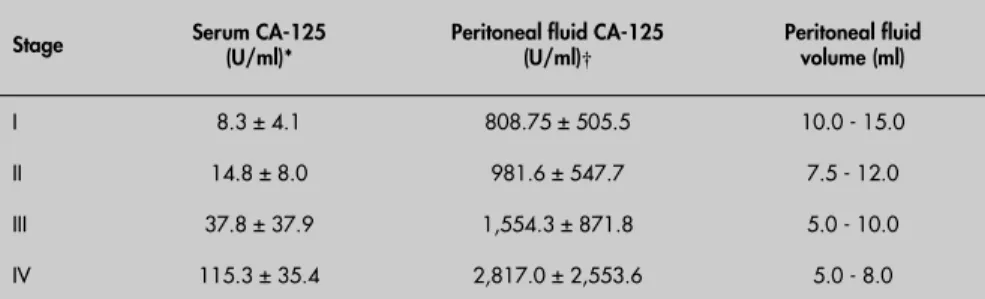
When the serum CA-125 levels of women with pelvic endometriosis were com-pared with disease staging, women with more advanced degrees of endometriosis showed higher CA-125 levels in both serum and peritoneal fl uid (p = 0.0001). The analysis of peritoneal fluid volume demonstrated that the more advanced the disease was,
Figure 1.Mean CA-125 levels in serum (S) and peritoneal fl uid (PF) from women with and without pelvic endometriosis.
Figure 2. Correlation between CA-125 levels in serum (S) and peritoneal fl uid (PF) from women with pelvic endometriosis.
CA- 125-S CA- 125-PF CA- 125-S CA- 125-PF 1
10 100 1000 10000
Endometriosis group Control group
U/
ml
r = 0.4880 r2
= 0.2381
Y = 14.293 + 0.01831X
CA
125
P
F
the smaller the volume of peritoneal fl uid detected at laparoscopy was (Table 1).
The lesions suggestive of endometriosis viewed during laparoscopy were divided into typical or pigmented (black, suggestive of chronic disease) and atypical or non-pig-mented (red, suggesting active disease). There was no signifi cant correlation between these lesions (typical and atypical) and serum or peritoneal fl uid CA-125 levels (Table 2).
DISCUSSION Increasing incidence of pelvic endome-triosis has been observed over recent decades.12
Superfi cial endometriosis has been described as a cyclical and normal phenomenon in the life of a woman, with the development and evolution of this disease occurring in some women as a result of immunological alterations.1 Today,
superfi cial endometriosis is thought to be a physiological and intermittent condition in women during their reproductive years, whereas evolving disease characterized as deep infi ltra-tive endometriosis and endometrial ovarian cysts is considered to be the true disease.12
Divergences persist regarding the natural history of endometriosis, its symptoms, extent, location and staging.13 The incidence of pelvic
pain, especially dysmenorrhea in women with endometriosis, plus infertility and dyspareu-nia, are the triad that characterize the disease. The severity of the disease and the intensity of symptoms presented by women with this disease are controversial subjects.12 In the
present study, we observed the presence of dys-menorrhea as the major symptom, but none of the symptoms observed was correla ted with the severity of the disease, as also reported by Fedele et al.14 In our study, we also found no
correlation between the number of endome-triotic implants and severity of pelvic pain or dysmenorrhea and the disease staging accord-ing to the ASRM15 (not shown). The behavior
of disease that solely develops at this site has characteristics that are distinct from peritoneal or rectovaginal septal disease.16
The low specificity of the diagnostic methods available, with respect to the sever-ity of the disease, has motivated new studies. CA-125 has been described as one of the possible markers for endometriosis follow-up. The high levels of CA-125 in the blood stream observed in the presence of an endometriotic ovarian cyst and/or endometriosis with deep infi ltration suggest that this antigen may pass into the circulation from endometrial cells of patients with endometriosis.2,3,17 The
CA-125 released by the endometrium may reach the blood stream and lymphatic circulation
by the peritoneal route, starting from retro-grade menstruation, thereby allowing contact with local infl ammatory reactions and thus re-leasing celomic CA-125.17,18 Another
explana-tion for the increase in CA-125 in blood could be its access to the abdominal cavity through tubal refl ux, resulting in absorption by peri-toneal lymphatic vessels.18 Despite the
mecha-nisms proposed, doubts still persist about the real mechanism for CA-125 release into the circulation, since retrograde menstruation is still controversial and the levels of this marker are altered during the postmenopausal period, as is the case for the utilization of CA-125 in the diagnosis of ovarian cancer.18
In the present study, evaluation of serum CA-125 levels showed that the mean levels of this marker in women with pelvic endome-triosis were higher than in normal women, thus confi rming data from other authors19-21
When calculating the correlation between CA-125 levels in serum and peritoneal fl uid and the disease staging we noted a signifi cant trend towards a linear increase in this marker with disease severity. Several investigators have detected this correlation between increased serum CA-125 levels and severity of the dis-ease, thus indicating the diagnostic potential of this marker for patients with stage III and IV pelvic endometriosis.2,11,22 Some studies
have detected higher CA-125 levels in the presence of stage I and II endometriosis,23,24
while others have reported increased CA-125 levels in stage III and IV endometriosis,2,25,26
as we also observed in our study.
The peritoneal fluid of women with endometriosis shows a more marked increase in macrophage numbers during the follicular phase.27 This increase causes higher local
secretion of various products. Among these are growth factors and cytokines, which may be involved in the mechanism for the im-plantation and subsequent development and proliferation of endometriotic implants. These alterations contribute towards the mechanism for infertility due to intraperitoneal exudate of unknown cause, even in the presence of normal ovulatory function. In the present study, we confi rmed that the volume of peri-toneal fl uid had an inverse correlation with the disease stage in patients with endometriosis, whereas in the control group we often had to exclude patients from the study due to the absence of peritoneal fl uid. This negative corre-lation has also been observed by others.28
Pittaway et al.3 evaluated the serum
variations of CA-125 and observed that they corresponded to the surface involved by endometriosis, thus suggesting a 10- to 100-fold increase in the levels of this marker in peritoneal fl uid. Progressive elevation of this marker has also been observed in perito-neal fl uid, similar to that in serum.14,17 This
feature was confi rmed in the present study, in which peritoneal fl uid CA-125 levels were 100- to 1000-fold higher than serum levels in women with pelvic endometriosis. We also observed a correlation between serum and peritoneal fl uid CA-125 levels, thus suggesting peritoneal production and
Table 2. Correlation between serum and peritoneal fl uid CA-125 levels and type of endometriotic lesion viewed in laparoscopy, i.e. typical or pigmented (black) and atypical or non-pigmented (red)
CA-125 (U/ml)
Type of lesion
p Atypical (n = 16) Typical (n = 19)
Serum 22.5 ± 25.7 53.0 ± 54.4 > 0.05 Peritoneal fl uid 1,627.2 ± 1,704.3 1,282.1 ± 760.8 > 0.05 Table 1. Mean serum and peritoneal fl uid CA-125 levels and staging of endome triosis in the endometriosis group, and range of peritoneal fl uid volume compared with disease staging
Stage Serum CA-125 (U/ml)* Peritoneal fl uid CA-125 (U/ml)† Peritoneal fl uid volume (ml)
I 8.3 ± 4.1 808.75 ± 505.5 10.0 - 15.0
II 14.8 ± 8.0 981.6 ± 547.7 7.5 - 12.0
III 37.8 ± 37.9 1,554.3 ± 871.8 5.0 - 10.0
IV 115.3 ± 35.4 2,817.0 ± 2,553.6 5.0 - 8.0
1. Abrão MS, Podgaec S, Filho BM, Ramos LO, Pinotti JA, de Oliveira RM. The use of biochemical markers in the diagnosis
of pelvic endometriosis. Hum Reprod.1997;12(11):2523-7.
2. Barbieri RL, Niloff JM, Bast RC Jr, Scaetzl E, Kistner RW, Knapp RC. Elevated serum concentrations of CA-125 in patients with
advanced endometriosis. Fertil Steril.1986;45(5):630-4.
3. Pittaway DE, Rondinone D, Miller KA, Barnes K. Clinical evaluation of CA-125 concentrations as a prognostic factor for pregnancy in infertile women with surgically treated endome-triosis. Fertil Steril.1995;64(2):321-4.
4. Koninckx PR. Is mild endometriosis a condition occurring inter-mittently in all women? Hum Reprod. 1994;9(12):2202-5. 5. Niloff JM, Knapp RC, Schaetzl E, Reynolds C, Bast RC Jr.
CA125 antigen levels in obstetric and gynecologic patients. Obstet Gynecol. 1984;64(5):703-7.
6. Koninckx PR, Martin DC. Deep endometriosis: a consequence of infi ltration or retraction or possibly adenomyosis externa? Fertil Steril.1992;58(5):924-8.
7. Moen MH, Hagen B, Onsrud M. CA 125 in perito-neal fl uid from patients with endometriosis. Hum Reprod. 1991;6(10):1400-3.
8. Williams RS, Rao CV, Yussman MA. Interference in the measurement of CA-125 in peritoneal fluid. Fertil Steril. 1988;49(3):547-50.
9. Barbati A, Cosmi EV, Spaziani R, Ventura R, Montanino G. Serum and peritoneal fl uid CA-125 levels in patients with endometriosis. Fertil Steril.1994;61(3):438-42.
10. Krasnicki D. Ocena stezenia CA-125 w plynie otrzewnowym i surowicy u kobiet z endometrioza. [Serum and peritoneal fl uid CA-125 concentration in women with endometriosis]. Ginekol
Pol.2001;72(12A):1365-9.
11. Matalliotakis IM, Goumenou AG, Mulayim N, Karkavitsas N, Koumantakis EE. High concentrations of the CA-125, CA 19-9 and CA 15-3 in the peritoneal fl uid between patients with and
without endometriosis. Arch Gynecol Obstet.2005;271(1):40-5.
12. Vercellini P, Trespidi L, De Giorgi O, Cortesi I, Parazzini F, Cro-signani PG. Endometriosis and pelvic pain: relation to disease stage and localization. FertilSteril.1996;65(2):299-304. 13. Redwine DB. Age-related evolution in color appearance of
endometriosis. Fertil Steril.1987;48(6):1062-3.
14. Fedele L, Bianchi S, Bocciolone L, Di Nola G, Parazzini F. Pain symptoms associated with endometriosis. Obstet Gynecol. 1992;79(5(Pt 1)):767-9.
15. Revised American Society for Reproductive Medicine classifi ca-tion of endometriosis: 1996. Fertil Steril.1997;67(5):817-21. 16. Abrão MS, Neme RM, Averbach M. Endometriose de septo
retovaginal: doença de diagnóstico e tratamento específi cos. [Rec-tovaginal septum endometriosis: a disease with specifi c diagnosis and treatment]. Arq Gastroenterol. 2003;40(3):192-7. 17. Koninckx PR, Muyldermans M, Meuleman C, Cornillie FJ. CA
125 in the management of endometriosis. Eur J Obstet Gynecol Reprod Biol. 1993;49(1-2):109-13.
18. Bon GG, Kenemans P, Dekker JJ, et al. Fluctuations in CA 125 and CA 15-3 serum concentrations during spontaneous
ovulatory cycles. Hum Reprod.1999;14(2):566-70.
19. Kafali H, Artuc H, Demir N. Use of CA125 fl uctuation during the menstrual cycle as a tool in the clinical diagnosis of endo-metriosis; a preliminary report. Eur J Obstet Gynecol Reprod Biol. 2004;116(1):85-8.
20. Somigliana E, Viganò P, Tirelli AS, et al. Use of the concomitant serum dosage of CA 125, CA 19-9 and interleukin-6 to detect the presence of endometriosis. Results from a series of reproduc-tive age women undergoing laparoscopic surgery for benign gynaecological conditions. Hum Reprod. 2004;19(8):1871-6. 21. Xavier P, Beires J, Belo L, et al. Are we employing the most effec-tive CA 125 and CA 19-9 cut-off values to detect endometriosis? Eur J Obstet Gynecol Reprod Biol. 2005;123(2):254-5. 22. O’Shaughnessy A, Check JH, Nowroozi K, Lurie D. CA 125 levels
measured in different phases of the menstrual cycle in screening
for endometriosis.Obstet Gynecol.1993;81(1):99-103.
23. Eisermann J, Collins JL. Enzyme immune assay determination of CA-125 in serum, peritoneal fl uid, and follicular fl uid from women with minimal endometriosis after ovarian hyperstimula-tion. Fertil Steril.1989;51(2):344-7.
24. Gürgan T, Kisnisçi H, Yarali H, Aksu T, Zeyneloglu H, Develio-glu O. Serum and peritoneal fl uid CA-125 levels in early stage
endometriosis. Gynecol Obstet Invest.1990;30(2):105-8.
25. Mol BW, Bayram N, Lijmer JG, et al. The performance of CA-125 measurement in the detection of endometriosis: a meta-analysis. Fertil Steril.1998;70(6):1101-8.
26. Kitawaki J, Ishihara H, Koshiba H, et al. Usefulness and limits of CA-125 in diagnosis of endometriosis without associated ovarian endometriomas. Hum Reprod. 2005;20(7):1999-2003. 27. Arici A, Oral E, Attar E, Tazuke SI, Olive DL. Monocyte
chemotactic protein-1 concentration in peritoneal fl uid of women with endometriosis and its modulation of expression in mesothelial cells. Fertil Steril.1997;67(6):1065-72. 28. Haney AF, Handwerger S, Weinberg JB. Peritoneal fl uid
pro-lactin in infertile women with endometriosis: lack of evidence of secretory activity by endometrial implants. Fertil Steril. 1984;42(6):935-8.
Sources of funding: Not declared
Confl ict of interest: Not declared
Date of fi rst submission: November 16, 2005
Last received: November 18, 2005
Accepted: July 4, 2006
REFERENCES hematogenic absorption. Koninckx &
Mar-tin,6 after evaluating CA-125, concluded
that superfi cial disease causes its elevation in peritoneal fl uid, whereas deep disease causes its elevation in blood. We could not confi rm these fi ndings in our study, since CA-125 levels increased with deeper infi l-tration of the disease, both in blood and in peritoneal fl uid, from women with pelvic endometriosis, in comparison with to
nor-mal patients. This confi rmatory paper may help in understanding the physiopathology of this enigmatic disease and may be used for future meta-analysis, since there are several controversial points regarding CA-125 levels in endometriotic patients.
CONCLUSION We found high concentrations of CA-125 in peritoneal fl uid from women with and
AUTHOR INFORMATION
Vivian Ferreira do Amaral, MD. Gynecology and Obstetrics Department, Faculdade de Medicina de Ribeirão Preto, Universidade de São Paulo, Ribeirão Preto, São Paulo; and postgraduate program in Health Sciences, Centro de Ciências Biológicas e da Saúde, Pontifi ca Universidade Católica do Paraná (CCBS-PUCPR), Curitiba, Paraná, Brazil.
Rui Alberto Ferriani, MD. Gynecology and Obstetrics Department, Faculdade de Medicina de Ribeirão Preto, Universidade de São Paulo, Ribeirão Preto, São Paulo, Brazil.
Marcos Felipe Silva de Sá, MD.Gynecology and Obstetrics Department, Faculdade de Medicina de Ribeirão Preto, Universidade de São Paulo, Ribeirão Preto, São Paulo, Brazil.
Antonio Alberto Nogueira, MD. Gynecology and Obstetrics Department, Faculdade de Medicina de Ribeirão Preto, Universidade de São Paulo, Ribeirão Preto, São Paulo, Brazil.
Julio César Rosa e Silva, MD. Gynecology and Obste-trics Department, Faculdade de Medicina de Ribeirão Preto, Universidade de São Paulo, Ribeirão Preto, São Paulo, Brazil.
Ana Carolina Japur de Sá Rosa e Silva, MD. Gyneco-logy and Obstetrics Department, Faculdade de Medicina de Ribeirão Preto, Universidade de São Paulo, Ribeirão Preto, São Paulo, Brazil.
Marcos Dias de Moura, MD.Gynecology and Obste-trics Department, Faculdade de Medicina de Ribeirão Preto, Universidade de São Paulo, Ribeirão Preto, São Paulo, Brazil.
Address for correspondence:
Rui Alberto Ferriani
Av. Bandeirantes, 3900 — 8o andar Ribeirão Preto (SP) — Brasil — CEP 14049-900 Tel. (+55 16) 602-2804 — Fax (+55 16) 633-0946 E-mail: [email protected]
E-mail: [email protected]
Copyright © 2006, Associação Paulista de Medicina
Resumo
Correlação positiva entre os níveis séricos e no fl uido peritonial de CA-125 em mulheres com endometriose pélvica
CONTEXTO E OBJETIVO:Um dos marcadores diagnósticos de endometriose é o CA-125, e seus níveis elevados são devidos à alta concentração no endométrio ectópico. O objetivo deste estudo foi correlacionar os níveis de CA-125 no soro e fl uido peritonial de mulheres com e sem endometriose pélvica.
TIPO DE ESTUDO E LOCAL:Estudo prospectivo, longitudinal, controlado, de pacientes consecutivas subme-tidas a laparoscopia por infertilidade, dor pélvica ou laqueadura tubária, durante a fase folicular precoce no Hospital Universitário da Faculdade de Medicina de Ribeirão Preto.
MÉTODOS:Cinqüenta e duas pacientes foram divididas em dois grupos: grupo endometriose, com 35 pacientes com biópsia confi rmada de endometriose pélvica, e grupo controle, com 17 pacientes sem endometriose. Níveis de CA-125 em amostras no soro e fl uido peritonial foram determinadas por quimi-luminescência.
RESULTADOS:Os níveis de CA-125 no soro e fl uido peritonial foram mais altos nas pacientes com en-dometriose pélvica avançada (média 39,1 ± 45,8 U/ml versus 10,5 ± 5,9 U/ml no soro, p < 0,005, 1469,4 ± 1350,4 U/ml versus 888,7 ± 784,3 U/ml no fl uido peritonial, p < 0,05), e o estudo mostrou uma correlação positiva entre eles (coefi ciente de correlação = 0,4880). Mulheres com estágios mais avançados de endometriose mostraram níveis de CA-125 maiores em ambos soro e fl uido peritonial (p = 0,0001).
CONCLUSÃO:Há uma correlação positiva entre os valores de CA-125 no soro e no fl uido peritonial em pacientes com e sem endometriose e seus níveis são maiores no fl uido peritonial. Endometriose avançada é relacionada com níveis mais altos de CA-125 em ambos soro e fl uido peritonial.