Low sensitivity of polymerase chain reaction for diagnosis
of tuberculous meningitis in southeastern Brazil
Baixa sensibilidade da reação em cadeia da polimerase para o diagnóstico
de meningite tuberculosa no sudeste do Brasil
Vânia Maria Sabadoto Brienze1, Ângela Pedroso Tonon1, Fabrício José Tarelho Pereira1, Elisabete Liso1, Waldir Antônio Tognola1, Manoel Armando Azevedo dos Santos2 and Marcelo Urbano Ferreira2
Abstract Two polymerase chain reaction (PCR) protocols showed low sensitivity (36% and 53% for TB
AMPLICOR and MPB64 nested PCR, respectively), when compared with classic microbiological methods (73% and 54% for Ziehl-Neelsen staining and culture, respectively), in the diagnosis of tuberculous meningitis in 91 patients in southeastern Brazil. Only three PCR-positive, microbiologically negative patients were found. Analysis of sequential cerebrospinal fluid samples by nested PCR detected Mycobacterium tuberculosis DNA up to 29 days after the introduction of antituberculosis chemotherapy.
Key-words: Diagnosis of tuberculous meningitis. PCR. Mycobacterium tuberculosis.
Resumo Dois protocolos de reação em cadeia da polimerase (PCR) apresentaram baixa sensibilidade
(36% e 53%, respectivamente), para TB AMPLICOR e PCR aninhado baseado no gene MPB64), quando comparados aos métodos microbiológicos clássicos (73% e 54% respectivamente para baciloscopia e cultura), no diagnóstico de meningite tuberculosa em 91 pacientes do sudeste do Brasil. Somente três pacientes apresentaram PCR positiva e microbiologia negativa. A análise de amostras seqüenciais de líquor com a PCR aninhada detectou DNA de Mycobacterium tuberculosis até 29 dias após a introdução de tratamento.
Palavras-chaves: Diagnóstico da meningite tuberculosa. PCR. Mycobacterium tuberculosis.
1. Departamento de Doenças Infecciosas e Parasitárias da Faculdade de Medicina de São José do Rio Preto, São José do Rio Preto, SP; 2. Laboratório de Parasitologia do Instituto de Ciências Biomédicas da Universidade de São Paulo, São Paulo, SP.
Financial support: Fundação Faculdade Regional de Medicina (FUNFARME), UNDP/World Bank/World Health Organization Special Program for Research and Training in Tropical Diseases, CNPq and Fundação de Amparo à Pesquisa do Estado de São Paulo (FAPESP).
Address to: Dr. Marcelo U. Ferreira. Deptº de Parasitologia/ICB/USP. Av. Prof. Lineu Prestes 1374, 05508-900 São Paulo, SP, Brazil. Tel: 55 11 3818-7273: Fax: 55 11 3818-7417.
e-mail: [email protected]
Recebido para publicação em 2/8/2000.
Tuberculosis is responsible for over two million deaths each year across the world, and Brazil is one of the 22 high-burden countries that together report 80% of new cases world-wide. The situation is further complicated by the spread of HIV/AIDS and the emergence of multi-drug resistance among local Mycobacterium tuberculosis strains16. Around 90,000 new cases are
reported yearly in Brazil, more than half of them involving patients living in the southeastern region of the Country. Almost 15% of all patients with newly diagnosed M. tuberculosis infection in São Paulo, southeast Brazil, are HIV-infected17. Pulmonary tuberculosis is by far the
most common clinical presentation of symptomatic M. tuberculosis infection, but extrapulmonar y involvement, such as meningitis, remains a significant cause of morbidity, especially among HIV-infected patients9. Bacteriological diagnosis of tuberculous
meningitis relies on the microscopic identification of acid-fast bacilli (AFB) on smears, and culture of mycobacteria on solid or liquid media. Microscopy is a simple and rapid screening test, but lacks sensitivity. Routine culture takes four to eight weeks to yield positive results. The radiometric BACTEC 460 TB system (Becton Dickinson) represents an important alternative to standard culture, since it requires only two weeks to detect most positive specimens1, but the high cost limits its availability in most
referral hospitals in developing countries.
The need for a rapid diagnosis has led to the assessment of several polymerase chain reaction (PCR)-based procedures for detection of M. tuberculosis DNA in cerebrospinal fluid (CSF) samples. The sensitivity of these molecular methods has been reported to vary between 33%15 and 90.5%13, and therefore the role of
available kit (TB AMPLICOR, Roche Diagnostic Systems) and a nested PCR described by Liu and colleagues13, in
the diagnosis of tuberculous meningitis in a tertiary care hospital in southeastern Brazil.
This study was performed at the Hospital de Base, a 600-bed teaching hospital situated in São José do Rio Preto, São Paulo State. A total of 105 CSF samples were obtained prospectively, between April 1996 and February 2000, from 91 patients with suspected meningitis (mean age, 33 years; range: 3 months-61 years). More than one CSF sample was available for 10 patients, and for 5 of them samples were also collected after the introduction of chemotherapy for tuberculosis. Sixty-six (73%) patients had serologically confirmed HIV infection (mean CD4 cell count, 81.2/ml; range: 2-384/ ml). All patients were grouped according to a set of clinical and laboratorial criteria, essentially as described and validated by Ahuja and colleagues2: (A) presence
of fever and headache lasting more than 14 days, with or without vomiting, alteration of sensorium or focal deficit; (B) presence of acid-fast bacilli (AFB) on CSF smears stained with Ziehl-Neelsen (ZN) or culture (B1), and absence of other bacteria and fungi, as well as malignant cells, in CSF (B2); (C) CSF pleocytosis with more than 10 cells/ml (more than 60% lymphocytes) (C1), and CSF concentrations of protein greater than 0.6mg/ml (C2) and of glucose less than 60% of the corresponding blood level (C3); (D) evidence of extraneural tuberculosis. Five groups were defined as different combinations of these criteria: (1) definite tuberculous meningitis (A + B1 + B2), (2) highly probable tuberculous meningitis (A + B2 + C1 + C2 + C3 + D), (3) probable tuberculous meningitis (A + B2 + two of the criteria C1, C2, C3 or D), (4) possible tuberculous meningitis (A + B2 + one of the criteria C1, C2, C3 or D), (5) other disease (absence of A or B2).
CSF samples (about 12ml) were divided into three portions. One aliquot was sent to the CSF laboratory of Hospital de Base for cytology, biochemistr y, bacterioscopy and culture for M. tuberculosis and other bacteria using standard methods3. The second portion
was sent to the Laboratory of Mycobacteria, at the Department of Microbiology of the University of São Paulo, in São Paulo, for DNA extraction and amplification with the TB AMPLICOR kit. This test targets a species-specific sequence within the small sub-unit (16S) ribosomal RNA (rRNA) gene from M. tuberculosis. CSF samples were stored at –20°C, and DNA extraction was performed as recommended for respiratory specimens, according to the manufacturer’s instructions. Briefly, 500ml of wash solution were added to 100-ml CSF aliquots, the mixture was vortexed and centrifuged (12,500g for 10 min). Supernatants were aspirated, and 100ml of lysis reagent was added to the pellet. The samples were then vortexed and incubated for 45 min at 60°C. Next, the samples were centrifuged (12,500g
DNA templates were used in PCR. Amplification was perfor med exactly as recommended by the manufacturer, using a GeneAmp System 9600 thermal cycler (Perkin-Elmer). The amplification products were incubated with 100ml of denaturation solution, and 25-ml aliquots of denaturated samples were transferred to the wells of microtiter plates coated with a probe that targets a M. tuberculosis-specific sequence within the PCR product. Internal positive and negative controls were included in all experiments. Due to budget limitations, only 46 samples were tested with AMPLICOR kits, and no sample was re-tested, regardless of the occurrence of discordant results between PCR-based and microbiological diagnoses.
The third CSF aliquot (200-500ml) was sent to the Laboratory of Molecular Parasitology, at the Faculty of Medicine of São José do Rio Preto, for DNA extraction and amplification by a nested PCR protocol that targets the species-specific MPB 64 protein gene from M. tuberculosis13. CSF samples were stored at –20°C
until DNA extraction. Template DNA was prepared from 200-ml aliquots of CSF, which were incubated with protease K (20mg/ml in a buffer containing 12mM-Tris-HCl pH 8.0, 6mM EDTA and 0.6% SDS) for 3 hours at 56°C, and purified in GFX columns (Amersham Pharmacia Biotech) according to the manufacturer’s instructions. Two rounds of PCR amplification were performed in a Genius thermal cycler (Techne). The first round used the pair of external primers (here named MPB1 and MPB2) originally described by Shankar and colleagues20, while the second round used the internal
primers (here named MPB3 and MPB4) designed by Liu and colleagues13. All primers were supplied by Gibco,
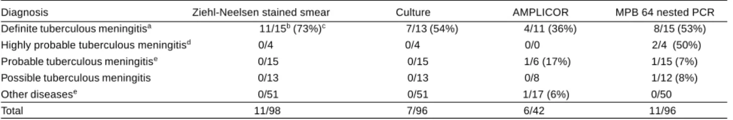
templates. A given sample was considered positive if M. tuberculosis DNA was detected in at least one test. Table 1 shows that both PCR-based approaches had a relatively low sensitivity in detecting M. tuberculosis (respectively 36% and 53% for AMPLICOR and nested PCR), when compared with routine microbiological methods (respectively 73% and 54% for ZN staining and
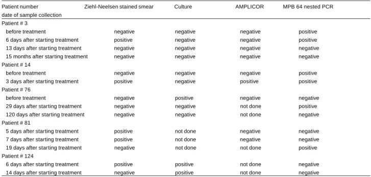
culture). Additionally, nested PCR was positive for one HIV-infected patient classified as having highly probable tuberculous meningitis (two CSF samples obtained on different occasions yielded concordant nested PCR results). Both nested PCR and AMPLICOR were also positive for an HIV-negative patient with probable tuberculous meningitis who was empirically given
Table 1- Proportion of cerebrospinal fluid samples that were positive by standard microbiological methods and polymerase chain reaction (AMPLICOR and MPB 64 nested PCR), according to the clinical diagnosis.
Diagnosis Ziehl-Neelsen stained smear Culture AMPLICOR MPB 64 nested PCR Definite tuberculous meningitisa 11/15b (73%)c 7/13 (54%) 4/11 (36%) 8/15 (53%)
Highly probable tuberculous meningitisd 0/4 0/4 0/0 2/4 (50%)
Probable tuberculous meningitise 0/15 0/15 1/6 (17%) 1/15 (7%)
Possible tuberculous meningitis 0/13 0/13 0/8 1/12 (8%) Other diseasese 0/51 0/51 1/17 (6%) 0/50
Total 11/98 7/96 6/42 11/96
aTwo patients provided two microbiologically positive samples. Microbiologically negative samples from patients with proven tuberculosis were not included
in this analysis (see Table 2); bNumber of positive/number of tested samples; cPercentage of positive samples within each clinical diagnosis category; dOne
patient provided two samples, that were both positive by MPB 64 nested PCR alone; eTwo patients provided two samples, that were negative by all methods.
specific chemotherapy. Furthermore, an HIV-negative patient with confirmed pulmonary tuberculosis and possible tuberculous meningitis was positive by nested PCR only. Thus, three PCR-positive and microbiologically negative patients were identified, suggesting that PCR was able to identify a quite limited number of additional cases missed by microbiological methods. We interpreted this relatively poor diagnostic performance as resulting from: (1) low number of organisms present in CSF samples, that were below the detection threshold of our tests; (2) presence of DNA polymerase inhibitors in CSF samples21, or (3) presence
of mycobacteria other than M. tuberculosis in some AFB-positive CSF samples, especially among HIV-infected patients. To examine the first hypothesis, we assessed the detection threshold of nested PCR by testing serial dilutions of purified DNA from M. tuberculosis culture. In these experiments, we were able to detect 0.2 pg of M. tuberculosis DNA, which corresponds to approximately 40 organisms18.
G i ve n t h e s a m p l e vo l u m e u s e d i n P C R , t h i s corresponds to approximately 800 organisms per ml of CSF. Microscopical detection of AFB on standard ZN-stained smears usually requires between 5,000 to 10,000 organisms per ml of CSF, and a successful culture requires usually the presence of 10 to 100 mycobacteria in the sample5. However, a previous
study7 showed that most culture-positive CSF
samples had fewer than 10 mycobacteria per ml. These data suggest that substantially larger sample volumes should be processed in order to improve t h e s e n s i t i v i t y o f P C R p r o t o c o l s1 1, i n c l u d i n g
AMPLICOR, which has been usually performed with 100ml of CSF6 (and this study).
To address the second hypothesis, pur ified M. tuberculosis DNA was diluted in PCR templates prepared with CSF from 17 patients with either clinically
suspected or microbiologically confirmed infection, but with negative nested PCR results. In all cases, it was possible to amplify M. tuberculosis DNA in these samples, suggesting that the presence of polymerase inhibitors played no major role in determining negative results. The authors have not performed a similar analysis for AMPLICOR-negative samples, but equivalent results for CSF samples had been reported in a previous analysis of this kind21, suggesting that the
presence of polymerase inhibitors in the CSF does not represent as major limiting factor for amplification of M. tuberculosis DNA. The third hypothesis regards atypical mycobacter ia, that have occasionally been detected as a cause of meningitis in immunocompromised patients8 10. Since the study
population included a large proportion of HIV-infected patients with low CD4 cell counts, and the target sequences of both PCR procedures are narrowly species-specific, the authors could not rule out the possibility that some false-negative results originated from the presence of atypical mycobacteria in AFB-positive patients. Further microbiological studies are required to address this issue.
At least one AMPLICOR-positive sample may be classified as a false-positive result (Table 1), since the patient did not meet clinical diagnostic criteria for tuberculous meningitis and fully recovered without specific chemotherapy. This patient was negative by nested PCR. This false-positive result may have originated from either carry-over contamination during PCR or from cross-reaction with non-mycobacterial DNA. No result may be surely considered as false-positive among those obtained with our nested PCR. Interestingly, a recent comparison12 showed that a PCR
empirical treatment and standard microbiological tests ware negative. Despite this, the role of PCR in the diagnosis of tuberculous meningitis in resource-poor countries remains controversial6 19. To the authors’
knowledge, only one previous paper on this subject had been published in Brazil. Machado and colleagues
volumes used for PCR analysis were not described14.
Further studies are clearly needed to evaluate the performance of PCR in different clinical settings, including empirically treated patients, before the role of this technique in routine diagnosis of tuberculous meningitis in Brazil may be established.
Table 2- Results of standard microbiological analyses and polymerase chain reaction (AMPLICOR and MPB 64 nested PCR) in sequential cerebrospinal fluid samples collected from five patients with proven tuberculous meningitis who were given specific chemotherapy.
Patient number Ziehl-Neelsen stained smear Culture AMPLICOR MPB 64 nested PCR date of sample collection
Patient # 3
before treatment negative negative negative positive
6 days after starting treatment positive negative negative positive
13 days after starting treatment negative negative negative negative
15 months after starting treatment negative negative negative negative
Patient # 14
before treatment negative negative negative positive
3 days after starting treatment positive negative positive positive
Patient # 76
before treatment negative positive negative negative
29 days after starting treatment negative negative not done positive
120 days after starting treatment negative negative not done negative
Patient # 81
5 days after starting treatment positive not done negative negative
7 days after starting treatment positive not done negative negative
19 days after starting treatment negative not done not done positive
Patient # 124
6 days after starting treatment positive positive not done negative
14 days after starting treatment negative positive not done negative
REFERENCES
1. Abe C, Hosojima S, Fucosawa Y, Kazumi Y, Takahashi M, Hirano K, Mori T. Comparison of MB-Check, BACTEC, and egg-based media for recover y of mycobacter ia. Jour nal of Clinical Microbiology 30: 878-881, 1992.
2. Ahuja GK, Mohan KK, Prasad K, Behari M. Diagnostic criteria for tuberculous meningitis and their validation. Tubercle and Lung Disease 75: 149-152, 1994.
3. Amer ican Thoracic Society. Diagnostic standards and
classification of tuberculosis. American Review of Respiratory Diseases 142: 725-735, 1990.
4. Amer ican Thoracic Society. Rapid diagnostic tests for tuberculosis: what is the appropriate use? American Journal of Respiratory and Critical Care Medicine 155: 1804-1814, 1996.
5. Bates JH. Diagnosis of tuberculosis. Chest 76 (suppl): 757-763, 1979.
6. Bonington A, Strang JIG, Klapper PE, Hood SV, Rubombora W, Penny M, Willers R, Wilkins EGL. Use of Roche AMPLICOR Mycobacter ium tuberculosis PCR in ear ly diagnosis of tuberculous meningitis. Journal of Clinical Microbiology 36: 1251-1254, 1998.
7. Davies LE, Rastogi KR, Lambert LC, Skipper BJ. Tuberculous meningitis in the southwest United States: a community-based study. Neurology 43: 1775-1778, 1993.
8. Gyure KA, Prayson RA, Estes ML, Hall GS. Symptomatic
Mycobacterium avium complex infection of the central nervous systems. A case report and review of the literature. Archives of Pathology and Laboratory Medicine 119: 836-839, 1995.
9. Kerr-Pontes LRS, Oliveira FAS, Freire CAM. Tuberculose associada à AIDS: situação de região do Nordeste brasileiro. Revista de Saúde Pública 31: 323-329, 1997.
10. Komachi H, Uchihara T, Saito Y, Takewaki S, Nagai R, Furukawa T. Successful treatment of atypical mycobacterial meningitis by fluoroquinolone. Journal of Neurological Sciences 142: 170-172, 1996.
11. Kox, LFF, Kuijper S, Kolk, AHJ. Early diagnosis of tuberculous meningitis by polymerase chain reaction. Neurology 45: 2228-2232, 1995.
12. Lee BW, Tan JAMA, Wong SC, Tan CB, Yap HK, Low PS, Chia JN, Tay JSH. DNA amplification by the polymerase chain reaction for the rapid diagnosis of tuberculous meningitis. Comparison of protocols involving three mycobacterial DNA sequences, IS6110, 65 kDa antigen, and MPB64. Journal of Neurological Sciences 123: 173-179, 1994.
14. Machado LR, Livramento JA, Bydlowski SP, Bendit I, Bravo LM, Spina-França A. Polymerase chain reaction in the diagnosis of tuberculous meningitis. Arquivos de Neoropsiquiatria 52: 445-446, 1994.
15. Nguyen LN, Kox LFF, Pham LD, Kuijper S, Kolk AHJ. The potential contribution of the polymerase chain reaction to the diagnosis of tuberculous meningitis. Archives of Neurology 53: 771-776, 1996.
16. Pinto WP, Hadad DJ, Palhares MC, Ferrazoli L, Telles MA, Ueki SY, Santos MT, Placco AL, Sauaia N, Palaci M. Drug resistance of M. tuberculosis isolated from patients with HIV infection seen at an AIDS Reference Center in São Paulo, Brazil. Revista do Instituto de Medicina Tropical de São Paulo 38: 15-21, 1996.
17. Secretar ia Estadual da Saúde, Centro de Vigilância Epidemiológica, Divisão de Tuberculose. Tuberculose: uma emergência mundial, São Paulo, 1995.
18. Sepkowitz KA, Raffalli J, Riley L, Kiehn TE, Armstrong D. Tuberculosis in the AIDS era. Clinical Microbiology Reviews 8: 180-199, 1995.
19. Seth P, Ahuja GK, Bhanu NV, Behari M, Bhowmik S, Broor S, Dar L, Chakraborty M. Evaluation of polymerase chain reaction for rapid diagnosis of clinically suspected tuberculous meningitis. Tubercle and Lung Disease 77: 353-357, 1996.
20. Shankar P, Manjunath N, Mohan KK, Prasad K, Behari M, Shriniwas, Ahuja GK. Rapid diagnosis of tuberculous meningitis by polymerase chain reaction. Lancet 337: 5-7, 1991.