Globulin and Campath-1H: Similar Effects Independent
of Specificity
Diana Stauch1, Annelie Dernier1, Elizabeth Sarmiento Marchese2, Kristina Kunert1, Hans-Dieter Volk1, Johann Pratschke3, Katja Kotsch1*
1Institute of Medical Immunology, Charite´ Universita¨tsmedizin Berlin, Campus Mitte, Berlin, Germany,2Immunology Department, Hospital General Universitario Gregorio Maran˜on, Madrid, Spain,3Department of Surgery, Charite´ Universita¨tsmedizin, Campus Virchow, Berlin, Germany
Abstract
T cell depleting strategies are an integral part of immunosuppressive regimens widely used in the hematological and solid organ transplant setting. Although it is known to induce lymphocytopenia, little is known about the effects of the polyclonal rabbit antithymocyte globulin (rATG) or the monoclonal anti-CD52 antibody alemtuzumab on Natural Killer (NK) cells in detail. Here, we demonstrate that induction therapy with rATG following kidney/pancreas transplantation results in a rapid depletion of NK cells. Treatment of NK cells with rATG and alemtuzumab in vitro leads to impairment of cytotoxicity and induction of apoptosis even at a 10-fold lower concentration (0.1mg/ml) compared with T and B cells. By generating
Fc-parts of rATG and alemtuzumab we illustrate that their ligation to FccRIII (CD16) is sufficient for the significant induction of degranulation, apoptosis and inflammatory cytokine release (FasL, TNFa and IFNc) exclusively in CD32CD56dim NK cells
whereas application of rATG and alemtuzumab F(ab) fragments abolishes these effects. These findings are of general importance as our data suggest that NK cells are also mediators of the clinically relevant cytokine release syndrome and that their targeting by therapeutic antibodies should be considered as they are functionally relevant for the effective clearance of opportunistic viral infections and anti-tumor activity posttransplantation.
Citation:Stauch D, Dernier A, Sarmiento Marchese E, Kunert K, Volk H-D, et al. (2009) Targeting of Natural Killer Cells by Rabbit Antithymocyte Globulin and Campath-1H: Similar Effects Independent of Specificity. PLoS ONE 4(3): e4709. doi:10.1371/journal.pone.0004709
Editor:Graham Pockley, University of Sheffield, United Kingdom
ReceivedOctober 7, 2008;AcceptedJanuary 7, 2009;PublishedMarch 5, 2009
Copyright:ß2009 Stauch et al. This is an open-access article distributed under the terms of the Creative Commons Attribution License, which permits unrestricted use, distribution, and reproduction in any medium, provided the original author and source are credited.
Funding:This study was supported in part by research funding from Genzyme to K.K. and a grant from the Deutsche Forschungsgemeinschaft (DFG PR 578/2-3) to J.P. E.S. was funded by the Instituto de Salud Carlos III, Madrid, Spain. The funders had no role in study design, data collection and analysis, decision to publish, or preparation of the manuscript.
Competing Interests:The authors have declared that no competing interests exist.
* E-mail: katja.kotsch@charite.de
Introduction
Antibodies raised against certain T cell antigens are increasingly used in patients undergoing hematopoietic stem cell transplan-tation (HSCT) or solid organ transplantransplan-tation (SOT) in order to prevent acute graft-versus-host disease (GvHD) or acute steroid-resistant graft rejection [1]. The polyclonal antithymocyte globulin (ATG) is a mixture of purified immunoglobulins M (IgM) and G (IgG) of sera derived from rabbits, horses, or goats immunized with human thymocytes or T cell lines. The most widely used preparations include rabbit ATG (rATG, IgG) which contains antibodies directed against a wide array of antigens involved in the immune response. These comprise selectin and integrin family members or immunoglobulin superfamily mole-cules expressed on the surface of T lymphocytes. Other cell types such as endothelial or B cells are also recognized by rATG due to shared epitopes with T cells [2,3]. However, the key mechanism of rATG action is T cell depletion [4,5], as it has been shown that CD3+
cell counts are lowered for years in patients treated with rATG [6–8]. Additionally, rather than T cell depletion as one of the key mechanisms, rATG has been demonstrated to affect dendritic cells [9] or to induce regulatory T cells in vitro and in vivo [10–12].
since anti-CD16 antibodies were able to inhibit ADCC and immune complex binding [21].
In recent years it has been shown that NK cell alloreactivity is beneficial following allo-HSCT because it mediates a graft-versus-leukemia (GvL) effect, eliminating residual malignant cells, removing host antigen-presenting cells (thereby reducing GvHD), and mediating immunity to viral pathogens directly through the cytolysis of virally infected tissues or indirectly by elaborating inflammatory cytokines, such as interferons (IFNs) [22,23]. The antiviral capacity of NK cells is even more important, as Epstein– Barr virus (EBV) or cytomegalovirus (CBV) infections, for example, are frequent complications of prolonged immune deficiency [24,25]. In this context, both rATG and alemtuzumab have been suggested to be associated with an elevated incidence of EBV/CMV reactivation and disease [26,27]. In general, the influence of different immunosuppressive drugs on NK cell function is of particular interest as it has recently been demonstrated that steroids and calcineurin inhibitors limit the function of IL-2-activated NK cells [28,29].
Given that both rATG and alemtuzumab mediate multiple immunomodulatory mechanisms in vitro and in vivo [9,30–32] we sought to extend these studies by investigating the effects of these antibodies on NK cells. In summary, we demonstrated that both rATG and alemtuzumab induce rapid apoptosis in NK cells and a strong induction of inflammatory cytokines, which is exclusively mediated via the binding of the IgG1 Fc-part to the low-affinity receptor for IgG, CD16 (FccRIII).
Materials and Methods
Patients
Between October 2007 and May 2008 eight simultaneous renal/pancreas patients received a non-HLA-identical allograft from deceased donors at the Departments of Surgery and Nephrology, Virchow-Clinic, Universita¨tsmedizin Charite´, Ber-lin. Transplant patients received initially 1.5 mg/kg body weight i.v. Thymoglobulin (Genzyme GmbH, Neu Isenburg, Germany) starting at day 0 followed by 4 further consecutive days in combination with tacrolimus, mycophenolate mofetil and steroids. Nine patients receiving renal allografts served as controls. These patients received two dosages of basiliximab (20 mg i.v. 2 hours before reperfusion an on day 4) which has been implemented as standard therapy in our clinic for prophylaxis of rejection as well as maintenance immunosuppres-sion consisting of tacrolimus, mycophenolate mofetil and prednisone. Blood drug concentrations of tacrolimus ranged between 8 and 10 ng/ml in both groups. All experiments using human material were approved by the Ethics Committee of the Charite´-Universita¨tsmedizin Berlin and all patients agreed to participate and signed an informed consent. Patient demograph-ics are summarized in Table 1.
Enrichment of NK cells and cell culture conditions Peripheral blood from transplant patients was collected pretransplant and three times/week/patient during hospitaliza-tion. Peripheral blood mononuclear cells (PBMCs) were isolated using ficoll density centrifugation with Bicoll Separating Solution (Biochrom, Berlin, Germany). For NK cell enrichment PBMCs were isolated from de-identified healthy donor buffy coats using ficoll density centrifugation with Bicoll Separating Solution (Biochrom, Berlin, Germany) and NK cells were obtained by negative purification using the NK Cell Isolation Kit II (Miltenyi Biotech, Bergisch Gladbach, Germany) according to the manu-facturer’s recommendation. The purity of the NK cells (CD32CD56+) was monitored by fluorescence-activated cell
sorting (FACS) and was more than 95%. Enriched NK cells were cultured in RPMI 1640 medium supplemented with 10% heat-inactivated fetal calf serum (FCS), 1% Glutamin and antibiotics (penicillin 100 U/ml, streptomycin 100mg/ml) in the presence of 200 IU/ml human recombinant IL-2 (Natutec, Frankfurt am Main, Germany). NK cell subsets (CD56bright/CD162, CD56dim
/ CD16+
) were enriched by flow cytometry (FACSAria, BD Biosciences), giving a purity of.99%. Rabbit ATG (Thymoglo-bulin, Genzyme, Neu Isenburg, Germany) was dissolved in sterile distilled water at a concentration of 5 mg/ml. Alemtuzumab (Campath-1H, MedacSchering, Mu¨nchen, Germany) was deliv-ered in a solution of 30 mg/ml. The monoclonal anti-CD3 antibody (Orthoclone OKT3, Janssen-Cilag BV, Tilburg, The Netherlands), the humanized anti-IL2Ra receptor (CD25) anti-body daclizumab (ZenapaxH; Hoffman La Roche, Basel, Switzer-land) and a polyclonal rabbit IgG (Bethyl Laboratories, Inc., Montgomery, Texas, USA) served as controls. Varying concen-trations of antibodies ranging from 0 to 100mg/ml were added to the NK cell cultures for 18 hours or as indicated in the figure legends. All experiments using human material were approved by the Ethical Committee of the Charite´-Universita¨tsmedizin Berlin.
Ex vivo whole blood (WB) assay
Whole blood of volunteer donors was collected into a BD vacutainer (Becton Dickinson, NJ, USA). One ml of fresh whole blood was co-incubated with 0, 1, 10 and 50mg/ml of intact antibody or Fc-parts of rATG and alemtuzumab for 2 hours at 37uC with 5% CO2. Red blood cells were lysed with lysis buffer (NH4Cl, 0.155 M; KHC03, 0,01 M; EDTA, 1 mM). Preparation of mRNA and quantitative real-time RT-PCR for FasL, TNFa, INFcand HPRT were performed as described below.
Antibodies and reagents
F(ab) fragments and Fc-parts of rATG, alemtuzumab and anti-CD16 (clone 3G8, a kind gift from Dr. Ofer Mandelboim, The Hebrew University of Jerusalem, Israel) were prepared by papain digestion and purified by exclusion chromatography on protein A
Table 1.Patient demographics.
female/ male (n)
LD/BD (n)
age (yrs)
(SD) 1. Tx re Tx
PRA Class I Class II
CI (hrs) mean (SD)
HLA-Mismatch
(SD) graft function aRx//no Rx
early delayed
ATG (n = 8) 2/6 0/9 48613 9 0 1/7 1/7 9.862.9 4.361.2 7 1 1/7
non ATG (n = 9) 7/2 0/9 52611 9 0 1/8 1/8 11.663.3 2.261.5 9 0 0/9
according to standard procedures. NK cells were analyzed by FACS using the following monoclonal antibodies (mAB): anti-CD3 FITC mAb (clone BW264/56, IgG2a), anti-CD3 PE mAb (clone BW264/56, IgG2a), anti-CD56 APC mAb (clone AF12-7H3, mouse IgG1), CD16 PE (clone VEP13, mouse IgM), anti-CD8 PE (clone BW 135/80, mouse IgG2a), anti-NKp30 PE (clone AF29-4D12, mouse IgG1), anti-NKp44 PE (clone 2.29, mouse IgG1), anti-NKp46 PE (clone 9E2, mouse IgG1), anti-NKG2D PE (clone BAT221, mouse IgG1), anti-KIR3DL1 PE (clone DX9, mouse IgG1), anti-IFNc FITC (clone 45-15, mouse IgG1) (Miltenyi Biotech, Bergisch Gladbach, Germany); anti-CD3 PeCy5 (clone UCHT1, mouse IgG1) (ImmunoTech, Marseille, France) and anti-KIR2DL1 FITC (clone 143211, IgG1) (R&D, Minneapolis, USA). Isotype controls conjugated with FITC, PE, APC and PeCy5 (Ebioscience, San Diego, USA) were used to discriminate unspecific binding. 26105 cells were incubated in
PBS supplemented with 1% fetal calf serum (FCS) and stained with the appropriate antibodies for 30 min at 4uC. For intracellular IFNc analysis expression, Brefeldin A (Sigma, St. Louis, USA) was added to the cell culture after 1 hour of incubation for further 2 hours and cells were surface-stained and fixed with freshly prepared 2% paraformaldehyde in PBS. 0.1% Saponin (Sigma, St. Louis, USA) was used for permeabilization. Cells were analyzed on a Becton Dickinson FACSCaliburH
interfaced with an Apple PowerMac using Cellquest Pro software. Cell culture supernatants were analyzed for the concentration of soluble cytokines TNFa and IFNc by Cytometric Bead Array System (CBA) from BD (Becton Dickinson, Heidelberg, Germany) according to the company’s instructions.
Analysis of apoptosis and necrosis
Apoptosis and necrosis of cells were determined by the 36 kD phospholipid-binding protein Annexin V FITC and propidium iodide (PI) according to the manufacturer’s instructions (Miltenyi Biotech, Bergisch Gladbach, Germany). In brief, cells were discriminated as intact (Annexin V FITC2/PI2), apoptotic (Annexin V FITC+/PI2) or necrotic (Annexin V FITC+/PI+).
Cytotoxicity assay
Assessment of the specific NK-cell-mediated killing was performed using the LIVE/DEAD cell-mediated cytotoxicity kit (Molecular Probes, Eugene, USA) with slight modifications. 16106
cells of the human MHC-deficient erythromyeloblastoid leukemia target cell line K562 (DSMZ, Braunschweig, Germany) were labeled with 30 nM of the membrane dye DiOC18(3) for 20 min and were subsequently washed with complete medium twice. A quantity of 16104labeled K562 cells were mixed with NK cells in a 96-well plate at effector-target cell (E/T) ratios of 10:1, 5:1 and 2.5:1 and were incubated for 3 hours. Afterwards propidium iodide (20 nM) was added to the cells. Samples were stored on ice until fluorescence measurements were performed. DiOC18(3) labeled cells were selected according to high FL1 green fluorescence intensity. Propidium iodide-positive dead target cells were identified by FL3 fluorescence and analysis was performed by dot plot. The specific killing percentage was calculated by the following equation: [% of dead (DiOc18(3)+PI+) target cells/(% of dead (DiOc18(3)+PI+) target cells+% of live (DiOc18(3)+PI2) target cells)]6100. The specific killing percentage was normalized
by subtracting the percentage of dead target cells without killing. Every sample was prepared and analyzed in triplicate.
Degranulation assay
CD107a lysosome-associated membrane protein-1 (LAMP-1) expression was used to measure NK cell degranulation, as
previously described [33]. NK cells were incubated in the absence or presence of K562 target cells at an E/T ratio of 2:1 for 3 hours. An anti-CD107a PeCy5 antibody (BD PharmingenTM, NJ, USA) was added to the cultures directly and Brefeldin A was added after 1 hour of incubation. Cells were then washed in PBS and stained with anti-CD56 and anti-CD3 for FACS analysis. The K562 specific NK cell degranulation was determined by subtracting the percentage of CD107a-positive cells in the cultures without K562 cells from the percentage of CD107a-positive NK cells co-incubated with K562 cells.
Quantitative real-time RT-PCR
Total RNA from cell lysates was isolated using RNeasyHMini Kit (Qiagen, Hilden, Germany). Real-time reverse transcriptase polymerase chain reaction (RT-PCR) for gene expression analysis of selected candidate genes was performed with the ABI PRISM 7500 Sequence Detection System (TaqManTM, Applied Biosys-tems, Darmstadt, Germany). All primers were designed using Primer Express software (Applied Biosystems) and validated at the Institute of Medical Immunology, Charite´-Universita¨tsmedizin Berlin (Table S1). In general, amplification primers were designed to span the exon borders to exclude cross-reactivity with genomic DNA. The PCR reaction was performed in a final volume of 25ml containing 1ml cDNA, 12.5ml Master Mix (TaqManTM Univer-sal PCR Master Mix, Applied Biosystems), 1ml fluorogenic hybridization probe, 6ml primer mix, and 5.5ml distilled water. The amplification took place in a two-step PCR (40 cycles; 15 s denaturation step at 95uC and 1 min annealing/extension step at 60uC). Specific gene expression was normalized to the house-keeping gene hypoxanthine-guanine phosphoribosyltransferase (HPRT) given by the formula 22DCt. The mean Ct values for the genes of interest (IFNc, TNFa, FasL) and HPRT were calculated from double determinations. Samples were considered negative if the Ct values exceeded 40 cycles.
Statistical analysis
Experimental and patient data are expressed as means6 stan-dard deviation (SD). Statistical analyses were performed with GraphPad Instat for Windows (version 3.06). One-way ANOVA was calculated for normally distributed data. Adjustments for multiple comparisons were done with the Tukey-Kramer Multiple Comparisons Test. For the statistical analysis of apoptosis and necrosis, cytotoxicity, degranulation and surface FACS staining rare data were used. The real-time RT-PCR data were log(10) transformed to stabilize the variance of the normally distributed data. Groups with p values less than or equal to 0.05 were considered to be statistically different.
Results
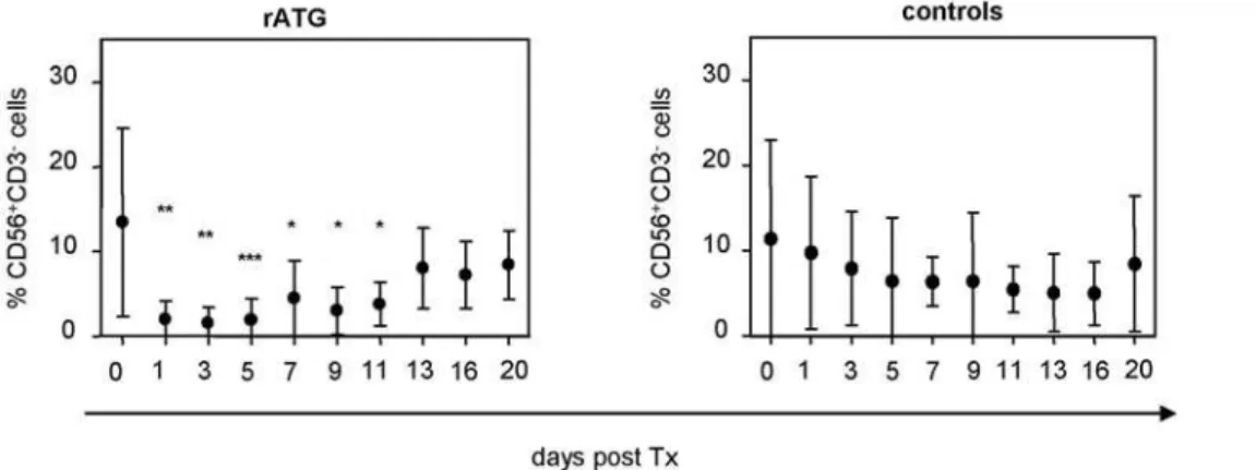
In vivo depletion of NK cells after ATG induction therapy following pancreas/kidney transplantation
Patients treated with rATG were found to demonstrate a significant decrease of CD32CD56+
rATG and alemtuzumab recognize the surface antigen CD16 on NK cells
NK cells derived from CD34+
hematopoietic stem cells undergo differentiation via NK cell precursors in the bone marrow through acquisition of functional surface receptors [34]. Therefore, certain NK cell specific receptors should not be targeted by rATG. As expected we could not detect any influence of rATG on the surface expression of the activating cytotoxicity receptors NKp30, NKp44, NKp46, NKG2D and killer-cell immunoglobulin-like receptors (KIRs) including KIR2DL1 and KIR3DL1 (data not shown). In contrast we confirmed previous data illustrating that rATG targets CD8 and CD16 and this effect was dose-dependent (Figure 2A) [35]. Targeting of CD16 was observed in the presence of 0.1mg rATG (67%610.8% versus 33.6%64.1%, p,0.001), and CD16 was further decreased to 1.2%60.6% at a concentration of 1mg/ ml rATG. In contrast, targeting of CD8 antigen required higher concentrations of rATG (e.g. 10mg, 38.6611.9% versus 9.664.3%, p,0.001). Whereas both rATG and alemtuzumab resulted in a significant and dose-dependent reduction of CD16 surface expression on CD56+
NK cells at a low dose concentration of 0.1mg/ml, no CD16 downmodulation could be observed for daclizumab, an anti-IL-2Ra(CD25) humanized antibody similar to alemtuzumab (Figure 2B). In contrast, higher concentrations of rabbit IgG (rIgG) led to a decrease of CD16 (e.g. 10mg, 80%66.3% versus 48.4%69.7% and 50mg/ml 22.1%64.3%, p,0.001).
Both rATG and alemtuzumab affect effector functions of peripheral blood CD32/CD56dimNK cells
Alemtuzumab is also used as induction therapy in clinical transplantation, inducing profound and prolonged lymphopenia. As recent reports suggest that alemtuzumab may enhance lymphocyte apoptosis in vitro in the absence of complement or immune effector cells [36], we ascertained its effects on NK cells. NK cells were isolated from healthy blood donors and cultured in the presence of human recombinant IL-2 (200 IU/ml) with increasing concentrations of antibodies overnight (18 hours). Treatment of NK cells with rATG resulted in a dose-dependent (0.1–100mg/ml) decrease of NK cytotoxicity directed against the target cell line K562, as illustrated in Figure 3A (e.g. 1mg/ml rATG resulted in 82.0%68.1% NK cell cytotoxicity versus 100% cytotoxicity of untreated controls, p,0.001) whereas control rIgG
Figure 1. Induction therapy of rATG in simultaneous kidney/pancreas transplantation results in a significant decrease of NK cells. Patients (n = 8) initially received 1.5 mg/kg body weight i.v. rATG (Thymoglobulin, Genzyme GmbH, Neu Isenburg, Germany) starting at day 0 followed by 4 further consecutive days posttransplantation in combination with tacrolimus, mycophenolate mofetil and steroids. We further enrolled nine patients who received a renal allograft as a control group. Control patients received two dosages of basiliximab (20 mg i.v., day 0 and day 4). Asterisks denote significant differences compared to pretransplant levels of CD32CD56+
NK cells. doi:10.1371/journal.pone.0004709.g001
Figure 2. Rabbit ATG and alemtuzumab target surface expression of CD16 on NK cells.Human NK cells cultured with IL-2 (IL-200 IU/ml) and different concentrations of rATG, alemtuzumab or control antibodies such as rIgG and daclizumab (18 hours) were harvested, washed and stained for CD3, CD56, CD8 and CD16.(A)The figure illustrates the average percentages of CD16 and CD8 staining on CD32CD56+
NK cells after co-incubation with varying concentrations of rATG. Results are displayed as means6SD (n = 5); asterisks (*) denote significant differences compared to untreated controls: ***p,0.001.(B) The mean percentage of CD16 on CD32CD56+ NK incubated with
affected NK cytotoxicity at higher concentrations (50mg/ml rIgG resulted in 81.1%611.1% versus 100% cytotoxicity of untreated controls, p,0.05). Co-incubation of NK cells with alemtuzumab also led to an impairment of cytotoxicity (1mg/ml alemtuzumab resulted in 75.9%616.6% versus 100% cytotoxicity of untreated controls, p,0.01) whereas daclizumab did not (Figure 3A). Based on their CD56 receptor expression density human NK cells can be distinguished as CD56dimor CD56bright NK cells. In peripheral blood the majority (.90%) are CD56dim demonstrating high expression of FccRIII (CD16), while the remaining 10% are CD56brightNK cells characterized by almost no or dim expression of CD16 [37]. As cytotoxicity was sharply targeted by rATG, we analyzed the intracellular protein expression of IFNcas an effector molecule in NK cells after co-incubation with K562 cells and detected a dose-dependent impairment exclusively in CD56dim
NK cells, whereas no influence was observed in CD56bright NK cells (Figure 3B, data not shown). Again, a significant decrease of IFNcwas observed even at a low dose concentration of 0.1mg/ml rATG (71.2%635.5% versus 100%, p,0.05) or alemtuzumab (65.4%610.1% versus 100%, p,0.001). Interestingly, higher dosages such as 50mg/ml of rIgG (61.5%63.0% versus 100%, p,0.001) or daclizumab (64.6%614.5% versus 100%, p,0.01) further led to decreased IFNcexpression (Figure 3B).
When we analyzed the degranulation capacity of NK cells preincubated with rATG by the measurement of CD107a, a marker significantly upregulated on the surface of NK cells following stimulation with MHC-devoid targets, we detected a significant impairment even at a low concentration of 0.1mg/ml rATG, and this was observed exclusively in CD56dim NK cells (56.8%611.6% versus 100%, p,0.05, Figure 4A). As was seen
Figure 3. Rabbit ATG and alemtuzumab decrease effector mechanisms of peripheral blood CD32CD56dimNK cells. (A)
Dose-dependent decrease of NK cell cytotoxicity after 18 hours pretreatment with rATG, alemtuzumab, daclizumab and rIgG (0–100mg). Killing assay for viable and antibody-depleted NK cells was performed using the target cell line K562 (E/T ratio was 10:1). Analysis of six independent experiments was performed by flow cytometry. Values demonstrate results normalized to untreated cells, which were set at 100%. Asterisks (*) indicate values that showed significantly less cytotoxicity compared to control: *p,0.05, **p,0.01, ***p,0.001.(B)K562 cells (E/T ratio 2:1) were added to NK cells cultured for 18 hours with IL-2 (200 IU/ml) and varying concentrations of rATG, alemtuzumab, daclizumab and rIgG. Cells were harvested, stained and analysis was performed for CD32CD56dimIFNc+
NK cells. Values demonstrate the mean of INFc+
/CD56dimNK cells normalized to untreated cells, which were set at 100%. Results are displayed as means6SD for 6 independent experiments. Asterisks (*) indicate values that showed significantly less IFNc expression compared to untreated controls: *p,0.05, **p,0.01, ***p,0.001.
with rATG, a significant decrease of degranulation capacity of CD56dim NK cells after pre-incubation with alemtuzumab was detected, even at a concentration of 0.1mg/ml (52.5%67.8% versus 100%, p,0.001, Figure 4A), in contrast to control antibodies (rIgG, daclizumab), which exhibit induction of degranulation at higher concentrations (50mg/ml–100mg/ml). In contrast to rATG, no dose-dependent effect was observed for
alemtuzumab (Figure 4A). Interestingly, both rATG and alemtu-zumab induced degranulation of NK cells without additional sensitization by adding K562 cells. In comparison with rATG a stronger CD107a induction in NK cells with alemtuzumab was detected (e.g. 0.1mg/ml alemtuzumab 18.3%66.6% versus 0.1mg/ml rATG 7.3%63.0%, p,0.001, Figure 4B,C). In contrast, induction of degranulation by rIgG and daclizumab
Figure 4. Rabbit ATG and alemtuzumab influence the degranulation of peripheral blood CD32CD56dimNK cells. (A)K562 cells (E/T
ratio 2:1) and CD107a antibody were added to NK cells cultured with IL-2 (200 IU/ml) and varying concentrations of rATG, alemtuzumab, daclizumab and rIgG. NK cells were harvested and stained for CD3 and CD56. For analysis the percentage of CD107a+
NK cells of controls (without K562) was subtracted from the CD107a+NK cells co-incubated with K562 cells. Values demonstrate CD107a expression on CD56dimNK cells normalized to untreated cells, which were set at 100%. Results are displayed as means6SD for 5 independent experiments. Asterisks (*) indicate values that showed significantly less degranulation compared to untreated controls: *p,0.05, ** p,0.01, *** p,0.001.(B)A degranulation assay was performed (A) without the addition of K562 cells. Results are displayed as the mean of CD107a+
cells on CD56dimNK cells6SD (n = 5). Asterisks (*) indicate values that showed significantly higher degranulation compared to untreated controls: *p,0.05, ** p,0.01, *** p,0.001.(C)Representative FACS dot plots of CD56+
CD32NK cells stained for CD107a. Treatment with 0.1mg/ml rATG and alemtuzumab produced a higher induction of CD107a in CD56dim NK cells compared to 0.1mg/ml daclizumab and rIgG.
was dose-dependent, while the latter antibody showed only moderate degranulation induction on NK cells (5.3%62.8% degranulation, 100mg/ml).
Induction of apoptosis in CD32/CD56dimNK cells It has already been demonstrated that rATG induces apoptosis in NK cells [30]. We could confirm these observations by showing that even low concentrations of rATG (0.1mg/ml) resulted in enhanced apoptosis and necrosis (30.3610.3, p,0.001, Figure 5A) and this effect was even more intensified in NK cells compared to T or B cells. However, higher concentrations of rATG (10– 100mg/ml) led to an increased rate of apoptosis and necrosis in T and B cells, whereas the rate of necrosis and apoptosis in NK cells could not be increased (Figure 5A). Next we investigated the induction of apoptosis following alemtuzumab pretreatment and also detected a significant induction of apoptosis comparable to rATG at a concentration of 0.1mg/ml (42.3616.6, p,0.001, Figure 5B). Induction of apoptosis was also detected with rIgG at a concentration of 50mg/ml. In order to verify the results observed in the bulk NK cell population we sorted CD56dimand CD56bright NK cells with a purity greater than 99% for each cell fraction. By analyzing FACS sorted CD56dim and CD56bright NK cells separately the main induction of necrosis/apoptosis was observed in the CD56dim subset (Figure 5C). Again, an impairment of CD107a expression was also exclusively confirmed for CD56dim NK cells.
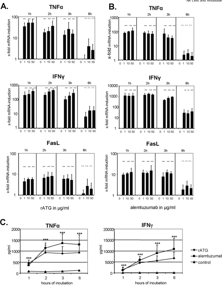
Induction of FasL, TNFaand IFNcin NK cells after treatment with rATG and alemtuzumab
The stimulation of CD16 (FccRIII) on NK cells can initiate autocrine programmed cell death by the up-regulation of, for example, Fas ligand (FasL) [38]. Whereas freshly isolated NK cells did not constitutively express FasL mRNA, incubation with rATG resulted in a rapid and dose-dependent mRNA induction within the first hour (p,0.001) which decreased after 6 hours of co-culture (Figure 6A). Furthermore it has been demonstrated that anti-T-cell therapy results in the cytokine release syndrome early after administration [39,40]. Similar to the induction of FasL mRNA, we detected a dose-dependent increase of TNFa and IFNc mRNA in NK cells within the first hour of co-incubation with rATG (p,0.001, Figure 6A). Additionally, the same induction of FasL, TNFa and IFNc mRNA was observed for alemtuzumab-treated NK cells (p,0.001, respectively), although the induction of IFNc and TNFa mRNA was more intense compared to rATG-pretreated NK cells (Figure 6B). Analysis of the control antibodies rIgG and daclizumab showed that both induced a cytokine induction at higher concentrations (10–50mg/ ml), which is clearly below the induction profile of rATG and alemtuzumab (Figure S1). We confirmed the rapid and significant induction of TNFa and IFNcat the protein level in cell culture
supernatants (p,0.001, respectively, Figure 6C), further demon-strating that alemtuzumab resulted in higher cytokine expression levels compared to rATG.
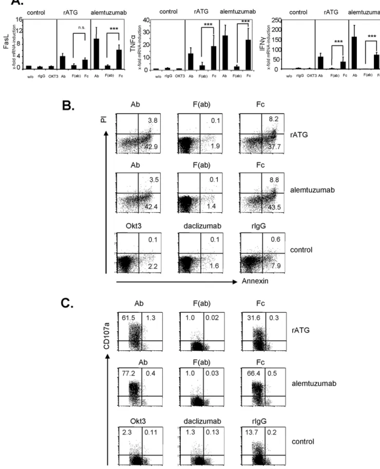
The Fc-part of rATG and alemtuzumab is sufficient to induce cytokine release, apoptosis and degranulation via FccRIII ligation
In order to investigate the influence of the unspecific binding of the Fc-part to FccRIII, we generated F(ab) fragments and Fc-parts of a CD16 blocking antibody (clone 3G8), rATG and alemtuzu-mab. Blocking FccRIII on NK cells by F(ab) fragments of an anti-CD16 resulted in a significant inhibition of FasL (p,0.001), TNFa
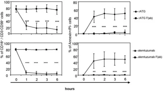
(p,0.05) and IFNc(p,0.001) mRNA induction. Additionally, the application of F(ab) fragments of either rATG or alemtuzumab further did not cause FasL, TNFa and IFNcmRNA induction (Figure S2). Next, we tested generated Fc-parts and could illustrate that the application of rATG and alemtuzumab Fc-parts only is sufficient to induce a significant cytokine induction and apoptosis in NK cells compared with F(ab) fragments. This induction showed a similar expression level compared with intact IgG antibodies. In contrast, the application of control rIgG, a monoclonal anti-CD3 antibody (OKT3, IgG2a isotype) or daclizumab (Figure S1, 1mg/ml for 1 hour) did not lead to induced cytokine levels and increased apoptosis (Figure 7A,B). Moreover, CD16 ligation by rATG and alemtuzumab Fc-parts resulted in degranulation of NK cells further emphasizing that antigen-specific crosslinking of antibodies is not necessary for the observed effector effects (Figure 7C). After the addition of rATG or alemtuzumab F(ab) fragments, targeting of CD16 on CD56+
CD16+
cells and induction of NK cell apoptosis was abolished compared to the application of intact IgG antibodies (p,0.001, Figure 8). Thus, our results clearly illustrate that FccRIII ligation with the Fc-part of rATG or alemtuzumab is sufficient to induce NK cell apoptosis and cytokine release and that this effect is independent of antibody specificity.
In order to ascertain whether high concentrations of serum immunoglobulins in whole blood may block the Fc-part-mediated cytokine release of rATG or alemtuzumab, we performed stimulation experiments in whole blood. Our results with co-incubation of whole blood with intact antibody or Fc-parts demonstrate that CD16 ligation is sufficient for TNFa, IFNcand FasL induction in this experimental setting (Figure 9).
Discussion
Rather than T cell depletion rATG mediates complement-related lysis or activation-associated apoptosis via the induction of Fas and FasL [41] and prevention of memory T cell migration [3,4]. Recent studies illustrate that the therapeutic effect of rATG might be due to the generation of regulatory T cells and
Figure 5. Induction of apoptosis and necrosis in CD32CD56dim NK cells after co-incubation with rATG or alemtuzumab. (A)
Magnetically isolated NK, T and B cells from healthy volunteer blood donors were treated with different concentrations of rATG for 18 hours. Annexin V+
and PI2cells were shown as apoptotic cells and Annexin V+
and PI+
cells as necrotic cells. Rabbit ATG induced apoptosis and necrosis of NK cells at lower concentrations (0.1mg) compared with T and B cells. Values demonstrate the results normalized to untreated cells and are displayed as means
of five independent experiments. Asterisks (*) indicate significant values: **p,0.01, ***p,0.001.(B)NK cells were cultured with IL-2 (200 IU/ml) and different concentrations of rATG, alemtuzumab, daclizumab and rIgG for 18 hours. Both rATG and alemtuzumab led to a rapid and significant induction of apoptosis of NK cells even at the low concentration of 0.1mg/ml. In contrast, rIgG resulted in NK cell apoptosis at higher concentrations
Figure 6. Rabbit ATG and alemtuzumab increase FasL, TNFaand IFNcmRNA in NK cells. (A)IL-2 (200 IU/ml) pre-activated NK cells cultured in the presence of rATG were analyzed for FasL, TNFaand IFNcmRNA after 1, 2, 3 and 6 hours of co-culture. rATG induced a rapid and dose-dependent induction of FasL, TNFaand IFNcmRNA in NK cells which decreased after 6 hours of co-incubation.(B)Similar to rATG, the application of alemtuzumab results in a rapid FasL, TNFaand IFNcmRNA induction within the first hour of co-incubation. Values demonstrate the results relativized to untreated controls (22DDct
) and are displayed as means of six independent experiments. Asterisks (*) indicate significant values compared to untreated controls: **p,0.01, ***p,0.001.(C)Induction of cytokines by rATG and alemtuzumab (10mg/ml) was further confirmed for TNFaand IFNc
polarization of monocyte-derived dendritic cells towards tolero-genic dendritic cells [10,42]. Although alemtuzumab has not been able to induce allograft tolerance as was initially hoped, it is assumed that both complement and non-complement-mediated mechanisms are similarly responsible for alemtuzumab-mediated killing of T cells. Recent reports suggest that alemtuzumab may enhance lymphocyte apoptosis in vitro in the absence of complement or immune effector cells, leading to cell death through a nonclassical caspase-independent pathway [43]. In addition it has been reported that alemtuzumab can indeed act through immunological mechanisms, such as complement-medi-ated (CDC) and/or ADCC by virtue of its IgG Fc region [44]. Despite the frequent use of rATG or alemtuzumab in clinical trials, detailed mechanistic studies to elucidate specific killing pathways in various lymphocyte subsets were still missing.
By analyzing patients after ATG induction therapy, treatment with rATG was shown to result in a significant decrease of CD32CD56+ NK cells within peripheral blood lymphocytes
following the first day posttransplantation. Although it has been mentioned that NK cell numbers are significantly higher in rATG treated liver transplanted patients compared with antibody-free treated patients in the long-term phase [45], our data indicate a normalization of NK cell frequencies starting at day 11 posttransplantation (Figure 1). In order to ascertain the in vitro effects on NK cells, we confirmed that surface expression of CD16 and CD8 is affected by low dose concentrations of rATG (0.1mg/ ml), and a similar observation was made for CD16 after treatment
of NK cells with alemtuzumab (Figure 2A,B). We further showed that preincubation of rATG and alemtuzumab resulted in decreased effector mechanisms in NK cells, including cytotoxicity, degranulation and intracellular IFNc production, exclusively in CD32CD56dim
cells (Figure 3A,B, 4A,C). Interestingly both antibodies led to degranulation of NK cells even in the absence of a sensitizing target, illustrating that CD56dim NK cells were more affected by alemtuzumab than by rATG (Figure 4B,C). Our results are in contrast of a recent publication [46], describing that the incubation of PBMCs with 10mg/ml alemtuzumab without target cells did not increase CD107a expression. Moreover the same authors observed enhanced degranulation capacity of NK cells after the infusion with rituximab (anti-CD20, IgG1) in non-Hodgkin’s lymphoma patients and attributed the observed anti-lymphoma effect to ADCC mediated by NK cells.
It has been shown that rATG induces apoptosis and necrosis in NK cells even at a low dose concentration (0.1mg/ml) [30]. We could confirm this observation and further demonstrate that this induction is exclusively restricted to CD56dimcells, whereas CD56high cells remain unaffected. In comparison, an induction of apoptosis in B cells was observed at 1mg/ml whereas higher concentrations in T cells were needed (10mg/ml) (Figure 5A,B). The induction of apoptosis in T cells may differ as higher concentrations are necessary for apoptosis induction in resting T cells [41]. Additionally, the application of 0.1mg/ml alemtuzumab also induced apoptosis in NK cells to a similar extent compared to rATG (p,0.001, respectively, antibody treated versus control, Figure 5C).
Figure 8. Application of F(ab) rATG and alemtuzumab fragments abolishes apoptosis and targeting of CD16 on CD56+CD16+NK
cells.NK cells were treated either with 1mg/ml rATG or alemtuzumab or with 1mg/ml F(ab) fragments of rATG or alemtuzumab for 1, 2, 3 and
6 hours. Controls remained untreated. Values of CD56+
CD16+
NK cells are displayed as means6SD (n = 5); asterisks (*) indicate significant values compared to controls (***p,0.001, left panel). F(ab) fragments of rATG or alemtuzumab did not induce apoptosis and did not target CD16. Annexin V+
/PI2CD56+
CD32cells were normalized to untreated cells. Values are displayed as means
6SD of five independent experiments. P values are related to the rATG/alemtuzumab treatment: ***p,0.001.
doi:10.1371/journal.pone.0004709.g008
mRNA demonstrate the results relativized to untreated controls (22DDct
) and are displayed as means6SD (n = 5): ***p.0.001.(B)Preactivated NK cells with IL-2 (200 IU/ml) were incubated with either 10mg/ml intact antibody, Fc-parts or F(ab) fragments of rATG and alemtuzumab, or OKT3 for
1 hour. The antibodies daclizumab and rIgG served as controls. FACS dot plots illustrate staining for Annexin V and PI of treated CD56+
CD32NK cells. One representative of four independent experiments is shown.(C)Preactivated NK cells with IL-2 (200 IU/ml) were incubated with 10mg/ml antibody
preparations and anti-CD107a mAb for 3 hours. FACS dot plots illustrate staining for CD107a of CD56+CD32 cells. One representative of four independent experiments is shown.
Patients treated with rATG or alemtuzumab may experience symptoms of cytokine release syndrome, reflected by elevated serum levels of TNFa, IFNcor IL-6 [47,48]. In the context of alemtuzumab it was demonstrated more than a decade ago in vitro that the induced cytokine release appears to be a consequence of IgG1-dependent CD16 ligation on NK cells [38]. Upon CD16-mediated activation, NK cells secrete cytokines, mediate ADCC and may undergo apoptosis as a consequence of FasL-induced cell death. We could demonstrate a rapid and significant induction of FasL, TNFaand IFNcmRNA in NK cells within the first hour of preincubation with rATG and alemtuzumab, which was further confirmed at the protein level (p,0.001, Figure 6). By incubating NK cells with F(ab) fragments of these antibodies mRNA cytokine and apoptosis induction was significantly inhibited (Figure 7A,B). It has been also reported, that both antibody specificity and the isotype are responsible for the cytokine release [38]. In contrast to previous data our results show that the Fc-part of rATG and alemtuzumab is sufficient not only for the induction of inflammatory cytokines, but also for the induction of apoptosis and degranulation in NK cells (Figure 7).
The cytokines TNFa, IFNc and FasL are induced after co-incubating whole blood with intact antibody or Fc-parts of rATG
and alemtuzumab (Figure 9), suggesting that high concentrations of immunoglobulins in patients’ sera may not block the induction of cytokine induction in vivo. However, it has to be considered in the experimental setting that cells other than NK cells are positive for CD16 (e.g. monocytes) [49], which might also contribute to enhanced cytokine induction. This might explain the different expression levels for TNFaand IFNcobserved in NK cell assays and whole blood assays. Additionally targeting of CD56+
CD16+
NK cells and induction of apoptosis was abolished by applying F(ab) fragments, corroborating the importance of the Fc-part of both antibodies (Figure 8). In this context, it is established that cross-linking of Fccreceptors is required for IgG-mediated cell activation. Since the Fc-portion is composed of two identical polypeptide chains that are related to each other by a two-fold axis of symmetry, each IgG molecule may potentially bind up to two Fccreceptors and initiate cellular responses even in the absence of a specific antigen [50]. Although it is still discussed that the application of OKT3 is associated with cytokine release [51], our data suggest that in contrast to rATG and alemtuzumab the application of OKT3 does not result in NK mediated induction of cytokines (Figure 7).
Our results illustrate that independent of antibody specificity, rATG and alemtuzumab affect the effector functions of NK cells by the ligation of CD16 via their Fc-part. In the clinical setting peak serum levels of rATG after induction therapy in vivo range between 60–100mg/ml for 5–7 days of treatment [47,52] and the presence of alemtuzumab after a total dosage of 100 mg was shown to be still detectable after 28 days of treatment (1mg/ml) in patient sera [53]. As induction of apoptosis and cytokines in NK cells was observed at a low dose concentration of 0.1mg/ml rATG and alemtuzumab in vitro, we suggest that targeting of NK cells by these antibodies in vivo might occur even at lower concentrations as currently used in the clinic. Furthermore the increased FasL expression may enhance apoptosis in a self-sustaining loop as CD16-induced up-regulation of FasL expands the capacity of NK cells to mediate autocrine killing through Fas/FasL interactions [38].
We assume that observed differences in the potency of degranulation induction or cytokine production (Figures 4C,6) in NK cells might be due to different binding affinities of the Fc-parts as alemtuzumab is a fully humanized antibody compared with rATG although both are of IgG1 specificity. We further included rIgG as control for rATG, demonstrating that rIgG applied at higher concentrations can also result in a downmodulation of CD16 whereas no effect was observed for daclizumab. In contrast to rATG and alemtuzumab both rIgG and daclizumab resulted in an impairment of NK cell killing capacity, IFNcproduction and induction of degranulation and cytokines only at higher concen-trations (10–100mg/ml). We conclude that rIgG contains various subclasses (IgG1–IgG3) and therefore displays lower specificity for CD16, in contrast to rATG, which consists of enriched IgG1 fractions. As the effector functions of antibodies are dependent on appropriate glycosylation of the antibody’s Fc-region and their affinity to FccR [54], we speculate that this aspect may be relevant for the different effector functions observed for alemtuzumab and daclizumab. Although daclizumab has been well characterized with respect to its glycosylation pattern, information about alemtuzumab is limited [55].
In summary, we demonstrated that the Fc-part of rabbit or humanized antibodies in contrast to murine Fc-parts (e.g. OKT3) is relevant and sufficient for FccRIII mediated effects such as cytokine release, degranulation and apoptosis of NK cells. As NK cells are functionally relevant for the effective clearance of opportunistic viral infections and anti-tumor activity this should be considered in defining the optimal treatment dosage in clinical
Figure 9. The Fc-part of rATG and alemtuzumab is sufficient to induce cytokine release in whole blood samples.Whole blood samples were treated for 2 hours with 1, 10 and 50mg/ml of intact antibody or Fc-parts of rATG and alemtuzumab. After red blood cell lysis induction of TNFa, IFNc and FasL mRNA was observed. Values demonstrate the results relativized to untreated controls (22DDct) and are displayed as means of four independent experiments. Asterisks (*) indicate significant values compared to untreated controls: * p,0.05, **p,0.01, ***p,0.001.
settings and for the generation of therapeutic antibodies in the future.
Supporting Information
Table S1
Found at: doi:10.1371/journal.pone.0004709.s001 (0.03 MB DOC)
Figure S1
Found at: doi:10.1371/journal.pone.0004709.s002 (0.18 MB DOC)
Figure S2
Found at: doi:10.1371/journal.pone.0004709.s003 (0.04 MB DOC)
Acknowledgments
We thank Dana Reinhardt from the Biochemistry Department at the Universita¨tsmedizin Charite´ for generating the Fc-parts and F(ab) fragments.
Author Contributions
Conceived and designed the experiments: DS KK. Performed the experiments: DS AD ESM KK. Analyzed the data: DS KK. Contributed reagents/materials/analysis tools: KK. Wrote the paper: DS KK. Approved the manuscript: HDV JP.
References
1. Remberger M, Aschan J, Barkholt L, Tollemar J, Ringde´n O (2001) Treatment of severe acute graft-versus-host disease with anti-thymocyte globulin. Clin Transplant 15: 147–153.
2. Bonnefoy-Berard N, Vincent C, Revillard JP (1991) Antibodies against functional leukocyte surface molecules in polyclonal antilymphocyte and antithymocyte globulins. Transplantation 51: 669–673.
3. Michallet MC, Preville X, Flacher M, Fournel S, Genestier L, et al. (2003) Functional antibodies to leukocyte adhesion molecules in antithymocyte globulins. Transplantation 75: 657–662.
4. Pre´ville X, Flacher M, LeMauff B, Beauchard S, Davelu P, et al. (2001) Mechanisms involved in antithymocyte globulin immunosuppressive activity in a nonhuman primate model. Transplantation 71: 460–468.
5. Zand MS, Vo T, Pellegrin T, Felgar R, Liesveld JL, et al. (2006) Apoptosis and complement-mediated lysis of myeloma cells by polyclonal rabbit antithymocyte globulin. Blood 107: 2895–2903.
6. Hardinger KL, Schnitzler MA, Miller B, Lowell JA, Shenoy S, et al. (2004) Five-year follow up of thymoglobulin versus ATGAM induction in adult renal transplantation. Transplantation 78: 136–141.
7. Esposito L, Kamar N, Durand D, Rostaing L (2005) Comparison of induction based on continuous vs discontinuous administration of antithymocyte globulins in renal transplant patients: Efficacy and long-term safety. Transplant Proc 37: 785–787.
8. Mu¨ller TF, Grebe SO, Neumann MC, Heymanns J, Radsak K, et al. (1997) Persistent long-term changes in lymphocyte subsets induced by polyclonal antibodies. Transplantation 64: 1432–1437.
9. Naujokat C, Berges C, Fuchs D, Sadeghi M, Opelz G, Daniel V (2007) Antithymocyte globulins suppress dendritic cell function by multiple mecha-nisms. Transplantation 83: 485–497.
10. Lopez M, Clarkson MR, Albin M, Sayegh MH, Najafian NA (2006) Novel mechanism of action for anti-thymocyte globulin: induction of CD4+CD25+ Foxp3+regulatory T cells. J Am Soc Nephrol 17: 2844–2853.
11. Ruzek MC, Waire JS, Hopkins D, Lacorcia G, Sullivan J, et al. (2008) Characterization of in vitro antimurine thymocyte globulin-induced regulatory T cells that inhibit graft-versus-host disease in vivo. Blood 111: 1726–1734. 12. Feng X, Kajigaya S, Solomou EE, Keyvanfar K, Xu X, et al. (2008) Rabbit
ATG but not horse ATG promotes expansion of functional CD4+ CD25high-FOXP3+regulatory T cells in vitro. Blood 111: 3675–3683.
13. Riechmann L, Clark M, Waldmann H, Winter G (1988) Reshaping human antibodies for therapy. Nature 332: 323–327.
14. Buggins AG, Mufti GJ, Salisbury J, Codd J, Westwood N, et al. (2002) Peripheral blood but not tissue dendritic cells express CD52 and are depleted by treatment with alemtuzumab. Blood 100: 1715–1720.
15. Knechtle SJ, Fernandez LA, Pirsch JD, Becker BN, Chin LT, et al. (2004) Campath-1H in renal transplantation: The University of Wisconsin experience. Surgery 136: 754–760.
16. Kaufman DB, Leventhal JR, Gallon LG, Parker MA (2006) Alemtuzumab induction and prednisone-free maintenance immunotherapy in simultaneous pancreas-kidney transplantation comparison with rabbit antithymocyte globulin induction – long-term results. Am J Transplant 6: 331–339.
17. Kaufman DB, Leventhal JR, Axelrod D, Gallon LG, Parker MA, et al. (2005) Alemtuzumab induction and prednisone-free maintenance immunotherapy in kidney transplantation: Comparison with basiliximab induction–long-term results. Am J Transplant 5: 2539–2548.
18. Hale G, Slavin S, Goldman JM, Mackinnon S, Giralt S, et al. (2002) Alemtuzumab (Campath-1H) for treatment of lymphoid malignancies in the age of nonmyeloablative conditioning? Bone Marrow Transplant 30: 797–804. 19. McSweeney PA, Nash RA, Sullivan KM, Storek J, Crofford LJ, et al. High-dose
immunosuppressive therapy for severe systemic sclerosis: initial outcomes. Blood 100: 1602–1610.
20. Moretta A, Biassoni R, Bottino C, Mingari MC, Moretta L (2000) Natural cytotoxicity receptors that trigger human NK-cell-mediated cytolysis. Immunol Today 21(5): 228–34.
21. Perussia B, Trinchieri G, Jackson A, Warner NL, Faust J, et al. (1984) The Fc receptor for IgG on human natural killer cells: phenotypic, functional, and comparative studies with monoclonal antibodies. J Immunol 133: 180–189. 22. Sentman CL, Barber MA, Barber A, Zhang T (2006) NK cell receptors as tools
in cancer immunotherapy. Adv Cancer Res 95: 249–292.
23. Ruggeri L, Capanni M, Urbani E, Perruccio K, Shlomchik WD, et al. (2002) Effectiveness of donor natural killer cell alloreactivity in mismatched hematopoietic transplants. Science 295: 2097–100.
24. van Esser JW, van der Holt B, Meijer E, Niesters HG, Trenschel R, et al. (2001) Epstein-Barr virus (EBV) reactivation is a frequent event after allogeneic stem cell transplantation (SCT) and quantitatively predicts EBV-lymphoproliferative disease following T-cell–depleted SCT. Blood 98: 972–978.
25. Scheinberg P, Fischer SH, Li L, Nunez O, Wu CO, et al. (2007) Distinct EBV and CMV reactivation patterns following antibody-based immunosuppressive regimens in patients with severe aplastic anemia. Blood 109: 3219–3224. 26. Chakrabarti S, Mackinnon S, Chopra R, Kottaridis PD, Peggs K, et al. (2002)
High incidence of cytomegalovirus infection after nonmyeloablative stem cell transplantation: potential role of Campath-1H in delaying immune reconstitu-tion. Blood 99: 4357–4363.
27. Calistri E, Tiribelli M, Battista M, Michelutti A, Corbellino M, et al. (2006) Epstein-Barr virus reactivation in a patient treated with anti-thymocyte globulin for severe aplastic anemia. Am J Hematol 81: 355–357.
28. Wang H, Grzywacz B, Sukovich D, McCullar V, Cao Q, et al. (2007) The unexpected effect of cyclosporin A on CD56+CD162 and CD56+CD16+ natural killer cell subpopulations. Blood 110: 1530–9.
29. Chiossone L, Vitale C, Cottalasso F, Moretti S, Azzarone BM, et al. (2007) Molecular analysis of the methylprednisolone-mediated inhibition of NK-cell function: evidence for different susceptibility of IL-2- versus IL-15-activated NK cells. Blood 109: 3767–3775.
30. Penack O, Fischer L, Gentilini C, Nogai A, Muessig AR, et al. (2007) The type of ATG matters – natural killer cells are influenced differentially by Thymoglobulin, Lymphoglobulin and ATG-Fresenius. Transpl Immunol 18: 85–87.
31. Bloom DD, Chang Z, Fechner JH, Dar W, Polster SP, et al. (2008) CD4+CD25+ FOXP3+regulatory T cells increase de novo in kidney transplant patients after Immunodepletion with Campath-1H. Am J Transplant 8: 793–802.
32. Haidinger M, Geyeregger R, Poglitsch M, Weichhart T, Zeyda M, et al. (2007) Antithymocyte globulin impairs T-cell/antigen-presenting cell interaction: disruption of immunological synapse and conjugate formation. Transplantation 84: 117–121.
33. Alter G, Malenfant JM, Altfeld M (2004) CD107a as a functional marker for the identification of natural killer cell activity. J Immunol Methods 294: 15–22. 34. Freud AG, Caligiuri MA (2006) Human natural killer cell development.
Immunol Rev 214: 56–72.
35. Mohty M (2007) Mechanisms of action of antithymocyte globulin: T-cell depletion and beyond. Leukemia 21: 1387–1394.
36. Nuckel H, Frey UH, Roth A, Du¨hrsen U, Siffert W (2005) Alemtuzumab induces enhanced apoptosis in vitro in B-cells from patients with chronic lymphocytic leukemia by antibody-dependent cellular cytotoxicity. Eur J Pharmacol 514: 217–224.
37. Cooper MA, Fehniger TA, Caligiuri MA (2001) The biology of human natural killer-cell subsets. Trends Immunol 22: 633–640.
38. Eischen CM, Schilling JD, Lynch DH, Krammer PH, Leibson PJ (1996) Fc receptor-induced expression of Fas ligand on activated NK cells facilitates cell-mediated cytotoxicity and subsequent autocrine NK cell apoptosis. J Immunol 156: 2693–2699.
39. Wing MG, Moreau T, Greenwood J, Smith RM, Hale G, et al. (1996) Mechanism of first-dose cytokine-release syndrome by CAMPATH 1-H: involvement of CD16 (FcgammaRIII) and CD11a/CD18 (LFA-1) on NK cells. J Clin Invest 98: 2819–2826.
41. Genestier L, Fournel S, Flacher M, Assossou O, Revillard JP, et al. (1098) Induction of Fas (Apo-1, CD95)-mediated apoptosis of activated lymphocytes by polyclonal antithymocyte globulins. Blood 91: 2360–2368.
42. Gillet-Hladky S, de Carvalho CM, Bernaud J, Bendahou C, Bloy C, et al. (2006) Rabbit antithymocyte globulin inhibits monocyte-derived dendritic cells maturation in vitro and polarizes monocyte-derived dendritic cells towards tolerogenic dendritic cells expressing indoleamine 2,3-dioxygenase. Transplan-tation 82: 965–974.
43. Xia MQ, Hale G, Lifely MR, Ferguson MA, Campbell D, et al. (1993) Structure of the CAMPATH-1 antigen, a glycosylphosphatidylinositol-anchored glyco-protein which is an exceptionally good target for complement lysis. Biochem J 293: 633–640.
44. Clynes RA, Towers TL, Presta LG, Ravetch JV (2000) Inhibitory Fc receptors modulate in vivo cytoxicity against tumor targets. Nat Med 6: 443–446. 45. Oertel M, Sack U, Kohlhaw K, Lehmann I, Emmrich F, et al. (2002) Induction
therapy including antithymocyte globulin induces marked alterations in T lymphocyte subpopulations after liver transplantation: results of a long-term study. Transpl Int 15: 463–471.
46. Fischer L, Penack O, Gentilini C, Nogai A, Muessig A, et al. (2006) The anti-lymphoma effect of antibody-mediated immunotherapy is based on an increased degranulation of peripheral blood natural killer (NK) cells. Exp Hematol 34: 753–759.
47. Guttmann RD, Caudrelier P, Alberici G, Touraine JL (1997) Pharmacokinetics, foreign protein immune response, cytokine release, and lymphocyte subsets in
patients receiving thymoglobuline and immunosuppression. Transplant Proc 29: 24S–26S.
48. Moreau T, Coles A, Wing M, Thorpe J, Miller D, et al. (1996) CAMPATH-IH in multiple sclerosis. Mult Scler 1: 357–365.
49. Belge KU, Dayyani F, Horelt A, et al. (2002) The proinflammatory CD14+CD16+DR++monocytes are a major source of TNF. J Immunol 168: 3536–42.
50. Cohen-Solal JF, Cassard L, Fridman WH, Saute`s-Fridman C (2004) Fc gamma receptors. Immunol Lett 15; 92: 199–205.
51. Nashan B (2005) Antibody induction therapy in renal transplant patients receiving calcineurin-inhibitor immunosuppressive regimens: a comparative review. BioDrugs 19: 39–46.
52. Regan JF, Lyonnais C, Campbell K, Smith LV, Buelow R, US Thymoglobulin Multi-Center Study Group (2001) Total and active thymoglobulin levels: effects of dose and sensitization on serum concentrations. Transpl Immunol 9: 29–36. 53. Morris EC, Rebello P, Thomson KJ, Peggs KS, Kyriakou C, et al. (2003) Pharmacokinetics of alemtuzumab used for in vivo and in vitro T-cell depletion in allogeneic transplantations: relevance for early adoptive immunotherapy and infectious complications. Blood 102: 404–406.
54. Nimmerjahn F, Ravetch JV (2005) Divergent immunoglobulin G subclass activity through selective Fc receptor binding. Science 310: 1510–1512. 55. Stadlmann J, Pabst M, Kolarich D, Kunert R, Altmann F (2008) Analysis of