Faculdade de Ciências M édicas (FCM ), Universidade Estadual de Campinas (UNICAM P), Campinas SP, Brasil:1Resident, Department of Neurology;2Postgraduate Student, Department of Neurology;3Resident, Department of Pathology;4Assistant Professor, Department of Pathology;5Assistant Professor, Department of Neurology. Received 6 August 2003. Accepted 14 November 2003.
Dr . M a r co n d es C. Fr a n ça Ju n i o r - Dep a r t a m en t o d e N eu r o l o g i a , FCM , U N I CA M P - 1 3 0 8 3 - 9 7 0 , Ca m p i n a s Sã o Pa u l o SP - Br a si l . FAX: 55 19 3788-7483. E-mail: [email protected]
WHIPPLE´S DISEASE WITH NEUROLOGICAL
M ANIFESTATIONS
Case report
M arcondes C. França Jr
1, Rafael de Castro
1, M árcio Luiz F. Balthazar
1,
George Linard S. M alveira
1, Clodoaldo Pirani Jr
2, Leonardo Deus-Silva
2,
Alexandre R. da Paz
3, Luciano S. Queiroz
4, Benito P. Damasceno
5ABSTRACT - Whipple´s disease (WD) is an uncommon multisystem condition caused by the bacillus Tropheryma whipplei. Central nervous system involvement is a classical feature of the disease observed in 20 to 40% of the patients. We report the case of a 62 yeards old man with WD that developed neurological manifestations during its course, and discuss the most usual signs and symp-toms focusing on recent diagnostic criteria and novel treatment regimens.
KEY WORDS: Whipple´s disease, inflammatory bowel disease, dementia, polyneuropathy.
Doença de Whipple com manifest ações neurológicas: relat o de caso
RESUMO - A doença de Whipple (DW) é distúrbio multissistêmico raro causado pelo bacilo Tropheryma whipplei. O envolvimento do sistema nervoso central é um aspecto clássico da doença, sendo observado em 20 a 40% dos pacientes. Relatamos o caso de homem de 62 anos com DW que desenvolveu manifestações neurológicas durante sua evolução, com o objetivo de discutir os sinais e sintomas mais comuns e destacar os critérios diagnósticos e propostas terapêuticas mais recentes.
PALAVRAS-CHAVE: doença de Whipple, doença inflamatória intestinal, demência, polineuropatia
Whipple´s disease (WD) or intestinal lypodystrophy was first described in 19071. It is a relatively rare multisystem disorder
caused by the gram-positive bacillus Tropheryma whipplei.The usual presenting complaints include pronounced w eight loss, mal-absorptive diarrhea (sometimes accompanied by abdom-inal cramping and bloody stool), recurrent non-deforming pol-yarthritis and longstanding low grade fever2. Cutaneous
hyper-pigmentation and lymphadenopathy are also common clini-cal signs. Central nervous system (CNS) involvement is a clas-sical feature of WD observed in 20 to 40% of cases. CNS man-ifestations are myriad and usually develop in later stages of the illness3, often with cranial nerve and cognitive complaints.
Approximately 5% of the patients follow an unusual presen-tation w ith isolated CNS symptoms4.
Available data on CNS WD are scant, consisting basically of isolated case reports. Gerard et al.3found only 122 reported
cases in the literature since 1960, most of them in Europe and
North America. To our know ledge, the follow ing case is the first to be reported in Brazil.
CASE
Sulfamethoxazole-trimethoprim (SMZ-TMP) double dose (160 mg/800 mg) was started, with excellent response.Abdominal symptoms com-pletely disappeared and he recovered weight. In 1992, treatment was interrupted.
Three years later, he presented with erectile dysfunction and lab tests showed hyperprolactinemia (87.6ng/ml) and low testosterone levels (13.7 pg/ml). Cranial computed tomography (CT) had no abnor-mal findings. Bromocriptine 2.5 mg/day was introduced leading to incomplete relief. In 1995, the patient developed progressive diplop-ia, gait disturbance, sensitive complaints and intense retro-ocular pain. Neurological examination showed an axial and left-sided cere-bellar ataxia, accompanied by slow voluntary vertical saccades with diminished abduction of the left eye.Tactile and painful sensation were also impaired in the right side of the body, but no pyramidal or cog-nitive deficits were found. Cerebrospinal fluid (CSF) showed pleocy-tosis with lymphocyte predominance (55cells/mm3) and 2
PAS-posi-tive cells. Serum folate and vitamin B12, thyroid function tests, anti-nuclear antibody titers,Venereal Disease Research Laboratory (VDRL) and human immunodeficiency virus (HIV) tests were all negative. Seven days after this first evaluation, lethargy and drowsiness devel-oped. Another lumbar puncture was performed and CSF analysis revealed 2986 cells/mm3(84% neutrophils), with increase in protein
concentration (215 mg/dl) and slight decrease in glucose concentra-tion. Gram´s stain, bacterial counterimmunoelectrophoresis and cul-tures were all negative. Ceftriaxone 1g b.i.d. was given for 14 days and there was marked improvement in the level of consciousness. Four weeks later, CSF analysis was normal and magnetic ressonance imag-ing (MRI) findimag-ings revealed bilateral ovoid images in the lentiform nuclei, with neighboring T1 hyperintense and FLAIR hypointense lesions, without contrast enhancement (Figure 2A - 2B). The patient was then discharged taking again SMZ-TMP.
Over the next few months, ophthalmoparesis w orsened, result-ing in complete loss of voluntary vertical eye movements associat-ed to massociat-edial deviation of the left ocular globe (left abducent nerve palsy). Vergence nystagmoid jerks of both eyes, w hich w ere syn-chronous w ith a soft chin tremor, soon developed, consistent w ith oculomasticatory myorhythmia (OMM).Additionally, primitive reflex-es (such as snout, sucking, and palmomental) began to be noticed. There were no changes in clinical or neurological features until 2001, w hen depressive and amnestic complaints appeared.
At present (M arch 2003), besides cerebellar ataxia, he has signs of polyneuropathy (abolished Achillean reflexes and decreased vibra-tory sensation in the feet). Neuropsychological and neuropsychiatric assessment using CAMCOG/CAMDEX batteries reveals slight
sive symptoms and mild dementia, w ith poor recall of 6 figures and 5 other items after a few minutes, impairment of constructional praxis and arithmetic calculation, but preserved time/place orienta-tion and basic language skills (except dysarthria and rare w ord find-ing difficulties, w ithout anomia). Sfind-ingle-photon emission computed tomography of the brain (99mTc ECD SPECT) show s diffuse and
het-erogeneous cortical hypoperfusion. The patient now takes SM Z-TM P, bromocriptine and fluoxetine on a regular basis. He keeps walking unassisted, w ithout major functional limitations, and his cognitive deficits have not significantly progressed.
DISCUSSION
WD usually affects caucasian middle-aged individuals (mean age at diagnosis around 50 years) and is about 8 times more frequent in men. Recent epidemiological studies, including sev-eral familial cases, point to an apparent genetic conditioned susceptibility related to some HLA loci2. This last finding
stron-gly suggests that a host factor (defective T-helper cells of type 1 immunity) plays a key role in pathogenic events. Systemic man-ifestations are generally the presenting features of WD.Abdominal complaints, recurrent arthropathy and longstanding fever are
particularly common and precede diagnosis for a few years. However, any organ can be involved and there are reports of myocarditis/endocarditis5,6, polyserositis2, hepatosplenomegaly2
and uveitis7. According to Louis et al.8, around 80% of CNS WD
patients have systemic signs or symptoms at diagnosis.Therefore, in the appropriate clinical setting, considering WD in a patient with neurological deficits can be important.
CNS involvement was first described in 19589and much
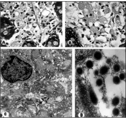
knowledge has emerged since then. Classical neuropatholog-ical features include generalized cerebral atrophy and dif-fusely scattered small chalky nodules in cortical and subependy-mal gray matter. In fact, these nodules are true granulomas that contain PAS-positive foamy macrophages and reactive astrocytes. Areas of intense demyelination (resembling mul-tiple sclerosis) and microinfarcts are also sometimes identi-fied in tissue specimens10.
Our patient first presented neurological manifestations two years after SMZ-TMP withdrawal. Such relapses of WD were frequently reported w hen initial antibiotic treatment had not been adequate and frequently included cerebral manifestations. Short-term use of SM Z-TM P or tetracycline (w hich does not cross the blood-brain barrier) were particularly related to CNS relapses11,12and a worse clinical course. Feldman et al.13
report-ed a patient disclosing acute meningoencephalitis during a relapse of the disease whose abnormalities closely recalled our ow n patient.
There are still conflicting data related to the real preva-lence of neurological signs and symptoms in WD. Recent review articles3,8suggest that almost half of WD patients have
indeed evidence of CNS dysfunction. These manifestations are quite diverse and traditionally include OM M , cognitive changes, voluntary gaze abnormalities, pyramidal signs, sen-sory deficits, hypothalamic manifestations, cranial nerve abnor-malities, ataxia and seizures. Besides such clinical variability, some patterns of presentation should clearly point to the diagnosis. One example is the classic triad of dementia, supranuclear ophthalmoparesis and myoclonus, w hich is observed in roughly 10% of cases. In this regard, OM M has not been described in any other neurological disturbance to date and therefore could be considered pathognomonic of CNS WD. It occurs in 20% of cases and consists in the synchronous coupling of rhythmic contractions of masticatory muscles and the pendular vergence oscillations of both eyes at a slow rate (1-2Hz). This unique sign of WD seems to be caused by brain stem lesioning and somew hat resembles segmental spinal myoclonus14.
Cognitive changes are perhaps the most common neuro-logical abnormalities in WD (71% of cases) and generally show incomplete response to the treatment. They tend to progress insidiously and are usually accompanied by depres-sion, personality and behavioral changes. Previous reports
identified disabling impairment in the domains of sustained attention, memory, executive function and constructional prax-is15. At later stages, these deficits completely fulfill criteria for
dementia (much like Alzheimer´s disease) and steadily w ors-en prognosis.
Voluntary gaze abnormalities preferentially affect vertical movements of the eyes and are noticed in half of the patients. There is usually progressive slowing of upward saccades, soon follow ed by dow nward saccades. Some affected individuals become virtually unable to look up or dow n at last and may receive a diagnosis of progressive supranuclear palsy (PSP). Isolated horizontal supranuclear gaze palsy is extremely uncommon, but combined impairment in both directions (hor-izontal and vertical) is frequently seen8. It is also interesting
that all cases presenting OM M had vertical supranuclear pal-sy.
Internuclear ophthalmoplegia, pupillary abnormalities and ptosis have been described in isolated WD patients. Oculomotor (III), trochlear (IV) and abducens (VI) palsies are not usual either, but reported in at least 5% of affected individuals7.
Together, cranial nerve abnormalities occur in 25% of cases.
Hypothalamic involvement has been clearly identified in pre-vious papers16-18. Sleep/arousal disturbances, hyperphagia and
polydipsia w ere reported in 31% of cases.
Similarly, endocrine deficits related to hypopituitarism w ere recognized in the setting of CNS WD19. These patients
presented with signs of hypothyroidism, complaints of erec-tile dysfunction (just like our case) or galactorrhoea and had good response to antimicrobial and hormonal replacement therapy.
Pyramidal and sensation deficits are other signs of CNS derangement recorded in WD, just as myoclonus and seizures, which are noticed in almost one fourth of patients.Additionally, gait and stance instability related to cerebellar damage occur in 20% of patients, being a major source of functional impair-ment8. A few cases presenting w ith rapidly evolving cranial
hypertension w ere reported, most of them caused by tumor-like lesions or acute hydrocephalus (Sylvius aqueduct steno-sis)3,11. Various forms of peripheral neuropathy have been
ob-served as in other chronic inflammatory bowel diseases, prob-ably associated to malabsorptive nutritional deficiencies20.
Conversely, spinal cord3,21and muscle3involvement are
exceed-ingly uncommon.
Once suspected, WD has been classically confirmed by intestinal biopsy and histopathological analysis (PAS staining and EM ) in at least 80% of affected individuals. Particularly in CNS WD, it is accurate in 70% of cases even if there are no abdominal complaints. Hence, patients with neurological defi-cits compatible w ith WD should initially have an upper diges-tive endoscopy and duodenal biopsy. Recently, polymerase chain-reaction (PCR) has been used to confirm the diagnosis
of WD2. This is emerging as a promising technique w ith
high-er sensibility and specificity22. Positive results of PCR assays
against Tropheryma whipplei have been obtained from sever-al tissues, including brain and CSF23. Therefore, PCR w ill
prob-ably become an interesting tool for WD diagnosis (particular-ly for CNS restricted disease) and long-term follow up. Microbial cultures and serology are of little diagnostic help yet.
Neuroimaging and CSF analysis are usually undertaken and can be diagnostically useful. Brain MRI findings in WD are large-ly non-specific24. Lesions tend to affect in decreasing order of
frequency, cerebral frontal cortex, basal ganglia, periventric-ular w hite matter, hypothalamus, temporal and parietal cor-tex3. Similarly, CSF non-specific abnormalities have been
iden-tified in at least half of patients8. M ost had slight CSF protein
elevation, lymphocyte pleocytosis and PAS-positive cells. Nevertheless, some authors report acute meningitic presen-tations with marked pleocytosis with polymorphonuclear pre-dominance25,26.
Diagnostic guidelines based on available data w ere ulti-mately proposed by Louis et al.8. These authors considered the
diagnosis of CNS WD definite in patients having at least one of the follow ing criteria: 1. OM M ; 2.positive tissue biopsy (PAS-positive cells); 3. positive PCR analysis.Additionally, if his-tological or PCR analysis w ere not performed on CNS tissue, then the patient must also demonstrate compatible neurolog-ical signs (supranuclear vertneurolog-ical gaze palsy, rhythmic myoclonus, dementia or hypothalamic manifestations). Our patient clear-ly matches these criteria, having laboratory and clinical evi-dence of CNS WD.
In the past, several antibiotic regimens w ere used for WD w ithout convincing results. The proper choice of an effective drug is now recognized as an essential part of the treatment, lessening the risks for future CNS relapses27,28. Oral SM Z-TM P
(160mg/800mg bid) is the currently recommended long-term therapy and should be prescribed for at least one year2,29. In
addition, a 2-week course of parenteral therapy consisting of ceftriaxone 2g/day is strongly suggested in severely ill patients and must precede maintenance therapeutics. Alternative approaches using penicillin plus streptomycin or minocycline have been used on an individual basis and can be applied in cases of sulfonamide intolerance.
These directions led to a lower rate of clinical relapses and a much better long-term outcome. SMZ-TMP was particularly effective for CNS WD patients, improving or at least stabiliz-ing their course3. Nevertheless, the precise duration of
antimi-crobial treatment and the best follow-up approach must still be determined. In some resistant cases, therapy is still a chal-lenge but recent reports2 on the use of supportive gamma
REFERENCES
1. Whipple GH. A hitherto undescribed disease characterized anatomi-cally by deposits of fat and fatty acids in the intestinal and mesenteric lymphatic tissues. Johns Hopkins Hosp Bull 1907;18:382-391. 2. Marth T, Raoult D. Whipple´s disease. Lancet 2003;36:239-246. 3. Gerard A, Sarrot-Reynauld F, Liozon E, et al. Neurologic presentation
of Whipple disease: report of 12 cases and review of the literature. Medicine (Baltimore) 2002;81:443-457.
4. Brown AP, Lane JC, Murayama S, Vollmer DG. Whipple´s disease pre-senting with isolated neurological symptoms: case report. J Neurosurg 1990;73:623-627.
5. Bostwick DG, Bensch KG, Burke JS, et al. Whipple´s disease present-ing as aortic insufficiency. N Engl J Med 1981;305:995-998.
6. Raoult D. A febrile, blood culture-negative endocarditis. Ann Intern Med 1999;131:144-146.
7. Chan RY, Yannuzzi LA, Foster CS. Ocular Whipple´s disease: earlier definitive diagnosis. Ophthalmology 2001;108:2225-2231.
8. Louis ED, Lynch T, Kaufmann P, Fahn S, Odel J. Diagnostic guidelines in central nervous system Whipple´s disease. Ann Neurol 1996;40:561-568. 9. Sieracki JC. Whipple’s disease: observations on systemic involvement.
Amer Med Asso Arch Pathol 1958;66:464-467.
10. Anderson M. Neurology of Whipple´s disease. J Neurol Neurosurg Psychiatry 2000;68:2-5.
11. De Coene B, Gilliard C, Indekeu P, et al. Whipple’s disease confined to the central nervous system. Neuroradiology 1996;38:325-327. 12. Verhagen WIM, Huygen PLM, Dalman JE, Schuurmans MMJ. Whipple’s
disease and the central nervous system: a case report and a review of the literature. Clin Neurol Neurosurg 1996;98:299-304.
13. Feldman M, Hendler RS, Morrison EB. Acute meningoencephalitis after withdrawal of antibiotics in Whipple’s disease. Ann Intern Med 1980;93:709-711.
14. Schwartz MA, Selhorst JB, Ochs AL, et al. Oculomasticatory myorhyth-mia: a unique movement disorder occurring in Whipple´s disease. Ann Neurol 1986;20:677-683.
15. Manzel K, Tranel D, Cooper G. Cognitive and behavioral abnormali-ties in a case of central nervous system Whipple disease. Arch Neurol 2000;57:399-403.
16. Halperin JJ, Landis DM, Kleinman GM. Whipple’s disease of the nerv-ous system. Neurology 1982;32:612-617
17. Feurle GE, Volk B, Waldherr R. Cerebral Whipple’s disease with neg-ative jejunal histology. N Engl J Med 1979;300:907-908.
18. Madoule P, Ciaudio-Lacroix C, Halimi P, et al. Osteoarticular lesions in Whipple’s disease. a propos of a destructive form and review of the literature. J Radiol 1985;66:345-350
19. Brändle M, Ammann P, Spinas GA, et al. Relapsing Whipple’s disease presenting with hypopituitarism. Clin Endocrinol 1999;50:399-403. 20. Topper R, Gartung C, Block F. Neurologic complications in
inflamma-tory bowel diseases. Nervenarzt 2002;73:489-499.
21. Clarke CE, Falope ZF, Abdelhadi HA, et al. Cervical myelopathy caused by Whipple’s disease. Neurology 1998;50:1505-1506.
22. Ramzan NN, Loftus E, Burgart LJ, et al. Diagnosis and monitoring of Whipple disease by polymerase chain reaction. Ann Intern Med 1997;126:520-527.
23. Von Herbay A, Ditton HJ, Scuhmacher F, et al. Whipple’s disease: stag-ing and monitorstag-ing by cytology and polymerase chain reaction analy-sis of cerebrospinal fluid. Gastroenterology 1997;113:434-441. 24. Kremer S, Besson G, Bonaz B, Pasquier B, Le Bas JF, Grand S. Diffuse
lesions in the CNS revealed by MR imaging in a case of Whipple dis-ease. Am J Neuroradiol 2001;22:493-495.
25. Romanul FC, Radvany J, Rosales RK. Whipple’s disease confined to the brain: a case studied clinically and pathologically. J Neurol Neurosurg Psychiatry 1977;40:901-909.
26. Thompson DG, Leidingham JM, Howard AJ, Brown CL. Meningitis in Whipple’s disease. BMJ 1978;2:14-15.
27. Feurle GE, Marth T. An evaluation of antimicrobial treatment for Whipple’s disease: tetracycline versus trimethoprim-sulfamethoxa-zole. Dig Dis Sci 1994;39:1642-1648.
28. Misbah SA, Mapstone NP. Whipple’s disease revisited. J Clin Pathol 2000;53:750-755.