Syntheses, crystal structure and spectroscopic characterization of
no
v
el
N
-R-sulfonyldithiocarbimate and triphenylphosphine nickel(II)
complexes
Marcelo R.L. Oli
v
eira
a,*
, Heulla P. Vieira
b, Geni
v
aldo J. Perpe´tuo
c, Jan Janezak
d,e,
Vito M. De Bellis
eaDepartamento de Quı´mica, Uni
versidade Federal de Vic¸osa, Vic¸osa, MG, CEP 36571-000, Brazil
bDepartamento de Quı´mica, Instituto de Cieˆncias Exatas e Biolo´gicas, Uni
versidade Federal de Ouro Preto, Ouro Preto, MG, CEP 35400-000, Brazil
cDepartamento de Fı´sica, Instituto de Cieˆncias Exatas e Biolo´gicas, Uni
versidade Federal de Ouro Preto, Ouro Preto, MG, CEP 35400-000, Brazil
dInstitute of Low Temperature and Structure Research, Polish Academy of Science, PO Box 1410, 50-950 Wroclaw, Poland
eDepartamento de Quı´mica, Instituto de Cieˆncias Exatas, Uni
versidade Federal de Minas Gerais, Belo Horizonte, MG, CEP 31270-901, Brazil
Received 8 April 2002; accepted 20 July 2002
Abstract
Three new complexes of the general formula: [Ni(PPh3)2(RSO2N/CS2)] where R/2-CH3C6H4(1), 4-CH3C6H4(2) and 4-BrC6H4
(3) were obtained in crystalline form by the reaction of the appropriate potassiumN-R-sulfonyldithiocarbimates (K2(RSO2N/CS2))
and triphenylphosphine with nickel(II) chloride in ethanol/water. The1complex crystallizes in the centrosymmetric space group of
the triclinic system with two molecules per unit cell, while2and3complexes crystallize in thePbcaspace group of the orthorhombic system with eight molecules per unit cell. The X-ray single-crystal analysis showed that all complexes present a similarly distorted square-planar configuration around the nickel atom due to the steric effect of the triphenylphosphine ligands and the didendate chelation by the two sulfur atoms of the dithiocarbimate ligand. The IR and UV/Vis spectral data are consistent with the formation
of almost square-planar nickel complexes. The 1H NMR, 13C NMR, 31P NMR spectra showed the expected signals for the triphenylphosphine and the dithiocarbimate moieties.
# 2002 Elsevier Science Ltd. All rights reserved.
Keywords: Dithiocarbimates; Triphenylphosphine; Nickel complexes; Crystal structures
1. Introduction
We became interested in the syntheses and character-ization of dithiocarbimate metal complexes due to their similarities with the dithiocarbamate compounds, which have a wide range of applications. For example, they are used in the rubber vulcanization process [1]as well as several dithiocarbamates salts and complexes have been used as fungicides [2]. Nickel(II) dithiocarbamates normally react with phosphines to form complexes with the NiS2P2chromophore[3]. Complexes containing the NiXS2P chromophore (X/halide) are usually
obtained by reacting NiX2 with dithiocarbamates and PR3 (R/alkyl/aryl) [4]. Complexes containing the NiXS2P chromophore have shown catalytic activity, especially for oligomerization of olefins [5/7]. Many complexes that involve nickel(II), phosphine and dithio-carbamate ligands, as [Ni(L)(PR3)2]
, [Ni(L)(P/P)]
, [NiX(L)(PR3)2] and [NiX(L)(P/P)] (L/several dithio-carbamates, R/many different groups, X/halides and pseudohalides and P/P/diphosphines) have been characterized[8,9].
Some complexes of palladium and platinum with dithiocarbimates and phosphines have been structurally characterized and using the quantum chemical calcula-tions (CNDO/2) the mechanism of the formation of the complexes has been investigated [10,11]. Additionally, dimeric nickel complexes [Ni(PR3)2(R?N/CS2)] (R and R? /alkyl/aryl) have been obtained by the reaction of
* Corresponding author. Tel.: /55-31-3899-3059; fax: /
55-31-3899-3065
E-mail address: [email protected](M.R.L. Oliveira).
www.elsevier.com/locate/poly
[Ni(NO2)(R?NHCS2)(PR3)] or [NiBr(R?NHCS2)(PR3)] with Lewis bases such as (CH3)3N and NH3 [12]. However, their structures were not determined by X-ray diffraction techniques.
Herein we investigate the three new bis(triphenylpho-sphine)N-R-sulfonyldithio-carbimatenickel(II) com-plexes (R/2-CH3C6H4, 4-CH3C6H4, 4-BrC6H4) that were obtained in the crystalline form by the reaction of NiCl2×/6H2O with triphenylphosphine and dithiocarbi-mate anions derived from sulfonamides. These com-plexes were characterized by UV/Vis, IR,
1
H, 13C and 31
P NMR, elemental analyses for C, H, N, Ni and by single crystal X-ray diffraction techniques.
2. Experimental
2.1. Methods and materials
The solvents were purchased from Merck and used without further purification. The sulfonamides, 4-bro-mophenylsulphonyl chloride, nickel(II) chloride hexa-hydrate and triphenylphosphine were purchased from Aldrich. Carbon disulfide and potassium hydroxide were purchased from Vetec. The phonamide was prepared from the 4-bromobenzenesul-phonyl chloride as described elsewhere [13]. The N -R-sulfonyldithiocarbimate potassium salts dihydrate were prepared in dimethylformamide from sulfonamides analogously as described in the literature[14,15]. These salts, which are soluble in water and insoluble in most of the organic solvents, were re-crystallized from hot ethanol/water. Melting points (m.p.) were determined with a Mettler FP5 equipment. Microanalyses for C, H and N were obtained from a Perkin/Elmer 2400 CHN. Nickel was analyzed by atomic absorption with a Hitachi Z-8200 Atomic Absorption Spectrophotometer. The IR spectra were recorded with a Perkin/Elmer 283 B infrared spectrophotometer using CsI pellets. The UV/Vis spectra were recorded with a Beckman DU 640 spectrometer using nujol mull suspension. The1H (400 MHz),13C (100 MHz) and31P (162 MHz) NMR spectra of the complexes were recorded on a Bruker Advance RX-400 spectrophotometer in CDCl3with TMS (H3PO4 for31P NMR spectra) as internal standard.
2.2. Syntheses
The syntheses of the nickel(II) complexes were performed according to the Scheme 1. A solution of
N-R-sulfonyldithiocarbimate dihydrate (1.0 mmol) in water (10 ml) was added to a solution of triphenylpho-sphine (2.0 mmol) in ethanol (40 ml). Nickel(II) chloride hexahydrate (1.0 mmol) was added to the suspension and the reaction mixture was stirred for 6 h at room temperature (r.t.). The color of the suspension changed from green to pink/red. The solid product of the reaction was filtered, washed with distilled water and ethanol, and dried under reduced pressure for 1 day yielding [Ni(PPh3)2(RSO2N/CS2)] (ca. 70%). Suitable crystals for X-ray structure analysis were obtained after slow evaporation of solutions of the compounds in dichloromethane/methanol and few drops of water.
2.2.1.
Bis(triphenylphosphine)2-methylphenyldithiocarbimatenickel(II) (1)
Elemental analysis: Found (Calc.): C, 63.39 (63.78); H, 4.37 (4.50); N, 1.70 (1.69); Ni, 7.01 (7.08)%. M.p. (8C): 165.5/166.0. UV/Vis (nm): 197; 236; 320; 426 and 557. IR (most intense bands) (cm1): 1440 n(C/N); 1300 nass(SO2); 1120 nsym(SO2); 920 nass(CS2) and 345 n(NiS). 1H NMR (d): 7.88/7.79 (m, 1H, H2? (R group)); 7.42/7.05 (m, 33H, H3?, H4?, H5?, R group and triphenylphosphine signals) and 2.63 (s, 3H, CH3). 13
C{1H} NMR (d): 197.35 (N/CS2); 140.77 (C1?); 137.98 (C2?); 131.49 (C3?); 129.02 (C5?); 127.94 (C6?); 125.09 (C4?) and 20.84 (CH3). Triphenylphosphine signals: 134.39 (t, J/7, C2ƒ and C6ƒ); 130.89 (s, C4ƒ); 129.35 (d, J/31, C1ƒ); 128.45 (t,J/7, C3ƒand C5ƒ). 31
P NMR (d): 31.69 (s).
2.2.2.
Bis(triphenylphosphine)4-methylphenyldithiocarbimatenickel(II) (2)
Elemental analysis: Found (Calc.): C, 63.91 (63.78); H, 4.39 (4.50) N, 1.72 (1.69); Ni, 7.11 (7.08)%. M.p. (8C): 166.5/167.0. UV/Vis (nm): 197; 234; 320; 429 and 557. IR (most intense bands) (cm1): 1445 n(C/N); 1300 nass(SO2); 1135 nsym(SO2); 910 nass(CS2) and 344 n(NiS).1H NMR (d): 7.69/7.57 (m, 2H, H2?and H6?(R group)); 7.44/7.20 (m, 30H triphenylphosphine signals); 7.17/7.15 (m, 2H, H3?and H5?(R group)) and 2.39 (s, 3H, CH3).13C{1H} NMR (d): 197.37 (N/CS2); 142.58 (C1?); 139.42 (C4?); 128.94 (C3? and C5?); 127.49 (C2? and C6?); 21.78 (CH3). Triphenylphosphine signals: 134.29 (t, J/6, C2ƒ and C6ƒ); 130.64 (s, C4ƒ); 129.25 (d, J/46, C1ƒ); 128.27 (t, J/5, C3ƒ and C5ƒ). 31P NMR (d): 30.47 (s).
2.2.3.
Bis(triphenylphosphine)4-bromophenyldithiocarbimatenickel(II) (3)
Found (Calc.): C, 57.29 (57.80); H, 3.79 (3.84); N, 1.60 (1.57); Ni, 6.61 (6.57)%. M.p. (8C): 169.0/170.5. UV/Vis (nm): 193; 247; 308; 426 and 557. IR (most intense bands) (cm1): 1435 n(C/N); 1310 nass(SO2); 1135nsym(SO2); 915nass(CS2) and 335n(NiS).
1
H NMR (d): 7.68/7.65 (m, 2H, H2? and H6? (R group)); 7.45/
7.19 (m, 32H, H3?, H5?R group and triphenylphosphine signals). 13C{1H} NMR (d): 145.00 (C1?); 131.93 (C3? and C5?); 128.88 (C2? and C6?); 127.50 (C4?). Triphe-nylphosphine signals: 134.29 (t, J/6, C2ƒ and C6ƒ); 130.75 (s, C4ƒ); 129.33 (d,J/54, C1ƒ); 128.33 (t,J/5, C3ƒand C5ƒ).31P NMR (d): 31.75 (s).
2.3. X-ray crystallography
X-ray intensity data for all crystals were collected using graphite monochromatic Mo Ka radiation on a four circle KUMA KM-4 diffractometer with a two-dimensional area CCD detector. Thev-scan technique with Dv/0.758 for each image was used for data collection. The 960 images for six different runs covered over 90% of the Ewald sphere were performed. Initially the lattice parameters were refined on about 200 reflections obtained from 40 images for eight runs with different orientation in the reciprocal space. Finally the lattice parameters were refined by least-squares method based on the all reflection withI/4s(F2). One image was used as standard for monitoring the data collection after every 40 images, and no correction on the relative intensity variation was necessary. Integration of the intensities, correction for Lorenz and polarization effects were performed using a KUMA KM-4 CCD
program system[16]. Total of 18 419 (9819 independent,
Rint/0.0294), 27 613 (10 354 independent, Rint/ 0.0750) and 55 811 (9891 independent, Rint/0.0773) reflections were collected for the complexes1/3, respec-tively. The face-indexed analytical absorption was calculated using theSHELXTLprogram[17].
The structures were solved by a Patterson heavy-atom method using theSHELXL-97 program [18]. The
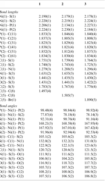
Patter-son map revealed the positions of the nickel, sulfur, bromide (3) and some of the P and C atoms. The remaining non-hydrogen atoms were located from difference Fourier synthesis. The structures were refined with anisotropic thermal parameters. Difference Fourier maps gave electron density concentrations approxi-mately located for all hydrogen atom positions; these positions were idealized (HFIX 43 for all hydrogen atoms of the phenyl rings with isotropic thermal parameters of 1.2 Ueq of the carbon atoms joined directly to the hydrogen atoms, and HFIX 137 for the CH3 group in the complexes 1 and 2 with isotropic thermal parameters of 1.5 Ueq of the methyl carbon atom). Final differences Fourier maps showed no peaks of chemically significance. Details of data collection parameters and final agreement factors are collected in Table 1. Selected bond lengths and angles are listed in Table 2.
3. Results and discussion
The compounds are quite stables at the ambient conditions. In contrast to the dithiocarbamate Ni-complexes with the general formula of [Ni(PR3)2(RN/ CS2)] which are soluble in THF, benzene and dichlor-omethane [12], these N-R-sulfonyldithiocarbimate Ni-complexes characterized in this paper are insoluble in most organic solvents and are slightly soluble in chloro-form and dichloromethane. These complexes are stable up to the melting point without decomposition. At-tempted to prepare the monotriphenylphosphine Ni-complexes like K[NiCl(PPh3)(N-RSO2N/CS2)] in the same conditions described above, with 1 equiv. of triphenylphosphine was unsuccessful.
The electronic spectra of the complexes show two shoulders (ca. 426 and 557) that are typical for square-planar nickel complexes and are assigned to d/d transitions [9]. The 190/300 nm region in the spectra of the complexes is dominated by three very intense bands of the triphenylphosphine and the dithiocarbi-mate ligands.
There are no strong or medium bands in the 1400/ 1600 cm1 region in the IR spectra of the potassium dithiocarbimates related to the complexes 1/3, the n(CN) band being observed around 1260 cm1
[14,19]. This low value indicates a great contribution of the canonical forms (a) and (b) for the resonance hybrid (Fig. 1). A strong band observed around 1455 cm1in the spectra of the complexes was assigned to the n(CN). In the spectrum of the complex [Ni(PPh3)2 (4-CH3C6H4N/CS2)] that has no SO2group linked to the nitrogen atom, this band is located at 1500 cm1[12]. In the case of the complex [Ni(4-CH3C6H4SO2N/CS2)2]2
this band is observed around 1345 cm1[20]. This fact is probably due to a major negative charge on the Ni atom in this anionic complex in comparison to the complexes here synthesized. The substitution of one dithiocarbimate ion by two triphenylphosphine mole-cules is expected to result in a greater drift of electrons from the remainder dithiocarbimate ion to the metal. This effect is also observed, for example when the spectrum of the complex [Ni(dtc)2] (dtc/N,N -diethyl-dithiocarbamate) (n(CN) band is observed in 1518 cm1) is compared with the spectrum of [Ni(dtc)(PPh3)2]
(n(CN) band is observed in 1530 cm1) [8]. The nass(CS2) were observed at higher frequency in the spectra of the potassium salts of dithiocarbimates (ca. 955 cm1) [19] than that in the spectra of the complexes here studied (ca. 915 cm1). The shifts observed in the nass(CS2) and n(CN) in the spectra of the complexes when compared with the spectra of the ligands, are consistent with the increased importance of the canonical form (c) after complexation (Fig. 1). The spectra of the complexes also show the expected medium band in the 300/400 cm
assigned to the NiS vibrations [21]. The n(NiP) band was not observed above 200 cm1.
The NMR spectra of 1/3 were typical for diamag-netic species. The 1H NMR spectra of the complexes showed the signals for the hydrogen atoms of the triphenylphosphine. The remaining signals could be assigned to the CH3group of the aromatic moiety and the other aromatic hydrogen atoms. The integration curves on the 1H NMR spectra were consistent with a 2:1 proportion between the triphenylphosphine ligands and the dithiocarbimate anion. The signals in the spectra of the free dithiocarbimates [19] and in the complexes show approximate the same chemical shifts. The chemical shifts of the aromatic carbon atoms of the dithiocarbimate anions in the complexes are similar to those of the corresponding sulfonamides [22]. The N/ CS2(C1) signal is shifted in the spectra of the complexes to higher field if compared with the spectra of the ligands (this signal is too weak and was not observed for 3). Although the solvents are, necessarily different, this shift is expected. If the canonical form (c) (Fig. 1) is more important for the complexes than for the ligands, then the C1 carbon atom is expected to be more shielded in the complexes. The C1 signal for the complex2is also shifted to higher field when compared with the spectrum of the correspondent anionic complex [Ni(4-CH3C6H4SO2N/CS2)2]2
[20]. Most of the triphenyl-phosphine signals in the13C NMR spectra appeared as pseudo triplets or doublets. This is consistent with
square-planar geometry for the complexes. Doublets or pseudo triplets are commonly observed in the 13C NMR spectra of bis-phosphine transition metal cis -complexes [23]. As expected, the 31P NMR spectra exhibited only one signal (ca. 30 d) indicating that all phosphorus atoms in the molecules are magnetically equivalent.
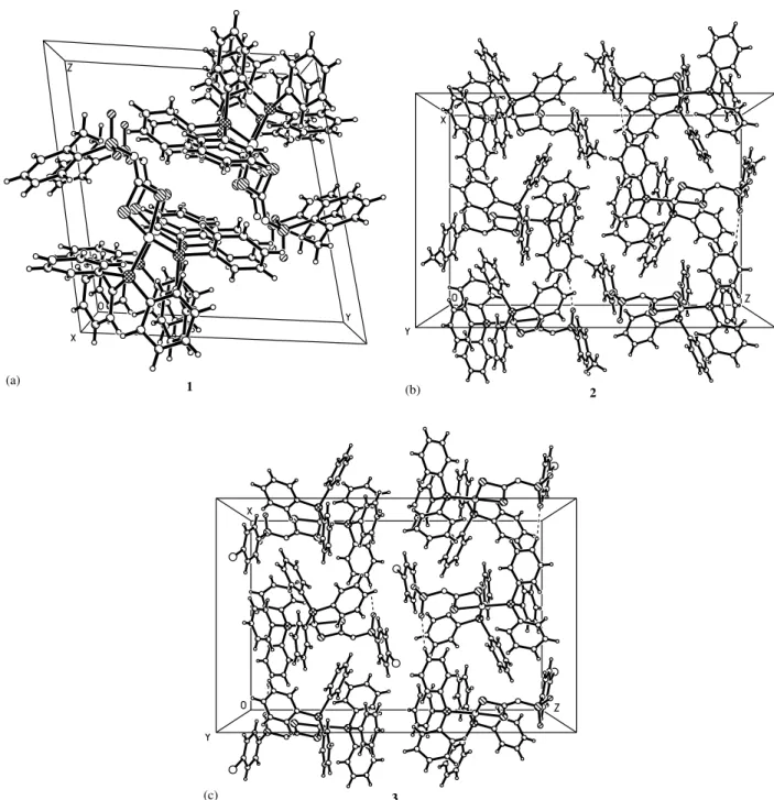
The X-ray molecular structures of the1/3complexes are illustrated in Fig. 2(a/c), respectively. In all com-plexes, the nickel cation is coordinated by two triphe-nylphosphine ligands and chelated by two sulfur atoms of the N-R-sulfonyldithiocarbimate anion into a simi-larly distorted square-planar configuration. The P(1)/ Ni(1)/S(1) and P(2)/Ni(1)/S(2) angles are less dis-torted from 908 than the P(1)/Ni(1)/P(2) and S(1)/ Ni(1)/S(2) angles. The P(1)/Ni(1)/P(2) angle is sig-nificantly greater than 908 in all complexes due to the steric effect of the large triphenylphosphine ligands and the S(1)/Ni(1)/S(2) angle is smaller than 908due to the chelation of the N-R-sulfonyldithiocarbimate ligand. The N-R-sulfonyldithiocarbimate anion (RSO2N/ CS2)2
as an asymmetric didentate ligand forms a stable four membered ring by chelation through the two sulfur atoms (NiS2C). The Ni(1)/S(1) bond lengths of 2.190(1) in 1, 2.179(1) in 2 and 2.178(1) in 3 A˚ are shorter than the Ni(1)/S(2) [2.220(1), 2.219(1) and 2.224(1) A˚ in1,2and3, respectively]. These deviations are more obvious than those found in the [Ni(CH3SO2N/CS2)2]2 [24]. This fact may be
ex-Table 1
Crystallographic data
Complex 1 2 3
Chemical formula C44H37NNiO2P2S3 C44H37NNiO2P2S3 C43H34BrNNiO2P2S3
Formula weight 828.61 828.61 893.48
Crystal dimensions (mm) 0.300.280.14 0.240.180.14 0.360.160.06 Crystal system triclinic orthorhombic orthorhombic
Space group P/1¯/ Pbcn Pbcn
a(A˚ ) 10.000(2) 16.556(3) 16.497(3)
b(A˚ ) 13.923(3) 18.333(4) 18.556(4)
c(A˚ ) 14.953(3) 25.412(5) 25.364(5)
a (8) 100.16(3)
b (8) 103.28(3)
g (8) 95.20(3)
V(A˚3) 1975.7(7) 7713(3) 7764(3)
Z 2 8 8
Dobsd(g cm3) 1.39 1.42 1.52
Dcalc(g cm3) 1.393 1.427 1.529
T(8C) 295 295 295
l(A˚ ) 0.71073 0.71073 0.71073
m(cm1) 0.769 0.788 1.812
Transmission factors: min, max 0.8020, 0.8999 0.8334, 0.8977 0.5616, 0.8991
R(F2) 0.0433 0.0540 0.0449
Rw(F
2
) 0.1104 0.1385 0.0639
S 1.015 1.140 1.004
Residual electron density (e A˚3) 0.524 and0.552 0.656 and0.611 0.819 and1.454
a
Function minimized:aw(½Fo½½Fc½) 2
, wherew4Fo 2
/s2(Fo 2
); [aw(½Fo½½Fc½) 2
/(NoNv)] 1/2
, whereNo, number of observations,Nv, number of
variables. The final discrepancy factors:Ra½Fo½½Fc½/a½Fo½andRw[aw(½Fo½½Fc½) 2
/awFo 2
plained by the stereochemistry affecting the phenyl ring of the N-R-sulfonyldithiocarbimate ligand, which is located in cis position to the S1 atom. The C1/N1 bond lengths of 1.279(3), 1.288(3) and 1.296(4) in1/3, respectively, are shorter than the normal single C(sp2)/ N(sp2) bond length (ca. 1.35 A˚ ), and similar to that of the double bond C/N (1.275/1.295 A˚ ) [25,26]. The C(1)/S(1) and C(1)/S(2) averaged bond lengths (1.745 A˚ ) are slightly shorter than the typical C/S single bond length (ca. 1.81 A˚ ) due to partialp-delocalization in the S/C/S group. The S(1)/C(1)/N(1) angles of 132.0(2)8,
Table 2
Selected bond lengths (A˚ ) and angles (8)
1 2 3
Bond lengths
Ni(1)S(1) 2.190(1) 2.179(1) 2.178(1) Ni(1)S(2) 2.220(1) 2.219(1) 2.224(1) Ni(1)P(1) 2.206(1) 2.134(1) 2.227(1) Ni(1)P(2) 2.224(1) 2.230(1) 2.184(1) P(1)C(11) 1.833(3) 1.846(4) 1.840(4) P(1)C(21) 1.837(3) 1.805(3) 1.808(3) P(1)C(31) 1.825(3) 1.829(4) 1.823(4) P(2)C(41) 1.838(3) 1.821(4) 1.820(3) P(2)C(51) 1.852(3) 1.812(4) 1.837(3) P(2)C(61) 1.834(3) 1.830(4) 1.834(4) C(1)S(1) 1.751(3) 1.739(4) 1.764(3) C(1)S(2) 1.740(3) 1.743(4) 1.725(3) C(1)N(1) 1.279(3) 1.288(5) 1.296(4) N(1)S(3) 1.631(2) 1.655(3) 1.626(3) O(1)S(3) 1.441(2) 1.435(3) 1.430(2) O(2)S(3) 1.431(2) 1.441(3) 1.422(2) C(2)S(3) 1.783(3) 1.767(4) 1.776(4) C(3)C(8) 1.497(4)
C(5)C(8) 1.505(7)
C(5)Br(1) 1.890(5)
Bond angles
P(1)Ni(1)P(2) 98.48(4) 98.84(4) 98.92(4) S(1)Ni(1)S(2) 77.87(4) 78.18(4) 78.14(3) S(1)Ni(1)P(1) 92.31(4) 90.76(4) 91.16(4) S(1)Ni(1)P(2) 168.21(3) 168.58(4) 167.95(4) S(2)Ni(1)P(1) 167.92(3) 167.01(4) 167.42(4) S(2)Ni(1)P(2) 91.96(4) 92.94(4) 92.53(4) S(1)C(1)S(2) 105.1(2) 105.6(2) 105.3(2) S(1)C(1)N(1) 132.0(2) 132.2(3) 131.0(3) S(2)C(1)N(1) 122.9(2) 122.1(3) 123.6(3) C(1)N(1)S(3) 120.7(2) 120.6(3) 121.5(2) N(1)S(3)O(1) 111.9(1) 111.6(2) 111.5(2) N(1)S(3)O(2) 106.0(1) 104.2(2) 105.8(2) O(1)S(3)O(2) 116.9(1) 118.7(2) 117.7(2) N(1)S(3)C(2) 105.7(1) 107.3(2) 107.7(2) O(1)S(3)C(2) 108.2(1) 108.0(2) 106.9(2) O(2)S(3)C(2) 107.5(1) 106.5(2) 106.8(2)
Fig. 1. Some possible canonical forms for theN -R-sulfonildithiocar-bimate anion.
132.2(3)8 and 131.0(3)8 in the complexes 1/3, respec-tively, are significantly greater than S(2)/C(1)/N(1) [122.9(2) in 1, 122.1(3)8 in2 and 123.6(3)8 in 3] due to the interaction between the SO2R group and the S(1) atom, which are joined incis position in relation to the C(1)/N(1) bond. The steric effect of the R-sulfonyl group is greater than the effect of the lone-pair of electron at the N(1) atom, since the C(1)/N(1)/S(3) angle is greater than 1208in all complexes. If R is a small group such as/SO2CH3[24]or /C/N[27]the repulsive interaction predicted by the valence-shell electron pair repulsion theory (VSEPR)[28]between the lone-pair of
electron at the N(1) atom and the S(2) atom is greater than the steric effect and the angle S(2)/C(1)/N(1) is greater than the angle S(1)/C(1)/N(1). In other dithio-carbimate complexes with larger R groups, the steric effect between S(1) and R group is more important and similarly as in the 1/3 complexes the angle S(1)/C(1)/ N(1) is greater than S(2)/C(1)/N(1)[29].
The C(1)/N(1)/S(3) fragment of the N -R-sulfonyl-dithiocarbimate ligand (Fig. 2(a/c)) is coplanar with the NiS2P2 fragment, thus the P(1)P(2)Ni(1)S(1)S(2)C(1)-N(1)S(3) atoms lie in plane. The phenyl ring of theN -R-sulfonyldithiocarbimate ligand is inclined by about
88.6(2)8in the complex 1, and 78.1(2)8 and 79.1(2)8in the isostructural crystals of the 2 and 3 complexes, respectively. The torsion angle of C(1)/N(1)/S(3)/C(2) describing the conformation of the ligand along the N(1)/S(3) bond is more similar in the complexes1and2 [/65.2(2)8 and /65.0(1)8], since both have a methyl group in the phenyl ring, than in3[/61.4(2)8], contain-ing a Br-substituted aromatic rcontain-ing. The S(3) atom has an expected slightly distorted tetrahedral geometry. The S(3)/O(1) and S(3)/O(2) bond lengths indicate their double bond character (S/O); a typical distance of the double S/O bond ranging from 1.431 to 1.442 A˚ [27]. The S(3)/C(2) bond with a distance ranging from 1.767(4) to 1.783(3) A˚ is well correlated with the CAr/ S bond length in the related complexes which comprise the CAr/SO2/N fragment [26]. The S(3)/N(1) bond length with avalue ranging from 1.626(3) to 1.655(3) A˚ indicates a single bond nature; the typicalvalue of the N(sp2)/S bond distance is 1.623/1.659 A˚ [26]. Both P atoms of the triphenylphosphine ligands have similar tetragonal geometry. The phenyl ring C(21)/C(26) of the P(1) triphenylphosphine is almost parallel to the phenyl ring C(41)/C(46) of the P(2) triphenylphosphine, the dihedral angle being equal to 9.3(2)8in the complex 1, and 15.2(2)8 and 15.8(2)8 in isostructural 2 and 3 complexes. Although the crystal of the complex1, is not isostructural with the crystals of 2 and 3 the structure analysis clearly shows that the geometry of the mole-cules of the compound1is quite similar to that of2and 3. The differences between the1and2and/or3 may be found in the molecular arrangement and the crystal packing. The complexes 1 and 2 differ only in the position of the methyl group substituted in the aromatic ring of the N-R-sulfonyldithiocarbimate ligands (1 in
orthoand2inparaposition). The crystal of the isomer2 is more compacted since the molecules interact more effectively than in compound 1. The arrangement of molecules in both2 and3 crystals is quite similar since both have para-substituents. These structures are slightly stabilized by weak C/H O intermolecular hydrogen interactions of neighbouring molecules to form a polymeric structure. The C/H O interactions with C O of approximately 3.37 and 3.36 A˚ distances in 2 and 3 compounds, respectively, lead to the formation of the pseudo mono-dimensional columns aligned along the a-axis in the crystal (Fig. 3(b/c)).
Although the intermolecular C/H O are weak, we suppose that they are important for the crystal packing, since this can be the reason for the more compacted arrangement of the molecules in2and in3in relation to the crystal of1, in which the C/H O interactions were not observed.
4. Conclusion
Three novel bis(triphenylphosphine)N -R-sulfonyl-dithiocarbimatenickel(II) complexes were prepared and characterized by UV/Vis, IR,
1
H, 13C and 31P NMR, elemental analyses for C, H, N, Ni and by single crystal X-ray diffraction techniques.
The spectroscopic and X-ray data for the complex [Ni(4-CH3C6H4SO2]2[20]show an interesting relation with the data obtained for compound 2(Table 3). The C(1)/N(1) bond length in 2 is shorter than in [Ni(4-CH3C6H4SO2N/CS2)2]2
[20]. The wavenumber for the n(CN) in the IR spectrum of2is greater than that in the spectrum of the related anionic complex and this band is observed in smaller wavenumber in the spectrum of the parent dithiocarbimate ligand [19]. The NMR spectra show that the carbon atom of the dithiocarbimate group of 2 is more shielded than that of the anionic complex and in the parent ligand. These facts are in accord with an increase of the contribution of the (c) canonical form (Fig. 1) to the resonance hybrid from the ligands to the complexes, with a consequent increase of the CN double bond character.
5. Supplementary material
Crystallographic data for the structural analysis have been deposited with the Cambridge Crystallographic Data Centre, CCDC Nos. 181695, 181696 and 181697 for the compounds 1/3, respectively. Copies of this information may be obtained free of charge from The Director, CCDC, 12 Union Road, Cambridge, CB2 1EZ, UK (fax: /44-1223-336033; e-mail: deposit@ ccdc.cam.ac.uk or www: http://www.ccdc.cam.ac.uk).
Table 3
Comparison between selected crystallographic and spectroscopic data for the CN bond in compound2and related compounds
Parameters 2 [Ni(4-CH3C6H4SO2NCS2)]2b (4-CH3C6H4SO2NCS2)2a
CN length (A˚ ) 1.288(5) 1.35(2)
n(CN) (cm1) 1445 1390 1260
13
C chemical shift (NCS2) (ppm) 197.37 210.07 225.19
Acknowledgements
This work has been supported by CNPq, CAPES and FAPEMIG (Brazil).
References
[1] P.J. Nieuwenhuizen, J. Reedijk, M. van Duin, W.J. McGill, Ruber Chem. Technol. 70 (1997) 368.
[2] D. Coucouvanis, Prog. Inorg. Chem. 11 (1969) 233.
[3] S. Thiumaran, K. Ramalingan, Transition Met. Chem. 25 (2000) 60.
[4] A. Manohar, L. Righi, K. Ramalingan, G. Bocelli, Inorg. Chim. Acta 314 (2001) 172.
[5] R.G. Cavell, B. Creed, L. Gelmini, D.J. Law, R. McDonald, A.R. Sanger, A. Somogyvari, Inorg. Chem. 37 (1998) 757.
[6] A.M.A. Bennet, D.A. Thorton, Spectroscopy 10 (1992) 85. [7] G.A. Foulds, A.M.A. Bennet, M.I. Niven, D.A. Thorton, K.J.
Cavell, S. Desjardins, E.J.J. Peacock, Mol. Catal. 87 (1994) 117. [8] K. Ramalingan, G. Avaramudan, M. Seshasayee, Inorg. Chim.
Acta 128 (1987) 231.
[9] R. Pastorek, Z. Tra´vnı´cˇek, J. Marek, D. Dastych, Z. .Sˇidela´r,
Polyhedron 19 (2000) 1713.
[10] G.A. Katsoulos, C.A. Tsipis, Inorg. Chim. Acta 84 (1984) 89. [11] J. Ahmed, K. Itoh, I. Matsuda, F. Veda, Y. Ishii, J.A. Ibers,
Inorg. Chem. 16 (1977) 620.
[12] C.A. Tsipis, I.P. Meleziadis, D.P. Kessissoglou, G.A. Katsoulos, Inorg. Chim. Acta 90 (1984) L19.
[13] A.I. Vogel, A Textbook of Practical Organic Chemistry Including Qualitative Organic Analysis, Longmans, Green, London, 1966. [14] K. Hartke, Arch. Pharm. 299 (1966) 164.
[15] H.U. Hummel, U.Z. Korn, Z. Naturforsch., Teil. B 44 (1989) 24. [16] Kuma Diffraction. KM-4 CCD Software, Ver. 163, Wroclaw,
Poland, 1999.
[17] G.M. Sheldrick,SHELXTL. Program System, Siemens Analytical X-ray Instruments, Madison, WI, 1990.
[18] G.M. Sheldrick, SHELXL97: Program for the Solution and
Refinement of Crystal Structures, Univesity of Go¨ttingen, Ger-many, 1997.
[19] M.R.L. Oliveira, V.M. De Bellis, Transition Met. Chem. 24 (1999) 127.
[20] M.R.L. Oliveira, J.E.J.C. Grau´do, N.L. Speziali, V.M. De Bellis, Struct. Chem. 10 (1999) 41.
[21] K. Nakamoto, Infrared and Raman of Inorganic and Coordina-tion Compounds, 3rd ed., Wiley, New York, 1978, p. 339. [22] M. Pomerantz, W.N. Chou, M.K. Witczak, C.G. Smith, J. Org.
Chem. 52 (1987) 159.
[23] D.A. Redfield, L.W. Cary, J.H. Nelson, Inorg. Chem. 14 (1975) 50.
[24] M.R.L. Oliveira, V.M. De Bellis, N.G. Fernandes, Struct. Chem. 8 (1997) 205.
[25] D. Coucouvanis, J.P. Frackler, Jr., Inorg. Chem. 6 (1967) 2047. [26] F.H. Allen, O. Kennard, D.G. Watson, L. Brammer, A.G.J.
Orpen, Chem. Soc., Perkin Trans. 2 (1987) S1. [27] F.A. Cotton, C.B. Harris, Inorg. Chem. 7 (1968) 2140. [28] R.J.J. Gillespie, Chem. Educ. 40 (1963) 295.