Radiol Bras. 2014 Jul/Ago;47(4):217–222 217
Optimization of a protocol for myocardial perfusion scintigraphy
by using an anthropomorphic phantom
*
Estudo de otimização de protocolo em cintilografia de perfusão miocárdica com a utilização de um simulador antropomórfico
Ramos SMO, Glavam AP, Kubo TTA, Sá LV. Optimization of a protocol for myocardial perfusion scintigraphy by using an anthropomorphic phantom. Radiol Bras. 2014 Jul/Ago;47(4):217–222.
Abstract
R e s u m o
Objective: To develop a study aiming at optimizing myocardial perfusion imaging.
Materials and Methods: Imaging of an anthropomorphic thorax phantom with a GE SPECT Ventri gamma camera, with varied activities and acquisition times, in order to evaluate the influence of these parameters on the quality of the reconstructed medical images. The 99mTc-sestamibi radiotracer was utilized, and then the images were clinically evaluated on the basis of data such as summed stress score, and on the technical image quality and perfusion. The software ImageJ was utilized in the data quantification.
Results: The results demonstrated that for the standard acquisition time utilized in the procedure (15 seconds per angle), the injected activity could be reduced by 33.34%. Additionally, even if the standard scan time is reduced by 53.34% (7 seconds per angle), the standard injected activity could still be reduced by 16.67%, without impairing the image quality and the diagnostic reliability.
Conclusion: The described method and respective results provide a basis for the development of a clinical trial of patients in an optimized protocol.
Keywords: Myocardial perfusion imaging; Optimization; Anthropomorphic phantom.
Objetivo: Realizar um estudo de otimização de exames de cintilografia de perfusão miocárdica.
Materiais e Métodos: Foram adquiridas imagens de um objeto simulador antropomórfico de tórax contendo coração, pulmões, fígado e coluna vertebral, em uma gama câmara SPECT GE modelo Ventri, utilizando-se diferentes atividades e variando-se os tempos de aquisição, de forma a verificar a influência destes parâmetros na qualidade da imagem clínica reconstruída. Foi utilizado o radiofármaco 99mTc-sestamibi e os testes realizados foram avaliados clinicamente a partir de notas, tanto para o summed stress score quanto para a qualidade técnica da imagem e perfusão. As quantificações foram realizadas pelo software ImageJ.
Resultados: Os resultados demonstraram que, para o tempo padrão utilizado na realização dos exames de 15 segundos por ângulo, a atividade injetada poderia ser reduzida em 33,34%. Além disso, se o tempo usual de exame for reduzido em 54,34% (7 segundos por ângulo), ainda assim a atividade padrão injetada poderia ser reduzida em 16,67%, sem prejudicar a qualidade da imagem e a confia-bilidade do diagnóstico.
Conclusão: O método desenvolvido e os resultados obtidos podem ser utilizados para o desenvolvimento de um estudo clínico de pacientes em um protocolo otimizado.
Unitermos: Cintilografia de perfusão miocárdica; Otimização; Simulador antropomórfico.
* Study developed at Instituto de Radioproteção e Dosimetria / Comissão Nacio-nal de Energia Nuclear (IRD/CNEN), Rio de Janeiro, RJ, Brazil. Financial support: Coordenação de Aperfeiçoamento de Pessoal de Nível Superior (Capes) / Comissão Nacional de Energia Nuclear (CNEN) – Master’s scholarship.
1. Biomedical Researcher, Master in Radioprotection and Dosimetry, Instituto de Radioproteção e Dosimetria / Comissão Nacional de Energia Nuclear (IRD/CNEN), Rio de Janeiro, RJ, Brazil.
2. Master in Cardiology, Nuclear Cardiologist, Clínica de Diagnóstico Por Imagem (CDPI/DASA), Rio de Janeiro, RJ, Brazil.
3. Master, Medical Physicist specialized in Images Post-processing and Nuclear Medicine, Clínica de Diagnóstico Por Imagem (CDPI/DASA), Hospital Unimed Rio and Phys RAD, Rio de Janeiro, RJ, Brazil.
4. PhD, Senior Technologist, Instituto de Radioproteção e Dosimetria / Comissão Nacional de Energia Nuclear (IRD/CNEN), Rio de Janeiro, RJ, Brazil.
Mailing Address: Susie Medeiros Oliveira Ramos. Instituto de Radioproteção e Dosimetria, Departamento de Física Médica. Avenida Salvador Allende, s/nº, Jaca-repaguá. Rio de Janeiro, RJ, Brazil, 22780-160. E-mail: [email protected].
Received September 29, 2013. Accepted after revision February 10, 2014.
INTRODUCTION
Coronary arterial disease (CAD) is the main cause of death among men and women in the world, according to the World Health Organization(1). Estimates indicate that
ap-proximately one individual dies every 40 seconds on account of cardiovascular diseases(2).
The greater prevalence of CAD in male individuals caused this disease to be underdiagnosed and undertreated in women, in spite of being an important morbimortality factor in this population. This is due to the fact that the great-est part of the studies in the literature approaching diagno-sis, treatment and prognosis of CAD has contemplated a relatively small number of women. Thus, the obtained
re-Susie Medeiros Oliveira Ramos1, Adriana Pereira Glavam2, Tadeu Takao Almodovar Kubo3, Lidia Vasconcellos
sults in such studies cannot always be extrapolated to this population.
The diagnosis of CAD in women is always a challenge. The clinical presentation is late (approximately 10 to 15 years after the men’s) and the symptoms are usually atypical. For being more elderly, women present with a greater number of comorbidities and worse prognosis. It is important to remind that the prevalence of CAD after the 7th decade of life is similar in men and women, and once it is diagnosed, the prognosis is worse in women. In developed countries, more than 50% of the women die on account of cardiovas-cular diseases, and sudden deaths are responsible for 35% of the mortality(3).
Myocardial perfusion scintigraphy (MPS) is a nonin-vasive imaging method with high diagnostic and prognostic value, and it is widely validated in clinical practice both for men and women(4–6). Such nuclear medicine imaging method
relies on the utilization of a gamma camera and a radiop-harmaceutical. Currently, 99m
Tc-sestamibi is the most used radiopharmaceutical in this procedure. The two-day proto-col includes recommended activities of 888 to 1,332 MBq, according to the American Society of Nuclear Cardiology(7),
and 600 to 900 MBq, as stated by the European Council on Nuclear Cardiology (ECNC), that also recommends a time of 25 seconds per acquisition as ideal for cardiac SPECT(8).
The present study was aimed to the optimization of 99m
Tc-sestamibi MPS by varying the activities and acquisition times, and verifying the influence of such parameters on the image quality in a cardiac anthropomorphic phantom.
MATERIALS AND METHODS Anthropomorphic phantom
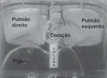
A Data Spectrum ECT/TOR/P model anthropomorphic phantom was utilized, simulating the upper part of a medium to large-sized individual’s chest. Such a phantom comprises heart simulators (with the possibility of attaching lesions of different sizes), lungs, liver, and dorsal spine (Figure 1).
Equipment and imaging protocol
The nuclear medicine imaging system was a GE Health-care Ventri model gamma camera with SPECT (single pho-ton emission tomography) comprising a fixed 90° dual-head system and an image processing software, Xeleris2 (release 2.151). Each detector has a 370 × 190 mm rectangular field of view (FOV) of a 9.5 mm-thick NaI(Tl) crystal.
The two-day protocol included the following character-istics:
• Simulated wait time for the performance of the scan: 60 minutes.
• Patient’s positioning: supine and prone. • Matrix: 64 × 64.
• Zoom: 1.0.
• Pixel size: 6.4 mm.
• Voxel size: 6.40 × 6.40 × 1 mm. • Orbit: 180°.
• Number of projections: 60 (30 per detector, with two detectors at a 90° geometry).
• Energy window: 140 keV ± 10%. • Contour: circular.
• Acquisition technique: step-and-shoot.
• Collimator: low energy high resolution (LEHR) paral-lel holes.
Different activities were utilized not only to reduce the doses received by patients, respecting the international ALARA (as low as reasonably achievable) concept, but also to meet possible restrictions in the radiopharmaceuticals supply, as it recently occurred during the so-called “genera-tor crisis” (2010/2011).
Image acquisitions were also performed with different projection times, in order to analyze whether the usual ac-quisition time could be optimized, both for the patient’s comfort as well as for the reduction of motion artifacts which may lead to scan repetition.
Simulated variations
• Injected activity: 555 to 1,110 MBq (15 to 30 mCi). • Time per projection: 7 to 20 seconds (standard: 15
sec-onds).
Image processing and reconstruction
The images were iteratively reconstructed with the aid of the software Evolution for Cardiac from GE Healthcare, a recently introduced algorithm for cardiac images recon-struction. Such algorithm incorporates RR (resolution recov-ery) and MAP (maximum a posteriori) type noise regular-ization, allowing the SPECT images to be acquired in half the time compared with standard OSEM (ordered subset ex-pectation maximization) algorithm, being known as HT (half time acquisition)(9).
Preparation of the anthropomorphic phantom
Measurements were performed with and without inser-tion of heart lesions, in order to simulate healthy individu-Figure 1. Anterior view of the ECT/TOR/P model anthropomorphic phantom, Data
als and the ones with CAD. For the studies that simulated hypo-uptake, a 1.0 cm-thick solid-type lesion with 2 cm in length was inserted in the lower basal region of the cardiac phantom.
The radioactivity concentration in the cardiac phantom followed the pharmaceutical biodistribution(10,11). The
de-tails of the activity in each organ are individually presented on Table 1. The concentration ratio (MBq/ml) between heart:liver:body was 12:8:1. According to the literature, the concentration ratio (MBq/ml) myocardium/liver is 1.3 ± 0.1(10–12).
tical uptake; 3 (three) = significant reduction in radiophar-maceutical uptake; 4 (four) = absence of radiopharmaceuti-cal uptake.
The sum of the values attributed to each representative segment of the stress phase is called SSS. The values be-tween 0 and 4 are considered as normal or equivocal (possi-bility of attenuation artifact) and SSS values > 4 are consid-ered as altconsid-ered.
Semi-quantitative analysis with software
The software analysis was performed with the aid of the software ImageJ. Short and long vertical axes images were selected for analysis resulting from processing, with 600% zoom. Customized regions of interest (ROI) were delimited for each one of the 17 segments. Such ROI were defined with the assistance of a nuclear cardiologist, being repeated in an identical manner in all heart images. The sum of the pixel count values in each ROI were graphically represented, com-paring the phantom with a patients’ data bank.
Validation of the method
In order to validate the data obtained with the phantom, the results were compared with those from a group of female patients with pre-test low probability of CAD, according with the criteria by Diamond et al.(12). A total of 40 studies were
used and originated a databank representative of the normal radiotracer uptake on each one of the analyzed 17 LV seg-ments. The mean age of the patients selected as standard databank was 57.3 years (36–69 years); mean body mass index of 25.40 (19–37); and mean injected activity of 811.78 MBq (699.30–1,013.80 MBq). Such data were collected from the databank of the clinic where the study was performed. Table 1—Activities injected into each organ individually for each simulation.
Phantom
Heart
Liver
Body
Total activity (MBq)
1,110 925 740 555
1,110 925 740 555
1,110 925 740 555
Activity injected in the organ (MBq)
17.76 ± 0.89 12.17 ± 0.61 8.69 ± 0.43 6.52 ± 0.33
124.54 ± 6.23 80.66 ± 4.03 52.98 ± 2.65 39.74 ± 1.99
153.55 ± 7.68 76.22 ± 3.81 56.61 ± 2.83 42.46 ± 2.12
Image evaluation method
Qualitative and semi-quantitative analyses: scores on medical evaluation
The clinical evaluation was blindly performed by an ex-perienced nuclear cardiologist, i.e., without any knowledge on the parameters utilized in the images acquisition. The scores considered three criteria, as follows:
• Technical quality of the images (evaluation of noise, ar-tifacts and contrast), based on the letters A, B and C: A – excellent quality, without any image characteristic that might impair interpretation of the study; B – good qual-ity, with presence of some characteristic(s) that might eventually impair the interpretation of the study; C – bad quality, impairing interpretation of the study. • Perfusion: normal – homogeneous radiotracer uptake;
abnormal – heterogeneous radiotracer uptake. • Summed stress score (SSS).
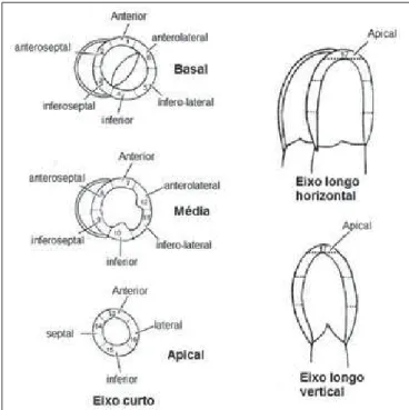
The semi-quantitative evaluation is aimed at standard-izing the segmental analysis of the left ventricle (LV) and the lower subjectivity in the interpretation. A system of scores for the 17 segments of the LV which considers three sections in the smaller axis (apical, median and basal) and a section on the long vertical axis, is utilized as shown on Figure 2.
Each one of the 17 segments is scored according to the radiopharmaceutical uptake, as follows: 0 (zero) = normal; 1 (one) = slight reduction in the radiopharmaceutical up-take; 2 (two) = moderate reduction in the
RESULTS
Initially, the activities and acquisition times were var-ied in the phantom without heart lesions. The scores from the evaluations performed by the cardiologist regarding tech-nical image quality, evaluation of perfusion and SSS value (observer-dependent) obtained for activities from 555 to 925 MBq and acquisition times from 7 to 20 seconds per projec-tion can be observed on Table 2.
The investigation performed with a lesion in the inferobasal region of the heart was not performed with 555 MBq (15 mCi) due to the results from the first part of the study. Likewise, the images were not acquired in the prone position with more than 7 seconds per projection. The car-diologist evaluation scores for simulation with lesion can be seen on Table 3.
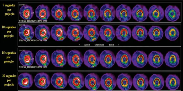
By simulating the usually administered activity of 1,110 MBq, the acquisition time could be reduced to 7 seconds per projection, which is a reduction of 53.34% of the time used in the standard protocol, without changing the possi-bility of identifying the lesion, as demonstrated on Figure 3. The ROI delineated for quantification at ImageJ can be observed on Figure 4. The counts for each segment were used to compare with the data from patients in the normal-ity databank.
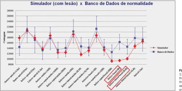
In order to validate the data, the counts for each seg-ment of the heart phantom were compared with those from the normality databank by the ImageJ software. The chart on Figure 5 shows such comparison.
The lesion positioned in the inferobasal region of the phantom was the only count with a value below those of the databank, as this comprised data from healthy patients. The injected activity in the phantom was 1,110 MBq, demonstrat-ing that this is the actual activity administered in the patients.
DISCUSSION
The results demonstrated that the scan cannot be per-formed with 555 MBq (15 mCi) of injected activity, as the technical quality of the image is not appropriate for an ac-curate diagnosis, according to the acquisition protocols adopted in the present study. In the images acquired with the phantom in ventral decubitus (prone), with 740, 925 and 1,110 MBq, no change was observed in the image quality, even for acquisitions with 7 seconds per projection.
It was observed that the injected activity could be reduced to 740 MBq (33.34% reduction) if the acquisition time re-mained the same as the usual time of 15 seconds per projec-tion. For a time shorter than this, the lesion may be masked by the lack of counting statistics. Nevertheless, the activity
Table 3—Scores given by medical evaluation, with the heart phantom without lesion.
740 MBq (with lesion)
925 MBq (with lesion)
1,110 MBq (with lesion)
Simulation Technical quality Perfusion SSS Simulation Technical quality Perfusion SSS Simulation Technical quality Perfusion SSS
Supine 7 s/p B Abnormal
4
Supine 7 s/p B Abnormal
2
Supine 7 s/p A Abnormal
3
Supine 10 s/p B Normal
0
Supine 10 s/p A Abnormal
2
Supine 10 s/p A Abnormal
2
Supine 15 s/p A Abnormal
2
Supine 15 s/p A Abnormal
2
Supine 15 s/p A Abnormal
2
Supine 20 s/p A Abnormal
2
Supine 20 s/p A Abnormal
2
Supine 20 s/p A Abnormal
2
Prone 7 s/p B Abnormal
2
Prone 7 s/p B Abnormal
2
Prone 7 s/p A Abnormal
2
s/p, seconds per projection.
Table 2—Medical evaluation scores, with the heart phantom without lesion.
555 MBq (without lesion) 740 MBq (without lesion) 925 MBq (without lesion) Simulation Technical quality Perfusion SSS Simulation Technical quality Perfusion SSS Simulation Technical quality Perfusion SSS Supine 7 s/p C Abnormal 2 Supine 7 s/p B Abnormal 1 Supine 7 s/p A Abnormal 1 Supine 10 s/p B Abnormal 3 Supine 10 s/p A Abnormal 2 Supine 10 s/p A Abnormal 2 Supine 15 s/p B Abnormal 2 Supine 15 s/p A Abnormal 1 Supine 15 s/p A Normal 0 Supine 20 s/p B Abnormal 2 Supine 20 s/p A Normal 0 Supine 20 s/p A Normal 0 Prone 7 s/p B Abnormal 1 Prone 7 s/p B Normal 0 Prone 7 s/p A Normal 0 Prone 10 s/p B Abnormal 1 Prone 10 s/p A Normal 0 Prone 10 s/p A Normal 0 Prone 15 s/p C Abnormal 2 Prone 15 s/p A Normal 0 Prone 15 s/p A Normal 0 Prone 20 s/p C Abnormal 1 Prone 20 s/p A Normal 0 Prone 20 s/p A Normal 0
could be reduced to 925 MBq (16.67% reduction) and 53.34% reduction in acquisition time with no change in image quality and with the lesion still being properly diag-nosed. With the usual injected activity of 1,110 MBq, the acquisition time could be reduced to 7 seconds per projec-tion both in the supine and in the prone posiprojec-tions (reduc-tion of 53.34%), without any change in image quality and in the diagnostic accuracy of the method.
The chart on Figure 5 shows the count on each one of the 17 heart segments for the normality databank,
consider-ing the count on the same segments of the heart phantom. The values observed were within the mean value of the counts considering the standard deviation, which confirms the va-lidity of the proposed phantom usage.
CONCLUSION
The present study demonstrated that the ECT/TOR/P Data Spectrum anthropomorphic phantom appropriately reproduces patients, within the mean and standard deviation of a randomly selected sample. After validation, activity and Figure 3. Processed images of the heart phantom with a lesion in the inferobasal segment, with times varying between 7 and 20 seconds per projection, highlighting that the protocol’s standard time is 15 seconds.
time parameters were varied for the two-day protocol study in MPS. The results demonstrated that for the same time currently used for the exam, the injected activity could be reduced in up to 33.34%. If the acquisition time is also re-duced (to 53.34% of the usual time), the activity could be reduced up to 16.67% of the usual injected activity, without any change in the image quality and in the diagnosis. The images acquired in ventral decubitus (prone) demonstrated improvement in relation to those acquired only in supine position, complementing the exam. A deeper study of opti-mization based on positioning is currently underway and should produce more results for the present investigation on the two-day protocol for this type of exam. Such results serve as grounds for the undertaking of clinical studies involving patients for definition of an optimized protocol.
REFERENCES
1. World Health Organization, World Heart Federation, World Stroke Organization. Global atlas on cardiovascular disease prevention and control. [acessado em 8 de agosto de 2013]. Disponível em: http:// www.who.int/cardiovascular_diseases/publications/atlas_cvd/en/. 2. Go AS, Mozaffarian D, Roger VL, et al. Heart disease and stroke
statistics – 2013 update: a report from the American Heart Associa-tion. [acessado em 2 de outubro de 2013]. Disponível em: http:// circ.ahajournals.org/content/127/1/e6.full.
3. Issa AFC, Pantoja MR. Valor prognóstico da cintigrafia de perfu-são miocárdica em mulheres comparado com homens com suspeita clínica de doença coronariana. Rev SOCERJ. 2006;19:9–19.
4. Henzlova MJ, Cerqueira MD, Hansen CL, et al. ASNC imaging guidelines for nuclear cardiology procedures: stress protocols and tracers. [acessado em 3 de junho de 2013]. Disponível em: https:// www.asnc.org/imageuploads/imagingguidelinesstressprotocols 021109.pdf.
5. Hesse B, Tägil K, Cuocolo A, et al. EANM/ESC procedural guide-lines for myocardial perfusion imaging in nuclear cardiology. Eur J Nucl Med Mol Imaging. 2005;32:855–97.
6. Ali I, Ruddy TD, Almgrahi A, et al. Half-time SPECT myocardial perfusion imaging with attenuation correction. J Nucl Med. 2009; 50:554–62.
7. Savi A, Gerundini P, Zoli P, et al. Biodistribution of Tc-99m methoxy-isobutyl-isonitrile (MIBI) in humans. Eur J Nucl Med. 1989;15:597–600.
8. Münch G, Neverve J, Matsunari I, et al. Myocardial technetium-99m-tetrofosmin and technetium-99m-sestamibi kinetics in nor-mal subjects and patients with coronary artery disease. J Nucl Med. 1997;38:428–32.
9. Zolle I. Technetium-99m pharmaceuticals: preparation and quality control in nuclear medicine. Berlin Heidelberg: Springer; 2007. 10. Bucerius J, Ahmadzadehfar H, Biersack HJ. 99
Tc-sestamibi: clinical applications. Berlin Heidelberg: Springer; 2012.
11. Cerqueira MD, Weissman NJ, Dilsizian V, et al. Standardized myo-cardial segmentation and nomenclature for tomographic imaging of the heart. A statement for healthcare professionals from the Cardiac Imaging Committee of the Council on Clinical Cardiol-ogy of the American Heart Association. Ciculation. 2002;105:539– 42.
12. Diamond GA, Forrester JS. Analysis of probability as an aid in the clinical diagnosis of coronary artery disease. N Engl J Med. 1979; 300:1350–8.