ISSN 0102-695X
http://dx.doi.org/10.1590/S0102-695X2012005000053 Received 24 Aug 2011 Accepted 2 Feb 2012 Available online 17 Apr 2012
cytotoxic activity
Jocélia P. C. Oliveira,
1Éverton L. F. Ferreira,
1Mariana H.
Chaves,
*,1Gardenia C. G. Militão,
2Gerardo M. V. Júnior,
3Arenice de M. Costa,
4Cláudia do Ó Pessoa,
4Manoel O. de
Moraes,
4Letícia V. Costa-Lotufo
41Departamento de Química, Universidade Federal do Piauí, Brazil,
2Campus Senador Helvídio Nunes de Barros, Universidade Federal do Piauí,
Brazil,
3Instituto de Ciências Naturais, Humanas e Sociais, Campus de Sinop,
Universidade Federal de Mato Grosso, Brazil,
4Laboratório de Oncologia Experimental, Universidade Federal do Ceará,
Brazil.
Abstract: The phytochemical investigation of the ethanol extract from leaves of Lecythis pisonis Cambess., Lecythidaceae, resulted in the isolation of seven triterpenes: α- and β-amyrin, uvaol and erythrodiol, ursolic and oleanolic acids and 3β-friedelinol (friedelan-3β-ol), as well as a mixture of sitosterol and stigmasterol steroids and a diterpene (E)-phytol. The structures of these compounds were
identified by 1H and 13C NMR spectral analysis and compared with literature data.
The mixture of triterpenes ursolic and oleanolic acids isolated from the active ethereal fraction showed moderate cytotoxic activity. This paper describes for the first time the phytochemical and cytotoxic study of Lecythis pisonis’ leaves.
Keywords: Lecythis pisonis Lecythidaceae triterpenes cytotoxicity
Introduction
Lecythidaceae, known as the “Brazil nut family”,
is considered the third most abundant tree of the Amazon
with about 25 genera and 489 species (Mori & Prance,
1990). Studies of plants in the Lecythidaceae family are still scarce and are restricted to 21 species in thirteen genera: Barringtonia, Bertholletia, Careya, Cariniana, Couroupita, Eschweilera, Foetidia, Grias, Gustavia, Lecythis, Napoleonaea, Planchonia and Petersianthus.
Barringtonia is the most explored genus with i ve species
(B. acutangula, B. racemosa, B. maunwongyathiae, B. asiatica and B. speciosa) (Oliveira, 2010).
The chemical constituents identii ed in plants of
the Lecythidaceae family include pentacyclic triterpenoids and their glycosides, neo-clerodane diterpenoids, sesquiterpenoids, monoterpenoids, steroids, alkaloids,
simple phenolic compounds, l avonoids, ellagic acid and derivatives, vitamin E, α-tocopherylquinone, 3-O -galloylepigallocatechin, epigallocatechin, sucrose, fatty
acid, ethyl esters and waxes (Oliveira, 2010).
Several pharmacological activities have been
report with plants of this family, such as antinociceptive, antibacterial, antitumor, anti-inl ammatory, antifungal,
antileishmanial, antioxidant, hepatoprotective and cytotoxic (Oliveira, 2010).
Lecythis pisonis Cambess, Lecythidaceae, is
popularly known as sapucaia or sapucaia nut which is
found among the states of Piauí, from Pernambuco to
São Paulo and in the Amazon region (Corrêa, 1978).
The leaves of sapucaia are popularly used in the form of baths for treatment of itching (pruritus) of the body and the oil extracted from the seeds is used as an emollient in
reducing muscle pain (Franco & Barros, 2006; Agra et al.,
2007).
Considering the scarcity of studies with plants
of the family Lecythidaceae, and above all, the lack of chemical investigation of the species L. pisonis, this
paper describes for the i rst time the phytochemical study
of the leaves of this species and the evaluation results of cytotoxicity.
Materials and Methods
General procedures
The NMR spectra of 1H and 13C were obtained on Varian Inova spectrometer and Brüker Avance DRX-500 model, operating at DRX-500 MHz (1H) and 125 MHz (13C). The chromatographic plates were prepared using a mixture of silica gel 60 G Vetec and 60 GF Fluka (1:1).
Spots on TLC were observed by spraying the plates with
solution of Ce(SO4)2, followed by heating at 100 °C. The
from 0.060 to 0.020 mm (Acros Organics) or Sephadex LH-20 (Pharmacia Biotech).
Plant material
The leaves of L. pisonis were collected in
Teresina municipality, Piauí State, Brazil (southern
latitude 05° 02' 53.2", western longitude 42° 47' 16.8", the sea level of 68 m), in July 2008. A voucher specimen (TEPB 26488) has been deposited in the Graziela
Barroso Herbarium at UFPI.
Extraction and isolation
The leaves of L. pisonis were dried at room
temperature and ground. The material (2 kg) was extracted exhaustively with ethanol six times, and each extraction had the duration of 48 h. The ethanol was removed under
vacuum and lyophilized, giving 272 g (13%) of ethanol
extract, which was suspended in a mixture of H2O/MeOH
(3:2) solution and extracted successively with n-hexane, diethyl ether and EtOAc, respectively, obtaining the
n-hexane (60 g, 30%), diethyl ether (24 g, 12%), EtOAc
(21 g, 10.5%) and water (70 g, 37.5%) fractions, and a
precipitate formed in the phase EtOAC (ppt-EtOAc, 10 g, 5%).
The diethyl ether fraction (10 g) was fractionated
by chromatography column on silica gel (53 x 5 cm, 250
g) eluted with CHCl3, CHCl3-MeOH in order of increasing polarity, yielding 103 fractions (240 mL each). The solvent
was removed under vacuum and analyzed by TLC. The fractions were combined into ifteen groups. Fraction F7 (242 mg) was suspended in n-hexane and yielding an amorphous precipitate corresponding to compound 7 (226 mg, 2.26%).
The group F17 (17-23, 603 mg) was suspended
in MeOH to yield a mixture of compounds 5 and 6 (475 mg, 4.75%).
The group F8 (8-15, 1.02 g) was fractionated on a silica gel column eluted with n-hexane, n-hexane-EtOAc in increasing order of polarity giving 162 fractions (50 mL each). After evaporation of the solvent and analysis by
TLC, the fractions were combined into fourteen groups. The groups G22 (33 mg) and G25 (25-45, 78 mg) were
rechromatographed on Sephadex LH-20 column (54 x 1.5 cm) using hexane-CH2Cl2 (1:4) as eluent, resulting in 20 mg (0.20%) of compounds 1, 2 and 8, and 13 mg (0.13%) of a mixture of sitosterol and stigmasterol. Group G92
(92-100, 54 mg) was puriied on Sephadex LH-20 column
using as eluent CH2Cl2- n-hexane (1:4) and CH2Cl2 -acetone (3:2) yielding 19 mg (0.19%) of a mixture of compounds 3 and 4.
Cytotoxicity studies
The cytotoxicity of the ethanol extract and diethyl ether fraction, compound 7 and mixture of compounds (5+6) was investigated against HL-60
(human leukemia), SF-295 (glioblastoma), HCT-8
(human colon carcinoma) and MDA-MB-435 (human melanoma) tumor cell strains (National Cancer Institute,
Bethesda, MD, USA). All lines were maintained in RPMI-1640 medium supplemented with 10% fetal
bovine serum, 2 mM glutamine, 100 U mL-1 penincillin
and 100 μg mL-1 streptomycin at 37 oC with 5% CO
2.
Cells were plated in 96-well plates (105 cells/well for
adherent cells or 0.3 x 10⁶ cells/well for suspend cells in 100 μL for medium). After 24 h, samples (0.39 to 25
μg mL-1) dissolved in DMSO (1%) were added to each
well and incubated for 72 h. Control groups received
the same amount of DMSO. Doxorubicin (0.01 to
0.58 μg mL-1) was used as positive control. Tumor cell
growth was quantified by the ability of living cells to reduce the yellow dye
3-(4,5-dimethyl-2-thiazolyl)-2,5-diphenyl-2H-tetrazolium bromide (MTT) to a blue
formazan product (Mosmann, 1983).
The percentage of cell growth inhibition (IC%) was calculated according to the report by Mahmoud et al. (2011). The IC50 values and their conidence intervals were obtained by nonlinear regression using the GraphPad program (Intuitive Software for Science, San Diego,
CA).
Results and Discussion
The phytochemical investigation of the ethanol extract from leaves of Lecythis pisonis Cambess, Lecythidaceae, resulted in the identification of seven
triterpenoids; a mixture of α- and β-amyrin (1+2) and the diterpenoid (E)-phytol (8), a mixture of uvaol and erythrodiol (3+4), a mixture of ursolic and oleanolic acids (5+6), friedelan-3β-ol (7), as well as a mixture of steroids sitosterol and stigmasterol. The structural
identification of these compounds was performed by
analysis of spectral data of 1H and 13C NMR (including
DEPT 135° and 90°), as well as by comparison of their spectral data with those reported in the literature (Olea & Roque, 1990; Ayres et al., 2008; Rahman & Ahmad, 1992; Mahato & Kundu, 1994; Junges et al., 2000; Salazar et al., 2000; De-Eknamkul & Potduang, 2003).
The 1H and 13C NMR spectra of mixtures (1+2+8), (3+4) and (5+6) showed signals between δ 3.11-3.16 (dd, J = 4.5-7.5 and 9.0-11.0 Hz, H-3), and
78.5-79.3 (C-3) characteristic of 3β-OH triterpenoids.
In addition, the 13C NMR spectra presented two pairs
of signals between δ 125.1-124.7, and 137.9-139.8; δ 122.0-122.4 and 143.6-145.4 assigned to olefinic
carbons C-12 and C-13 skeletons ursane and oleanane,
respectively (Olea & Roque, 1990).
HO
H
H R3
R1
R2
H
1R1=R3=CH3; R2=H
2R1=H; R2=R3=CH3
3R1=CH3; R2=H; R3=CH2OH 4R1=H; R2=CH3; R3=CH2OH
5R1=CH3; R2=H; R3=CO2H 6R1=H; R2=CH3; R3=CO2H
HO
H
7
H H
OH
8
determined by comparing the integrations of signals from oximethinic hydrogen (H-3) of triterpenoids, steroids or oximethilenics of 8 with oleinic hydrogens (Ayres et al.,
2008).
The mixture of triterpenoids α-amyrin (1),
β-amyrin (2) and the diterpenoid (E)-phytol (8) was
identiied at a ratio of 40:36:24. (E)-phytol was evidenced
in the 1H NMR spectrum by signals at δ 4.16 (dd, J = 0.5 and 7.0 Hz) assigned to the hydrogens oximethilenics
and allylics, and δ 5.42 (tq, J = 7.0 and 1.5 Hz), referring
to the oleinic hydrogen. The presence of (E)-phytol was
conirmed by observation in the 13C NMR spectrum of the
signals in δ 59.1 (CH2O), 123.4 and 140.5 (oleinic CH
and C) and by comparison with literature data (Rahman & Ahmad, 1992; Ayres et al., 2008).
The triterpenoid mixture (3+4) was identiied
at a ratio of 66:34 and their structures were determined
by the additional presence in the 1H NMR spectrum of
two doublet with J = 11.0 Hz, located in δ 3.14 and 3.56,
assigned H-28a and H-28b, in accordance with the signals
at δ 69.7 and 69.9 in the 13C NMR spectrum assigned to
C-28 of both substances. The NMR data obtained were consistent with those reported for uvaol (3) and erythrodiol (4) (Mahato & Kundu, 1994).
The mixture of compounds 5 and 6 was
evidenced by the signals at δ 180.4 and 180.6, in the 13C
NMR spectrum, assigned to carboxylic carbon (C-28), compared to the spectrum of mixture of α-and β-amyrin (1 and 2). The NMR data obtained were consistent with
those reported for ursolic (5) and oleanolic (6) acid at a ratio (59:41) (Junges et al., 2000).
The analysis of 13C NMR and DEPT 135° and
90° of the substance 7 showed signals corresponding to six non-hydrogenated carbons, ive methine, eleven
methylene and eight methyl groups. We also observed a
characteristic signal at δ 72.8 from oximethinic carbon
(C-3), consistent with the signal in δ 3.67 (H-3) in the 1H NMR spectrum. The NMR data obtained are in agreement
with the structure of friedelan-3β-ol (7) (Salazar et al., 2000).
The 1H NMR spectrum of the mixture of steroids
sitosterol and stigmasterol showed signals between δ
0.6 and 0.9 corresponding to methyl groups of steroids,
a multiplet in δ 3.53 (H-3) corresponding to oximethinic carbon and a singlet in δ 5.35 assigned to H-6 in steroid-Δ5. In the 13C NMR spectrum signals in δ 72.0 (C-3)
of oximethinic carbon, δ 121.9 and 141.0 referring to oleinic carbons C-6 and C-5 were observed, respectively. The presence of two signals of lower intensity at δ 129.5 and 138.5 assigned to C-23 and C-22 of the stigmasterol conirmed the presence of this substance. The evidences above allowed identiication of the steroids: sitosterol and stigmasterol (De-Eknamkul & Potduang, 2003) at a ratio of 85:15.
The substances α-amyrin (1) and β-amyrin (2),
friedelan-3β-ol (7), ursolic (5) and oleanolic (6) acid, sitosterol and stigmasterol had been reported in species
of Lecythidaceae, however triterpenoids uvaol (3), erythrodiol (4) and diterpenoid (E)-phytol (8) have been
reported for the irst time in this family.
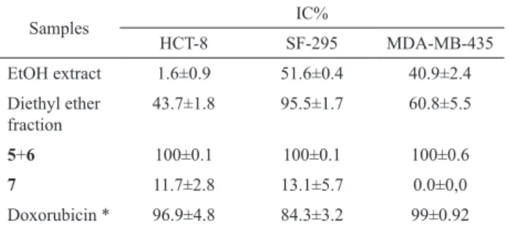
In the test with tumor cells, the EtOH extract
and triterpenoid 7 (25 µg mL-1), showed a low percentage
of cell growth inhibition (IC%), while the diethyl ether
fraction had values ranging from 43.7 to 95.5% (Table 1). The mixture composed of ursolic (5) and oleanolic acid (6) isolated from the diethyl ether fraction, showed
100% inhibition for the strains of HC-T8 (human colon
carcinoma), SF-295 (glioblastoma) and MDA-MB-435
(human melanoma) when tested in the concentration
compared to doxorubicin, used as positive control (Table 2).
Table 1. Percentage of growth inhibition (IC%) in the panel of
three cancer cell lines after treatment for 72 h with samples of
Lecythis pisonis concentration of 25 µg mL-1. The results are
expressed as mean and standard deviation.
Samples IC%
HCT-8 SF-295 MDA-MB-435
EtOH extract 1.6±0.9 51.6±0.4 40.9±2.4
Diethyl ether fraction
43.7±1.8 95.5±1.7 60.8±5.5
5+6 100±0.1 100±0.1 100±0.6
7 11.7±2.8 13.1±5.7 0.0±0,0
Doxorubicin * 96.9±4.8 84.3±3.2 99±0.92
HL-60: human leukemia; HCT-8: human colon; SF-295: glioblastoma;
MDA-MB-435: human melanoma * positive control.
Table 2. Growth inhibition (IC50 in µg mL-1) with its respective
conidence interval in tumor cell lines treated for 72 h by mixing
5+6.
Samples IC50
HL-60 HCT-8 SF-295 MDA-MB-435
5+6 3.4 1.6-7.0
7.1
5.5-8.9 1.5-3.92.4
10.6
8.3-13.5
Doxorubicin 0.04 0.03-0.05
0.02 0.02-0.03
0.48
0.34-0.72
0.96
0.68-1.32 HL-60: human leukemia; HCT-8: human colon; SF-295: glioblastoma;
MDA-MB-435: Human Melanoma * positive control.
In the literature there are reports of tests with the cell line HCT-15 (human colon carcinoma), with
inhibitory concentration of 30 µM to ursolic acid and 60 µM to oleanolic acid (Vechia et al., 2009). The M4Beu
cell line (human melanoma) was tested only with ursolic
acid and the IC50 value ranged from 12.5 to 15.0 µM (Vechia et al., 2009). There are no reports in the literature
on the cytotoxicity of these triterpene acids with strains of
human glioblastoma, a type of the most lethal malignant
brain tumors. Patients with this type of tumor have a median survival of ifteen months after diagnosis (Horvath et al., 2006), justifying the search for new molecules with
potential antitumor activity.
In conclusion, we believe that the above mentioned
data are of importance from the chemotaxonomy and
pharmacological point of view. On the other hand, further
phytochemical studies on L. pisonis are necessary to
obtain better knowledge of this species.
Acknowledgment
The authors are grateful to CNPq, CAPES and
FINEP for the fellowships and inancial support. They
are also grateful to Dr. G. M. Sousa, Graziela Barroso
Herbarium, UFPI, for the identiication of botanical
material.
References
Agra MF, Freitas PF de, Barbosa Filho JM 2007. Synopsis of the plants known as medicinal and poisonous in Northeast of Brazil. Rev Bras Farmacogn 17: 116-155.
Ayres MCC, Escórcio SP, Costa DA, Chaves MH, Vieira Júnior GM, Cavalheiro AJ 2008. Constituintes químicos das folhas de Qualea grandiflora: atribuição dos dados de RMN de dois lavonóides glicosilados acilados diastereoisoméricos. Quim Nova 31: 1481-1484.
Corrêa MP 1978. Dicionário das plantas úteis do Brasil e exóticas cultivadas. Rio de Janeiro: Imprensa nacional. De-Eknamkul W, Potduang B 2003. Biosynthesis of β-sitosterol
and stigmasterol in Croton sublyratus proceeds via a mixed origin of isoprene units. Phytochemistry 62:
389-398.
Franco EAP, Barros RFM 2006. Uso e diversidade de plantas medicinais no Quilombo Olho D’água dos Pires, Esperantina, Piauí. Rev Bras Pl Med 8: 78-88.
Horvath S, Zhang B, Carlson M, Lu KV, Zhu S, Felciano RM, Laurance MF, Zhao W, Qif S, Chen Z, Lee Y, Scheck AC, Liau LM, Wu H, Geschwind DH, Febbo PG, Kornblum HI, Cloughesy TF, Nelson SF, Mischel OS 2006. Analysis of oncogenic signaling networks in glioblastoma identiies ASPM as a molecular target.
Proc Natl Acad Sci USA 103: 17402-17407.
Junges MJ, Fernandes JB, Vieira PC, Fernandes MFGS, Rodrigues-Filho E, Frühauf M, Barañano AG 2000. Triterpenos ursânicos e oleanânicos isolados do caule
de Eugenia florida DC. Revista de Pesquisa e
Pós-graduação da Universidade Regional Integrada do Alto Uruguai e das Missões 1: 13-29
Mahato SB, Kundu AP 1994. 13C spectra of pentacyclic
triterpenoids - a campilation and some salient features.
Phytochemistry 37: 1517-1575.
Mahmoud TS, Marques MR, Pessoa CO, Lotufo LVC, Magalhães HIF, Moraes MO, Lima DP, Tininis AG, Oliveira JE 2011. In vitro cytotoxic activity of Brazilian Middle West plant extracts. Rev Bras Farmacogn 21: 456-464. Mori SA, Prance GT 1990. Lecythidaceae. Part II. The
zygomorphic-lowered New World genera (Couroupita, Corythophora, Bertholletia, Couratari, Eschweilera & Lecythis). Flora Neotropica 21: 1-346.
Mosmann T 1983. Rapid colorimetric assay for cellular growth and survival:application to proliferation and cytotoxicity assays. J Immunol Methods 65: 55-63.
Olea RSG, Roque NF 1990. Análise de misturas de triterpenos por RMN de 13C. Quim Nova 13: 278-281.
Rahman A, Ahmad VU 1992. 13C-NMR of Natural Products:
Diterpenes. New York: Plenum Press.
Salazar GCM, Silva GDF, Duarte LP, Vieira Filho SA, Lula IS 2000. Two epimeric friedelane triterpenes isolated from
Maytenus truncata Reiss: 1H and 13C chemical shift
assignments. Magn Reson Chem 38: 977-980.
Vechia LD, Gnoatto SCB, Gosmann G 2009. Derivados oleananos e ursanos e sua importância na descoberta de novos fármacos Com atividade antitumoral,
anti-inlamatória e antioxidante. Quim Nova 32: 1245.
*Correspondence
Mariana Helena Chaves
Departamento de Química, Universidade Federal do Piauí 64049-550 Teresina-PI, Brazil