Plane-parallel ionization chamber for X-radiation
of conventional radiography and mammography*
Câmara de ionização de placas paralelas para radiação-X de radiografia convencional e mamografia
Alessandro Martins da Costa1, Linda V. E. Caldas2
OBJECTIVE: To develop a double-faced plane-parallel ionization chamber for measurement of air kerma and air kerma rate in X-radiation fields utilized in conventional radiography and mammography. MATERIALS AND METHODS: The chamber has entrance windows made of aluminized polyester film, inner and guard elec-trodes of aluminum at one side (face A) and graphite at the other side (face G). The present study evaluated operational characteristics as regards response linearity, angular and energy dependence. RESULTS: The linearity of response was 0.86% for face A and 0.92% for face G. For radiation incidence angles, of 0° to ± 5°, the response variation was less than 0.8% for both faces of the chamber. The energy dependence of response was 0.8% for face A on X-ray qualities for conventional radiography, and 2.4% for the face G of the chamber on X-ray qualities for mammography. CONCLUSION: This chamber can be utilized on a routine basis for measurement of air kerma and air kerma rate in X-ray beams utilized in conventional radiography and mammography.
Keywords: Conventional radiography; Mammography; Quality control; Ionization chamber.
OBJETIVO: Desenvolver uma câmara de ionização de placas paralelas de dupla face para determinação de kerma no ar e taxa de kerma no ar em campos de radiação-X utilizados em radiografia convencional e mamo-grafia. MATERIAIS E MÉTODOS: A câmara desenvolvida tem janelas de entrada de poliéster aluminizado, elétrodos internos e anéis de guarda de alumínio em uma face (face A) e de grafite na outra (face G). Neste trabalho foram testadas as características operacionais de linearidade, dependência angular e energética de resposta. RESULTADOS: A linearidade de resposta foi de 0,86% para a face A e de 0,92% para a face G. Para ângulos de incidência da radiação de 0° a ± 5°, a variação da resposta relativa foi menor que 0,8% para ambas as faces da câmara. A dependência energética de resposta foi de 0,8% para a face A nas qua-lidades de raios-X para diagnóstico convencional e de 2,4% para a face G da câmara nas quaqua-lidades de raios-X para mamografia. CONCLUSÃO: Esta câmara pode ser utilizada rotineiramente na determinação de kerma no ar e taxa de kerma no ar em feixes de raios-X utilizados em radiografia convencional e mamografia.
Unitermos: Raio-X convencional; Mamografia; Controle da qualidade; Câmara de ionização.
Abstract
Resumo
* Study developed at Instituto de Pesquisas Energéticas e Nucleares – Comissão Nacional de Energia Nuclear (IPEN-CNEN), São Paulo, SP, Brazil.
1. PhD in Nuclear Technology, Doctor Professor, Department of Physics and Mathematics at Faculdade de Filosofia, Ciências e Letras de Ribeirão Preto da Universidade de São Paulo (FFCLRP-USP), Ribeirão Preto, SP, Brazil.
2. PhD in Nuclear Physics, Researcher at Instituto de Pesquisas Energéticas e Nucleares – Comissão Nacional de Energia Nuclear (IPEN-CNEN), São Paulo, SP, Brazil.
Mailing address: Dr. Alessandro Martins da Costa. Departamento de Física e Matemática, Faculdade de Filosofia, Ciências e Letras de Ribeirão Preto, Universidade de São Paulo. Avenida Bandeirantes, 3900. Ribeirão Preto, SP, Brazil, 14040-901. E-mail: [email protected]
Received May 11, 2007. Accepted after revision June 19, 2007.
reference levels in radiology are still to be regulated, but must be considered as a prac-tical mechanism to promote a better local quality control.
An accurate estimation of the skin en-trance dose requires a precise measurement of the incident air kerma (5) at the entrance
skin plane, and also of the beam half-value layer (HVL) (6). The incident air kerma is
converted into skin entrance dose by the application of an appropriate backscatter factor(7,8). Also, the management of the
following parameters is particularly impor-tant: tube voltage (kVp); air kerma repro-ducibility and linearity with the tube cur-rent product-exposure time (mAs). These parameters characteristics may vary over the time; therefore tests should be per-Costa AM, Caldas LVE. Câmara de ionização de placas paralelas para radiação-X de radiografia convencional e mamografia. Radiol Bras. 2008;41(1):39–43.
mal radiation exposure to the patient(1–4).
Radiodiagnosis quality guarantee includes, among other aspects, evaluation of imag-ing quality, film rejection analyses, evalu-ation of the dose to patients, and evaluevalu-ation of physical parameters of the different X-ray system components. Several tests for quality control are required for guarantee-ing the correct X-ray equipment operation. The tests for quality control of conven-tional and mammographic X-ray equip-ment must include a representative estima-tion of the skin entrance dose practiced in the radiological clinic and the correspond-ing values must be compared with the di-agnostic reference levels in radiology es-tablished by the Brazilian standards(3). It
should be emphasized that the diagnostic INTRODUCTION
mini-formed at regular intervals. Consequently, it is necessary to understand how an image is influenced by these parameters, and how their characteristics could be measured with appropriate tools.
Incident air kerma, HVL, reproducibil-ity and linearreproducibil-ity of air kerma rate as a func-tion of the mAs are generally determined by the utilization of calibrated ionization chambers(6). The knowledge about
ioniza-tion chamber limitaioniza-tions is essential for its correct utilization(9). The specifications for
an ionization chamber design must be care-fully understood by the user and correctly taken into consideration before any mea-surement is accepted as valid(10).
In Brazil there has been an increasing interest in designing and constructing low-cost ionization chambers, demonstrating the feasibility of constructing radiation detectors with materials available in the domestic market.
Initially, ionization chambers were de-veloped for X-radiation(11) and
beta-radia-tion(12), respectively by Instituto de
Radio-proteção e Dosimetria, Rio de Janeiro, RJ, and Escola de Engenharia da Universidade Federal de Minas Gerais, Belo Horizonte, MG.
Plane-parallel ionization chambers for low-energy X-radiation and beta-radia-tion(13), high-energy electrons(14–16),
com-puted tomography(17,18), radioprotection(19),
extrapolation chambers for X-radiation and beta-radiation of dermatologic and oph-thalmologic applications utilized in bra-chytherapy(20,21) have been constructed by
Instituto de Pesquisas Energéticas e Nu-cleares.
The present study was aimed at devel-oping a double-faced plane-parallel ioniza-tion chamber for determining air kerma and air kerma rate in X-radiation fields utilized in conventional diagnosis and mammogra-phy. One face of this ionization chamber is appropriate for measurements in conven-tional radiography (face A); the other (face G) is appropriate for measurements in mammography.
The ionization chamber developed in this study was calibrated for standard X-ray beams and submitted to tests according to the international recommendations(22–24).
Operational characteristics of linearity, angular and energy dependence were tested
according to the procedures applied for other ionization chambers(13,14,17,19–21,25,26).
MATERIALS AND METHODS
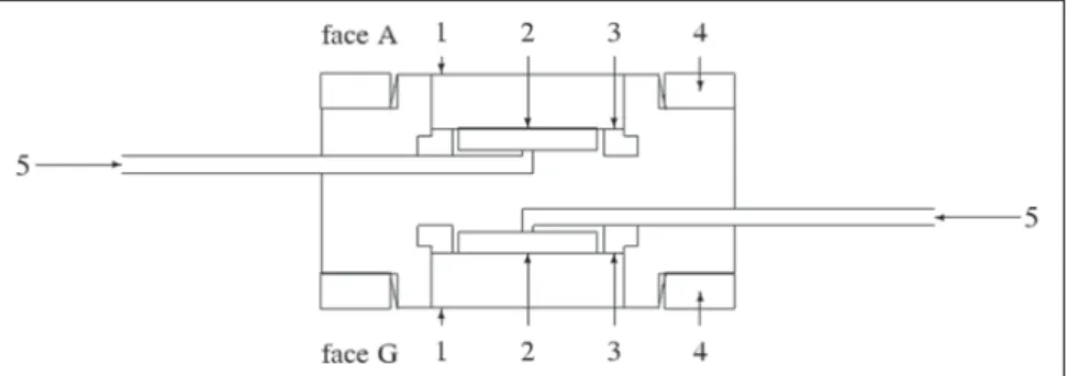
A double-faced plane-parallel ioniza-tion chamber for X-radiaioniza-tion beams uti-lized in conventional radiography and mammography was designed and con-structed. The chamber is comprised of alu-minized polyester entrance windows (1.7 mg/cm²), aluminium (face A) and graphite (face G) inner electrodes and guard rings (face A) and sensitive volume of air 2.5 cm³. Figure 1 shows a schematic diagram of this chamber.
Ionization currents were measured with electrodes whose relevant characteristics ser shown on Table 1.
Tests were performed with the ioniza-tion chamber coupled with a PTW Unidos electrometer. The polarization voltage was +400 V. The efficiency of the ions collec-tion is > 99% at +400 V for both faces of the chamber(27–29). The leakage current is <
2 fA and the accuracy is > ±0.05% for both faces of the chamber.
The following irradiation systems were utilized: Rigaku Denki, Geigerflex model X-ray equipment with constant potential, with a Philips tube model PW 2184/00, beryllium 1 mm window and tungsten tar-get, operating up to 60 kV; Medicor Mövek Röntgengyara, model Neo-Diagnomax X-ray single-phase equipment, full wave rec-tification, with tungsten target, operating up to 125 kV in radiographic mode, and up to 100 kV in fluoroscopic mode.
The X-ray systems characteristics are shown on Tables 2 and 3.
Table 4 shows the characteristics of the reference standard systems utilized in the ionization chamber calibration.
Considering that the ionization cham-bers utilized in the present study are un-sealed, all the measurements were cor-rected for reference temperature and pres-sure conditions (20 °C and 101.3 kPa).
The measurement uncertainties evalua-tion and expression were performed
ac-Figure 1. Schematic diagram of the ionization chamber developed in the present study: (1) entrance windows (external electrodes), (2) collector electrodes (inner electrodes), (3) guard rings, (4) entrance windows fixation rings, (5) cables. The sensitive volume of air is 2.5 cm³.
Table 1 Electrometers characteristics.
Manufacturer
PTW-Freiburg Radcal Corporation
Model
Unidos 9015
Chamber voltage
+ 400 V + 260 V
Measurements accuracy
≤ 0.5% reading + 1 digit 4% reading + 1 digit
Table 2 Rigaku-Denki system features for mammography qualities; inherent filtration: 1 mmBe; addi-tional filtration: 0.06 mmMo. Focus-chamber distance:100 cm.
Quality
RXM25 RXM28 RXM30 RXM35
Generation potnential (kV)
25 28 30 35
Half-value layer (mmAl)
cording to the “Guidelines for measure-ment uncertainty expression” (30).
RESULTS
The linear relation between the ioniza-tion current and the air kerma rate was de-termined by sequential irradiation of the both faces of the chamber in the RXM35 quality of mammography X-ray (Table 2) with variable tube current. The chamber was positioned at a 100 cm distance from the source, taking the entrance windows surface center as a reference. The air kerma rates were determined according to the standard system for mammography X-rays (Table 4). The data obtained are shown on
air kerma rate, 100 cm focus-chamber dis-tance, taking the entrance windows surface as reference. The response was measured with the incidence angle ranging between 0° and ±5°, where 0° corresponds to a fron-tal irradiation. The results obtained are shown on Table 5. The responses were normalized for 0° corresponding to the average of five successive measurements. It may be observed that the chamber is in compliance with the IEC standard require-ments(23): response variation up to ±3,0%
because of incidence angle variation of ±5°. Maximum response variation was 0.8%.
X-ray qualities utilized in the chamber calibration are shown on Tables 2 and 3. The chamber was irradiated in the air, and positioned at the calibration distance tak-ing the entrance window surface as refer-ence. The calibration coefficients were obtained with the standard systems for each energy range (Table 4). Calibration coeffi-cients obtained for the face A of the cham-ber in the qualities of conventional diag-nostic X-rays, and for the face G in the qualities of mammography X-radiation are presented as a function of the half-value
Table 3 Medicor Mövek Röntgenyara system features for conventional diagnostic X-rays qualities: inherent filtration: 0.8 mmAl; total filtration: 2.5 mmAl. Focus-chamber distance: 50 cm.
Quality
RQR 3 RQR 5 RQR 7
Generation potential (kV)
50 70 90
Half-value layer (mmAl)
1,8 2,4 3,1
Table 4 Features of the standard systems utilized.
Quality
Mammography
Conventional X-rays
Chamber
Plane-parallel Radcal 10x5-6M (Conversor 9060)
Plane-parallel PTW 77334
Volume (cm³)
6.0
1.0
Electrometer
Radcal 9015
PTW Unidos
Figure 2.A: Response linearity, face A, in the RXM35 quality of mammography X-ray. The straight line is a result from a linear adjustment of data. Uncertainties in ionization current values are < 2%. B: Response linearity, face G, in the RXM35 quality of mammography X-ray. The straight line is a result from a linear adjustment of data. Uncertainties in ionization current values are < 2%.
A B
Table 5 Relative chamber response as a function of the inclination angle.
Inclination angle (°)
– 5
0 + 5
Face A
1.002 ± 0.004
1 1.002 ± 0.004
Face G
0.992 ± 0.004
1 0.992 ± 0.004 Figure 2. The straight lines represent the
results from linear adjustments of these data. The uncertainty obtained for the an-gular coefficient, i.e., the uncertainty ob-tained for the response linearity was 0.86% for face A, and 0.92% for face G.
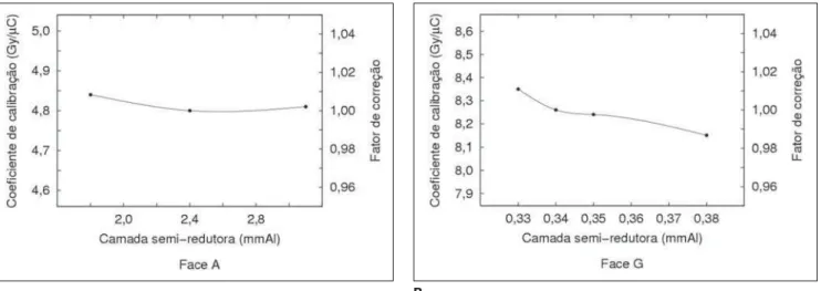
layer on Figures 3A and 3B. On these Fig-ures, the right vertical scales represent cor-rection factors normalized for reference qualities for each case.
The response energy dependence was 0.8% for face A in the qualities of conven-tional diagnostic X-rays, and 2.4% for the face G of the chamber in the qualities of mammography X-radiation. It may be ob-served that the chamber is in compliance with the IEC standard requirements(23):
re-sponse variation up to ±5% with the radia-tion quality.
DISCUSSION
For a consistent quality of the images produced by manual techniques, the air kerma rate linearity as a function of the mAs for a determined kVp must be ≤ 20%(6). It is important that the measured
linearity is a characteristic of the X-ray equipment and is not affected by the lack of response linearity or inaccuracy of the ionization chamber utilized for the mea-surement. The standard uncertainty for any mAs value can be reduced, by increasing the number of measurements, provided all the measurements uncertainties are random and normally distributed. The accuracy is > ±0,05% for both faces of the chamber developed in the present study, limiting the number of exposures required for the mea-surement of the air kerma rate linearity as a function of the mAs and is not significant for the measurement uncertainty. The un-certainty obtained from the chamber
re-sponse linearity is a systematic uncertainty and cannot be minimized by the measure-ments repetition. This uncertainty is small (< 1%) as compared with the criterion for an X-ray equipment performance (≤ 20%). So, the chamber response linearity does not affect the determination of the air kerma rate linearity as a function of the mAs of an X-ray equipment.
The air kerma rate for a determined kVp and mAs should not vary more than 10% across four consecutive exposures(6), i.e.
the air kerma rate must be ≤ 10%. The ac-curacy of the chamber developed in the present study is much higher than the one acceptable for the air kerma rate of an X-ray equipment, and has no adverse effect on the reproducibility measurement.
Plane-parallel ionization chambers are designed to be utilized with the entrance windows facing the radiation source and perpendicular to beam axis. The angular dependence of the chamber developed in the present study was evaluated, consider-ing the possibility of a little variation in the radiation incidence angle due the chamber positioning. It may be observed that the response variation caused by this little variation in the incidence angle does not result in a significant measurement uncer-tainty.
An ionization chamber response energy dependence is one of the main sources of uncertainty in the determination of an X-ray beam air kerma rate and HVL for a cer-tain kVp. The ionization chamber utilized should present a minimal response energy
dependence for each energy interval corsponding to the X-ray equipment. The re-sponse energy dependence is minimal both for the face A of the ionization chamber in the qualities of conventional diagnostic X-ray and for the face G in the qualities of mammography X-rays. The face A can be utilized for measuring the air kerma and the air kerma rate, and for determining the half-value layer in the qualities of conventional diagnostic X-rays, and face G in the quali-ties of mammography X-rays.
CONCLUSIONS
The performance tests demonstrated that the ionization chamber developed in the present study can be routinely utilized for determining the air kerma rate in a pro-gram for controlling the quality of X-ray equipment. The advantage of this chamber is that it can be utilized both for measure-ments in conventional radiography and mammography, provided the appropriate face is selected. Typically a specific type of ionization chamber is utilized for measure-ments in conventional radiography, and another type of chamber in mammography. The construction of this chamber cor-roborates the feasibility of producing radia-tion detectors with materials available in the domestic market.
Acknowledgements
Partial financial support for the devel-opment of the present study was granted by Conselho Nacional de Desenvolvimento
Figure 3.A: Calibration coefficients for the qualities of conventional diagnostic X-rays and correction factors normalized for 2.4 mmAl HVL. Uncertainties are < 3%. B: Calibration coefficients for the qualities of mammography X-rays and correction factors normalized for 0.34 mmAl HVL. Uncertainties are < 2%.
Científico e Tecnológico (CNPq) and Fun-dação de Amparo à Pesquisa do Estado de São Paulo (Fapesp).
REFERENCES
1. World Health Organization. Quality assurance in diagnostic radiology. Geneva: WHO; 1982. 2. Food and Agriculture Organization of the United
Nations, International Atomic Energy Agency, International Labour Organisation, Nuclear En-ergy Agency of the Organisation for Economic Co-operation and Development, Pan American Health Organization, World Health Organization. International Basic Safety Standards for Protec-tion Against Ionizing RadiaProtec-tion and for the Safety of Radiation Sources, Safety Series No. 115. Vienna: IAEA; 1996.
3. Brasil. Ministério da Saúde. Secretaria de Vigi-lância Sanitária. Diretrizes de proteção radioló-gica em radiodiagnóstico médico e odontológico. Portaria nº 453, 1 de junho de 1998. Diário Ofi-cial da União, Brasília, 2 de junho de 1998.
4. International Atomic Energy Agency, Pan Ameri-can Health Organization, World Health Organi-zation. Radiological protection for medical expo-sure to ionizing radiation. Safety Standards Se-ries No. RS-G-1.5. Vienna: IAEA; 2002. 5. International Commission on Radiation Units and
Measurements. ICRU Report 74: Patient dosim-etry for X rays used in medical imaging. J ICRU. 2005;5(2).
6. Brasil. Ministério da Saúde. Agência Nacional de Vigilância Sanitária. Radiodiagnóstico médico: segurança e desempenho de equipamentos. Bra-sília: Anvisa; 2005.
7. Petoussi-Henss N, Zankl M, Drexler G, et al. Cal-culation of backscatter factors for diagnostic ra-diology using Monte Carlo methods. Phys Med Biol. 1998;43:2237–50.
8. European Commission. European protocol on do-simetry in mammography. Report EUR 16263 EN. Luxembourg: EC; 1996.
9. Wagner LK, Cerra F, Conway B, et al. Energy and rate dependence of diagnostic x-ray exposure meters. Med Phys. 1988;15:749–53.
10. DeWerd LA, Wagner LK. Characteristics of ra-diation detectors for diagnostic radiology. Appl Radiat Isot. 1999;50:125–36.
11. Austerlitz C, Nette P, Cordilha A. Construção, calibração e teste de uma câmara de ionização para medidas de exposição X e gama entre 40 e 1250 keV. Rev Fís Aplic Instr. 1986;1:320–8. 12. Silva I. Projeto e construção de uma câmara de
extrapolação para dosimetria beta. (Dissertação de Mestrado). Belo Horizonte: Universidade Fe-deral de Minas Gerais; 1985.
13. Albuquerque MPP, Caldas LVE. New ionization chambers for beta and X-radiation. Nucl Instrum Meth A. 1989;280:310–3.
14. Souza CN, Caldas LVE, Sibata CH, et al. Two new parallel-plate ionization chambers for electron beam dosimetry. Radiat Meas. 1996;26:65–74. 15. Bulla RT, Caldas LVE. Determinação de dose
absorvida em feixes de elétrons utilizando câma-ras de ionização de placas paralelas. Radiol Bcâma-ras. 2004;37:171–7.
16. Bulla RT, Caldas LVE. Comparação entre fatores de calibração em termos de dose absorvida no ar para uma câmara de ionização de placas parale-las. Radiol Bras. 2006;39:203–7.
17. Maia AF, Caldas LVE. A new extended-length parallel-plate ionization chamber. Phys Med Biol. 2005;50:3837–47.
18. Maia AF, Caldas LVE. Calibração das câmaras de ionização para feixes de tomografia computado-rizada no Brasil: a realidade atual. Radiol Bras. 2006;39:209–13.
19. Vivolo V. Desenvolvimento de um sistema de referência para determinação do equivalente de dose pessoal e da constância de feixes de radia-ção-X. (Tese de Doutorado). São Paulo: Univer-sidade de São Paulo; 2006.
20. Dias SK, Caldas LVE. Development of an ex-trapolation chamber for the calibration of beta-ray applicators. IEEE T Nucl Sci. 1998;45:1666–9.
21. Oliveira ML, Caldas LVE. A special mini-extrapo-lation chamber for calibration of 90Sr+90Y
sources. Phys Med Biol. 2005;50:2929–36.
22. International Atomic Energy Agency. Calibration of dosemeters used in radiotherapy, Technical Reports Series No. 374. Vienna: IAEA; 1994. 23. International Electrotechnical Commission. IEC
61674: Medical electrical equipment dosimeters with ionization chambers and/or semi-conductor detectors as used in X-ray diagnostic imaging. Geneva: IEC; 1997.
24. International Electrotechnical Commission. IEC 61267: Medical diagnostic X-ray equipment: ra-diation conditions for use in the determination of characteristics. Geneva: IEC; 2005.
25. Caldas LVE. A sequential Tandem system of ionisation chambers for effective energy determi-nation of X-radiation fields. Radiat Prot Dosim. 1991;36:47–50.
26. Caldas LVE, Albuquerque MPP. Angular depen-dence of parallel-plate ionisation chambers. Radiat Prot Dosim. 1991;37:55–7.
27. Geleijns J, Broerse JJ, Zweers D. General ion recombination for ionization chambers used un-der irradiation conditions relevant for diagnostic radiology. Med Phys. 1995;22:17–22. 28. Das IJ, Akber SF. Ion recombination and
polar-ity effect of ionization chambers in kilovoltage x-ray exposure measurements. Med Phys 1998; 25:1751–7.
29. International Atomic Energy Agency. Absorbed dose determination in external beam radio-therapy: an international code of practice for do-simetry based on standards of absorbed dose to water. Technical Reports Series No. 398. Vienna: IAEA; 2002.