Impact of a program of diagnostic imaging quality
control in mammography centers of the Federal District,
Brazil*
Impacto de um programa de avaliação da qualidade da imagem nos serviços de mamografia do Distrito Federal
Rosangela da Silveira Corrêa1, João Emílio Peixoto2, Lynn Dee Silver3, Cintia Melazo Dias4, Maria do Socorro Nogueira5, Suy Ferreira Hwang6, Rubemar de Souza Ferreira7
OBJECTIVE: The present study was aimed at evaluating the quality and the impact of an intervention involving inspection and education in mammography centers of the Federal District, Brazil. MATERIALS AND METHODS: Forty one mammography centers in the Federal District were studied in the period between 2000 and 2002. The intervention involved an initial inspection followed by a training activity and notification of mammography centers by the Federal District Sanitary Vigilance authority. The imaging quality was compared before and after the intervention. RESULTS: None of the 36 centers which completed the study reached more than 90% compliance with the standard imaging quality prior to the interventions, whereas ten were above 90% afterwards. Major improvements were observed in chassis maintenance, breast compression and visualization of microcalcifications. CONCLUSION: Despite the availability of a great number of mammography centers in the Federal District, most of them did not meet the required quality standards. The intervention has shown to be effective for improving the imaging quality, however a continued action is required to solve the remaining problems and increase the impact of the program.
Keywords: Mammography; Quality control; Federal District; Impact.
OBJETIVO: Esta pesquisa visou a avaliar a qualidade dos serviços de mamografia do Distrito Federal e o impacto de uma intervenção de inspeção e capacitação. MATERIAIS E MÉTODOS: Foram estudados 41 serviços de mamografia no Distrito Federal no período de 2000 a 2002. A intervenção consistiu na inspeção inicial seguida de um treinamento e notificação oficial da Vigilância Sanitária. Os resultados de qualidade da imagem foram comparados “antes” e “depois” da intervenção. RESULTADOS: O estudo demonstrou que dos 36 serviços que completaram a pesquisa, nenhum estava acima de 90% de conformidade antes da in-tervenção. Após a intervenção, dez unidades atingiram mais de 90%. As principais melhorias foram em re-lação aos chassis, compressão da mama e visualização de microcalcificações. CONCLUSÃO: Apesar de o Distrito Federal dispor de muitos serviços, na sua maioria não eram de qualidade. A intervenção foi eficaz para a melhoria da qualidade, porém, torna-se necessária uma ação continuada para resolver os problemas restantes e aumentar o impacto.
Unitermos: Mamografia; Controle de qualidade; Distrito Federal; Impacto.
Abstract
Resumo
* Study developed by Centro Regional de Ciências Nucleares do Centro-Oeste – Comissão Nacional de Energia Nuclear (CRCN-CO/CNEN), Abadia de Goiás, GO, Brazil.
1. Master, Senior Technologist, Centro Regional de Ciências Nucleares do Centro-Oeste – Comissão Nacional de Energia Nuclear (CRCN-CO/CNEN), Abadia de Goiás, GO, Brazil.
2. PhD, Researcher, Universidade Federal do Rio de Janeiro (UFRJ), Rio de Janeiro, RJ, Brazil.
3. PhD, Sub-secretary for the New York Health Department, New York, NY, USA, Associate Professor, Universidade de Brasí-lia (UnB), BrasíBrasí-lia, DF, Brazil.
4. PhD, Technologist, Distrito do Planalto Central – Comissão Nacional de Energia Nuclear (Diplan/CNEN), Brasília, DF, Brazil. 5. PhD, Researcher, Centro de Desenvolvimento da Tecnolo-gia Nuclear – Comissão Nacional de EnerTecnolo-gia Nuclear (CDTN/ CNEN), Belo Horizonte, MG, Brazil.
6. Master, Researcher, Centro Regional de Ciências Nuclea-res do Nordeste – Comissão Nacional de Energia Nuclear (CRCN-NE/CNEN), Recife, PE, Brazil.
the other regions of the country. According to data from the Ministry of Health, breast cancer kills more Brazilian women in than any other type of cancer.
Breast cancer, among the 237,480 new cases estimated for 2006, would be the main type of neoplasm to affect the female population, possibly accounting for up to 48,930 new cases. The Federal District pre-sents an estimated gross incidence of 53 cases for each 100,000 women(1).
In the absence of effective primary pre-vention mechanisms efforts should be fo-cused on the early detection of this disease. Corrêa RS, Peixoto JE, Silver LD, Dias CM, Nogueira MS, Hwang SF, Ferreira RS. Impact of a program of diagnostic imaging quality control in mammography centers of the Federal District, Brazil. Radiol Bras. 2008;41(2):109–114.
INTRODUCTION
Two types of cancer stand out amongst women: uterine cervix cancer and breast cancer. The first one is the main cause for cancer deaths in the Center-Western and Northern regions, while breast cancer, in
7. PhD, Technologist, Centro Regional de Ciências Nucleares do Centro-Oeste – Comissão Nacional de Energia Nuclear (CRCN-CO/CNEN), Abadia de Goiás, GO, Brazil
Mailing address: Rosangela da Silveira Corrêa. Rodovia Br-060, km 174,5, Zona Rural. Abadia de Goiás, GO, Brazil, 75345-000. E-mail: [email protected]
Mammography is a method utilized for de-tection and diagnosis of breast diseases, and frequently is performed in healthy women who intend to maintain this sta-tus(2). However, this type of diagnostic method presents some limitations, and the screening may result in adverse conse-quences. A frequent problem is the false-positive result, where the suspicion for a malignant lesion is not confirmed after his-topathological studies. Another limitation is represented by false-negative results that may lead to the postponement of an appro-priate action in relation breast cancer.
Taking these problems into consider-ation, the development of quality control programs represents a necessity in terms of efficiency, and an obligation in ethical and moral terms(3). The imaging quality re-duces, although does not eliminate, the occurrence of positive and false-negative results.
Therefore, all mammography centers must be focused on a permanent improve-ment of their services in order to achieve a harmonious integration among the follow-ing areas: medical, technological, admin-istrative, economic, public health assis-tance, and, if applicable, academic and re-search.
The greatest risk for a woman who has been submitted to mammography is that a small breast cancer goes undetected be-cause of the low quality of mammographic images. This risk is ten times higher than the risk for a radiation-induced breast can-cer(4,5).
The present comparative study was de-veloped in mammography centers in the Federal District, Brazil, over the period from 2000 to 2002, including a chronologi-cal results (before and after-type analysis).
MATERIALS AND METHODS
The term “intervention”, in the present study, is defined as a sequence of actions involving: inspection (first and second technical visits), qualification courses for both radiologists and technicians, and no-tification from the Departamento de Fis-calização de Saúde do Distrito Federal (DpFS-DF) (Federal District Department of Health Inspection) to mammography cen-ters requesting irregularities correction.
The results evaluation was focused on the quality of images for early breast can-cer detection in compliance with the tech-nical requirements included in the Order (Portaria) no. 453(6) of the Brazilian Min-istry of Health, the European guidelines for quality assurance in mammography screen-ing(7) and the American College of Radiol-ogy guidelines for breast cancer screen-ing(8).
The performance of the whole mammo-grams production chain in mammography centers was quantitatively evaluated with the aid of a breast phantom comprised of test structures in several shapes, sizes and compositions, and producing images simi-lar to the anatomical structures of interest, besides an optical densities scale.
The radiologic breast phantom was de-veloped by the Unit of Radiology at Santa Casa da Misericórdia do Rio de Janeiro(9) and approved by Colégio Brasileiro de Radiologia (Brazilian College of Radiol-ogy)(10). The evaluation included the fol-lowing items:
a) Alignment between the X-ray field and the images receptor;
b) automatic exposure control perfor-mance testing;
c) breast compression rate;
d) alignment of the breast compression paddle;
e) evaluation of the images recording system (cassette holders – screen-film con-tact);
f) films processing quality; g) imaging quality:
– images definition (spatial resolution); – high-contrast detail;
– low-contrast threshold; – low-contrast linear detail;
– tumor-like masses; – background optical density.
RESULTS
Forty-one mammography centers in the Federal District were enrolled in the present study. However, only 36 of them were evaluated, considering that five units were excluded from the study right away at the first visit, for not meeting the inclusion criteria. This fact reflects the significance of the impact at the first phase of the inter-vention, considering that three of these centers (7.3%) were closed down because of quality-related issues.
Evaluation of imaging quality in mam-mography centers of the Federal District
Imaging quality indicators are gathered in two different sets of results: the first one includes technical and performance param-eters of mammographs and processors; the second one includes parameters regarding the final quality of the images from a logic breast phantom recorded on a radio-graphic film.
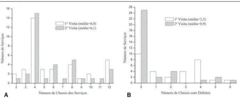
Figure 1A shows the distribution of cas-sette holders utilized in the mammography centers. It may be observed that this num-ber has not changed between the first and second technical visits. On the other hand, Figure 1B clearly demonstrates that the number of defective cassette holders in each center changed significantly, espe-cially regarding the number of centers with no defective cassette holder that changed from 10 at the first visit, to 25 at the second. As regards the performance of the mammographs technical parameters evalu-ated in the two visits, Figure 2A
demon-Figure 1. Cassette holders performance before and after the intervention.
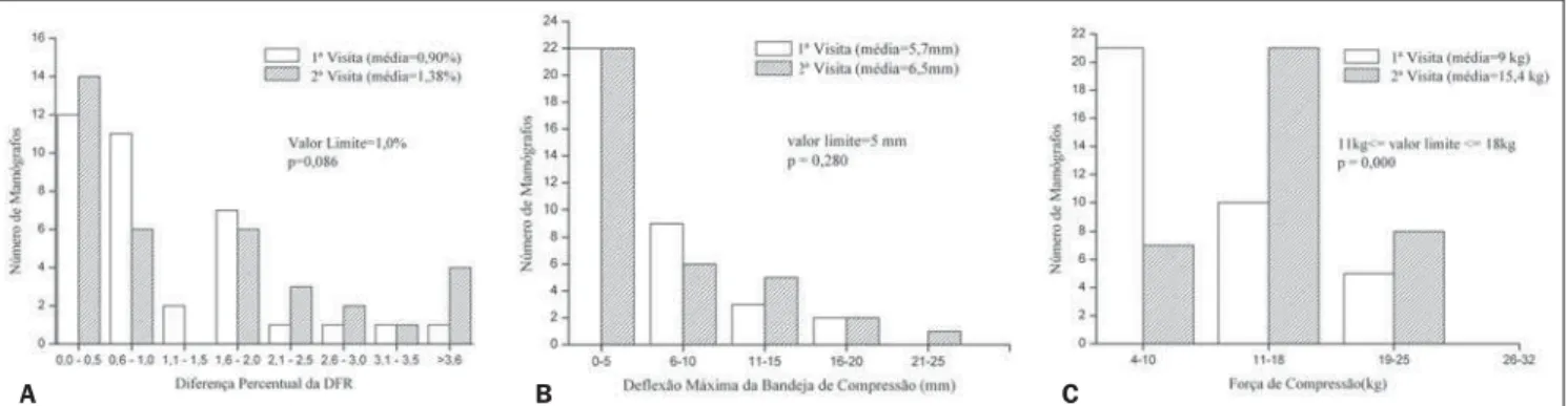
strates that the results from the measure-ments of alignment between X-ray field and the chest wall of the patients did not present a statistically significant improve-ment (p = 0.086). The same occurred in relation to the measurement of maximum compression paddle deflection, with a non-statistically significant difference (p = 0.280) between the two technical visits, as shown on Figure 2B. The third parameter evaluated — breast compression rate — presented a statistically significant differ-ence (p = 0.000) between the two evalua-tions. Figure 2C demonstrates that the number of mammographs in compliance with this parameter increased from seven to 20 between the first and the second evaluations.
The appropriate operation of the auto-matic exposure control system is essential in the practice of mammography and con-stitutes a feature of the images production chain evaluated in the present study. Fig-ure 3 shows the results from measFig-urements of the differences among optical densities for different breast thicknesses (5 cm, 4 cm, 3 cm and 2 cm).
Additionally, Figure 3 demonstrates a statistically significant improvement in the operation of automatic exposure control systems, most appropriately balancing the images darkening effect for 4 cm and 3 cm-thick breasts. However, no improvement was observed in the performance of this device for imaging 2 cm-thick breasts.
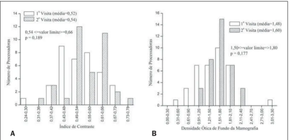
The performance of automatic films processors was evaluated by means of a sensitometric test measuring the optical densities at three different points on a gray scale with 21 optical steps. Measurement points were the following: the base+fog step, the speed step, and the contrast step. Figure 4A demonstrates that the base den-sity plus fog of mammograms, both in the first and second technical visits, was within the range recommended for this parameter in the greatest majority of centers, with no statistically significant difference (p = 0.340) between the two data set regarding this parameter. As regards the measurement of the speed step optical density, Figure 4B demonstrates a statistically significant variation (p = 0.034) between the first and second evaluations, and an increase in the
frequency, from six to 15, of the number of films processors operating within the range recommended for this parameter.
For the contrast step optical density, Fig-ure 4C shows that not only the difference between the two data sets was not statisti-cally significant (p = 0.480), but also there was no increase in the number of proces-sors in compliance with the reference limit for this parameter.
As regards the evaluation of final qual-ity parameters for the images of a logic breast phantom recorded on the radio-graphic film, Figure 5 shows the results for visualization of relevant imaging details in mammography. This figure demonstrates that, for microcalcifications, masses, metal grids, fibers, and low-contrast disks, the results presented a statistically significant improvement between the first and second evaluations.
Additionally, as regards other mam-mograms quality indicators, Figure 6 dem-onstrates that, although the results both for the image contrast index and background optical density have not presented a statis-tically significant difference (p > 0.05),
Figure 2. Mammographs performance before and after the intervention.
A B C
A B C
there was an increase in the frequency of processors in compliance with the ranges recommended for these parameters.
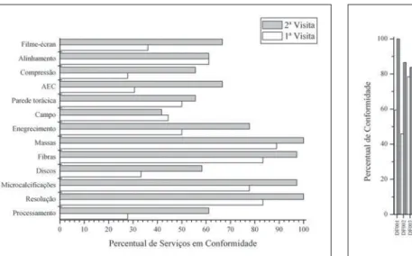
Considering the quality tests applied, Figure 7 shows the percentage of mam-mography centers in compliance with each of the parameters evaluated. As regards ra-diographic films processing, even with the increase (from 28% to 61%) in the number of compliant centers, this still remains as a critical issue in the mammographic images production chain. This is confirmed by the visualization of low-contrast disks that, by the time of the first technical visit was com-pliant in 33% of the centers, and in the
sec-A B C
Figure 4. Automatic processor performance before and after the intervention.
Figure 5. Imaging quality parameters before and after the intervention.
A B C
D E
B A
Figure 7. Percentage of mammography centers in the Federal District presenting compliance in relation to quality parameters in the first and second technical visits.
Figure 8. Imaging quality compliance rates before and after the interven-tion.
ond visit was compliant in 58%. This is the evidence that the reference limit for visu-alization of these testing objects is the most reliable imaging quality indicator reflect-ing the quality in the radiographic films processing.
Other results regarding imaging quality indicators show that metal grids and micro-calcifications imaging is poorly or not af-fected at all by the processing, considering their nature (high-contrast); and, also, that images of fibers and tumor-like masses relatively thick in terms of mammography are poorly affected by the processing.
As regards these four imaging quality indicators, the percentage of compliant mammography centers increased from about 80% in the first technical visit to about 100% in the second one.
Results regarding mammographs per-formance and evaluation of the images re-cording systems indicative of the opera-tional status of the radiographic cassette holder (screen/film contact) show that, despite the improvement from the first to the second technical visit, the percentage of compliant centers in these items re-mained below or around 60%.
Intervention impact on mammography centers
Figure 8 shows the compliance rate re-sulting from the tests described in the present study. In the evaluation of the in-tervention impact on the 36 mammography
centers of the Federal District, the mean rate of compliance regarding imaging qual-ity increased from 53.61% to 73.19% be-tween the first and second technical visits. According to the paired t test, at 5% signifi-cance level and with 35 freedom degrees, the difference between means was statisti-cally significant, with the intervention af-fecting the improvement of the imaging quality in mammography centers.
DISCUSSION
The performance of mammographs op-erational parameters most directly related to the imaging quality was evaluated by means of the previously mentioned tests. It should be emphasized that these tests were based on the requirements clearly estab-lished by the Order (Portaria) no. MS 453/ 98.
The most significant results for mam-mographs performance in the present study were those regarding automatic exposure control systems. Figure 8 shows that, de-spite de increase in the percentage of com-pliant centers in this item, from 30% in the first technical visit to 66% in the second one, several mammography devices in the Federal District still require technical ad-justments.
The higher disagreement was observed in the difference between optical densities of 2 cm-thick breasts and 5 cm-thick breasts. This same situation is reported in
the literature. LaFrance et al.(11), discussing similar results, have concluded that the in-appropriate performance of the automatic exposure control system may be associated with the change in the X-ray beam energies spectrum caused by breast tissues attenua-tion (beam hardening), failure in reciproc-ity (to maintain a constant darkening for increasingly shorter exposure times) of the radiographic film response or to a current generated in the circuit itself in the absence of radiation exposure. According to the authors, the prevalent effect is the beam hardening. This effect may be explained as follows: as the compressed breast thickness increases, the emerging X-ray beam pen-etration increases, and a high percentage of photons is transmitted through the intensi-fier screen and the radiographic film, fall-ing upon the sensor of the automatic com-pensation device. In summary, the higher the breast thickness, the greater is the beam hardening and the larger is the amount of energy absorbed by the sensor in relation to the energy absorbed by the cassette hold-ers. Therefore, the film darkening increases as the breast thickness decreases, and vice-versa.
It is important to emphasize that the ra-diologic breast phantom utilized in the present study demonstrated to be appropri-ate for evaluating the performance of this technical feature in mammographs. This appropriateness is due to the fact that this phantom is constituted by layers simulat-ing breasts of different thicknesses.
The other technical features of mam-mographs performance evaluated in the present study were the following: align-ment between the X-ray field and the patient´s chest wall, the maximum com-pression paddle deflection, and the breast compression rate. Results presented on Figures 3A,B and 8 indicate that the per-centage of compliant centers regarding alignment between field and compression paddle has not changed between the first and second technical visits, remaining in the range between 50% and 60%. The in-fluence of the non-compliance on these two technical features could not be estimated, considering that the radiologic breast phan-tom utilized did not require compression to be imaged, so the phantom image quality was not affected by the compression paddle. This also is valid for the breast compression rate. Although Figure 3C demonstrates that the number of compliant mammographs in relation to this parameter increased from seven units in the first tech-nical visit to20 units in the second visit, the phantom image could not demonstrate this improvement in the mammograph perfor-mance.
Based on the above considerations, it is clear that the phantom image should not be the sole element to be considered in the evaluation of mammographic images qual-ity, considering that several mammographs performance features related to breast com-pression and positioning cannot be evalu-ated by means of phantom images.
As regards the processors performance evaluation, the present study demonstrated
that, although the percentage of compliant centers has increased from 28% in the first technical visit to 61% in the second visit, this is the main source of images quality loss. Similar results can be found in the lit-erature. Hendrick et al.(12) have reported that about 47% of mammography centers lacking approval by the American College of Radiology Accreditation Program pre-sented non-compliance regarding proces-sors performance. Previously, Galkin et al.(13) had already reported similar results. Approximately 40% of the films processors evaluated presented an excessive variation in their performance over a 15-day period. According to the authors, this was the main reason for the variation in the images qual-ity and radiation doses among the facilities participating in a regional program devel-oped in the United States for early detec-tion of breast cancer.
CONCLUSION
After an intervention process in mam-mography centers of the Federal District, the impact on the imaging quality, although positive both in terms of quality and in terms of quantity, has shown to be beneath the desired target-conformity level of > 90%. Several factors may have contributed for this partial result, among others, the unsatisfactory performance of films pro-cessors, poorly adjusted devices for con-trolling exposure and other operational parameters of the mammograph. The re-sults from the intervention performed in the present study were similar to the ones achieved in interventions performed in other Brazilian states. And the experience acquired through other sanitary vigilance programs in mammography centers, par-ticularly the one developed in the state of Paraíba(14), shows that a compliance rate in the range of 90% only can be achieved by means a continued action.
REFERENCES
1. Estimativas da incidência e mortalidade por cân-cer no Brasil, 2006. Instituto Nacional do Cân-cer (INCA). [Acessado em 11 de abril de 2006]. Disponível em: http://www.inca.gov.br/estima-tiva2006
2. Bassett LW, Hendrick RE, Bassford TL, et al. Quality determinants of mammography: clinical practice guideline no. 13. AHCPR publication no. 95-0632. Rockville: Agency for Health Care Policy and Research, Public Health Service, U.S. Department of Health and Human Services; 1994. 3. Azevedo AC, Koch HA, Canella EO. Auditoria em centro de diagnóstico mamário para detecção precoce de câncer de mama. Radiol Bras. 2005; 38:431–4.
4. Hendrick RE. Mammography quality assurance cancer. Cancer. 1993;72:1466–74.
5. Caldas FAA, Isa HLVR, Trippia AC, et al. Con-trole de qualidade e artefatos em mamografia. Radiol Bras. 2005;38:295–300.
6. Ministério da Saúde. Secretaria de Vigilância Sanitária. Diretrizes de proteção radiológica em radiodiagnóstico médico e odontológico. Porta-ria nº 453. Diário Oficial da União, 1/6/1998.
7. Perry NN, Broeders M, Wolf C, et al., editors. European guidelines for quality assurance in mammography screening. 3rd ed. Luxembourg: European Comission, Europe Against Cancer; 2001.
8. American College of Radiology. Recommended specifications for new mammography equipment. Reston: American College of Radiology; 1993.
9. Pina DR, Morceli J, Duarte SB, et al. Otimização de imagens mamográficas. Radiol Bras. 2006;39: 351–4.
10. Colégio Brasileiro de Radiologia. Boletim do CBR nº 165, novembro 2001. p. 21.
11. LaFrance R, Gelskey DE, Barnes GT. A circuit modification that improves mamographic photo-timer performance. Radiology. 1988;166:773–6.
12. Hendrick RE, Smith RA, Wilcox PA. ACR Accreditation and legislative issues mammogra-phy. In: Haus AG, Yaffe MJ, editors. Syllabus: a categorical course in physics – technical aspects of breast imaging. Oak Brook: Radiological Society of North America; 1993. p. 137–49.
13. Galkin BM, Feig SA, Muir HD. The technical quality of mammography in centers participating in a regional breast cancer awareness program. Radiographics. 1988;8:133–45.