SCIENTIFIC ARTICLE
A Comparative Study among Three Techniques of General
Anesthesia for Ultrasound-Guided Transrectal Prostate Biopsy
Ricardo Antônio Guimarães Barbosa, TSA
1, Camerine Domingues da Silva
2, Mary Yumi Takei Torniziello
2,
Luciana Mendes de Olivera Cerri
3, Maria José Carvalho Carmona, TSA
4, Luiz Marcelo Sá Malbouisson, TSA
5Summary: Barbosa RAG, Silva CD, Torniziello MYT, Cerri LMO, Carmona MJC, Malbouisson LMS – A Comparative Study among Three Tech-niques of General Anesthesia for Ultrasound-Guided Transrectal Prostate Biopsy.
Background and objectives: Ultrasound-guided transrectal prostate biopsy is a reference in the diagnosis of prostate neoplasias. The higher the number of samples, the greater is the pain and discomfort. The objective of this study was to compare three anesthetic techniques in this group of patients.
Methods: Forty-five patients were included in this study. Patients were divided into three groups: 1 – Propofol; 2 – Propofol + Prostatic Plexus Block; 3 – Propofol + Fentanyl. Patients were monitored with non-invasive blood pressure, continuous electrocardioscopy, pulse oximetry (SpO2), and Bispectral Index. Patients did not receive pre-anesthetic medication. Intraoperative and postoperative hemodynamic parameters, intraopera-tive bispectral index, and postoperaintraopera-tive visual analogue scale (VAS) and the use of dypirone to treat postoperaintraopera-tive pain were evaluated. Results: A significant difference among the three groups was not observed for: anthropometric parameters, amount of propofol, number of fragments, and duration of the exam. Hemodynamic parameters and SpO2 presented similar behavior in all three groups. In group 1, the pain evaluated by the VAS was more severe and required more dypirone than in the other groups.
Conclusions: Sedation with propofol alone for biopsy is associated with greater postoperative pain and discomfort than in prostatic plexus block or systemic fentanyl. Besides hypnosis, intraoperative analgesia is required to guarantee postoperative comfort.
Keywords: ANESTHESIA, General; ANESTHESICS, Intravenous: propofol; DISEASES, Oncology: prostata neoplasia, PAIN, Postoperative; DIAGNOSIS EXAMS: prostata biopsy.
[Rev Bras Anestesiol 2010;60(5): 457-465] ©Elsevier Editora Ltda.
INTRODUCTION
Ultrasound-guided biopsy is a reference in the diagnosis of pros-tate tumors 1-3. Successful detection of prostate cancer is related
to the number of samples. On the other hand, the higher the number of samples greater the pain and discomfort 1.
Pain during the procedure results from: the introduction of the transrectal transducer and penetration of the needle in the prostate capsule, which stimulates pain receptors located in this region 4. The prostate is innervated by parasympathetic
Received from the Hospital das Clínicas da Faculdade de Medicina da Universidade de São Paulo – HCFMUSP.
1. PhD in Sciences from the Faculdade de Medicina da Universidade de São Paulo – FMUSP, Anesthesiology Professor of Faculdade de Ciências Médicas de Santos-UNILUS. Supervising Physician of the Anesthesiology Department of the Instituto de Radiologia do HCFMUSP.
2. Anesthesiologist of the Anesthesiology Department of FMUSP
3. PhD in Sciences from USP, Supervising Physician of the Ultrasound Department of the Instituto de Radiologia do HCFMUSP
4. Associated Anesthesiology Professor of the Faculdade de Medicina da USP. TSA, Direc-tor of the Anesthesiology Department of the Instituto Central do HCFMUSP
5. PhD in Sciences from USP. Intensive care specialist from AMIB, ICU Coordinator of the Anesthesiology Department of the Instituto Central do HCFMUSP
Submitted on March 25, 2010. Approved on May 3, 2010.
Correspondence to:
Dr. Luiz Marcelo Sá Malbouisson
Divisão de Anestesia do ICHC Av. Enéas Carvalho de Aguiar, 255 – 8º andar. Cerqueira César
05403-900 – São Paulo, SP, Brazil E-mail: [email protected]
fibers that originate in the pelvic splanchnic nerves (S2 to S4) and sympathetic fibers of the inferior hypogastric nerves 5.
Approximately 96% of the patients report pain, with a score of 2 to 6 in the VAS, and 20% of those patients consider the pain to be severe 1.
Due to the severity of the pain, an analgesia/anesthesia strategy is fundamental in prostate biopsies to decrease the discomfort for the patient and, consequently, increase the number of samples collected and the success of the proce-dure. Besides, many patients will need more than one inter-vention to collect biopsies in order to achieve a conclusive diagnosis 6.
Several anesthetic techniques have been described and investigated in prostate biopsies, but a consensus in the li-terature regarding the most effective technique, such as conscious sedation with midazolam, general anesthesia with spontaneous ventilation with fentanyl and propofol, spinal anesthesia, and pudendal nerve block, is lacking 6,7. Prostatic
plexus block, performed by the radiologist, with the injection of several local anesthetics, is the analgesic technique used more often 8-12. However, in this technique, the patient can
undergo important physical and emotional discomfort 2,3,8.
BARBOSA, SILVA, TORNIZIELLO ET AL.
458 Revista Brasileira de Anestesiologia
Vol. 60, No 5, September-October, 2010
study was to compare the sedation of isolated propofol, asso-ciated with prostatic plexus block, and with systemic opioids regarding the quality of hypnosis, hemodynamic parameters, and postoperative pain.
MATERIALS AND METHODS
This study was approved by the Scientific Commission of the Instituto de Radiologia and Medical Ethics Commission of the Hospital das Clínicas da Faculdade de Medicina da Universi-dade de São Paulo. Forty five patients with classification I or II of the American Society of Anesthesiologists, and with indi-cation of ultrasound-guided prostate biopsy, were randomly divided in three groups.
GROUP 1 (G1): Fifteen patients who received propofol, at an initial dose of 1 to 1.5 mg.kg-1, with supplementary doses as
needed;
GROUP 2 (G2): Fifteen patients who received propofol, at an initial dose of 1 to 1.5 mg.kg-1, with supplementary doses of
propofol as needed, associated with prostatic plexus block, performed by the ultrasonographist, with 40 mg of 1% lidocai-ne without adrenalilidocai-ne.
GROUP 3 (G3): Fifteen patients who received propofol at an initial dose of 1 to 1.5 mg.kg-1, with supplementary doses of
propofol as needed, associated with 0.5 µg.kg-1 of fentanyl.
This study was undertaken at the Instituto de Radiologia do Hospital das Clínicas da Faculdade de Medicina da Universi-dade de São Paulo; patients signed an informed consent and the presence of a companion was mandatory.
Patients were monitored with non-invasive blood pressure, continuous electrocardiogram, pulse oximetry, and bispectral index. Since all patients were from the outpatient clinic, pre-anesthetic medication was not used. During the procedure, patients remained on spontaneous ventilation. To maintain the peripheral oxygen saturation above 95%, oxygen, 5 L.min.-1,
was administered via a face mask.
The following parameters were evaluated:
Intraoperatively: Mean arterial, systolic, and diastolic pressu-res, heart rate, oxygen saturation, and bispectral index, were evaluated before the procedure, 5 minutes after beginning the procedure, and at the end. The total duration of the procedure and total dose of propofol were also recorded.
Postoperatively: Mean, systolic, and diastolic arterial pressures, heart rate, oxygen saturation, pain assessment using the verbal analogue scale, and the use of dypirone as rescue medication to treat postoperative pain. Those parameters were evaluated 15, 30, 45, and 60 minutes after the end of the procedure.
After the procedure, patients were transferred to the post-anesthetic recovery room, where they remained for 2 hours for observation of complications. Dypirone 2 g IV, was chosen as rescue medication in case of pain greater than 5 in the visual analogue scale.
The parameters were analyzed using the SPSS 16 (SPSS Inc., IL, USA) and Aabel 3 (Gigawiz Inc., USA) software. The
Kolmogorov-Smirnov test was used for the distribution of con-tinuous or subtle variables. Anthropometric data were compa-red by one-way Analysis of Variance, for age and BMI. Wei-ght, heiWei-ght, duration of the exam, total dose of propofol, ratio of propofol dose/weight, and the number of fragments were compared by the Kruskall-Wallis test. The ASA physical sta-tus distribution was compared among groups by the likelihood ratio test. Two-way Analysis of Variance for repetitive measu-rements was used to compare hemodynamic parameters, bis-pectral index, and oxygen saturation along time, followed by the Newmann-Keuls test when indicated for multiple compari-sons. Pain levels according to the visual analogue scale were analyzed by the Friedman test, with intergroup comparison by the Kruskall-Wallis test, followed by the Dunn test in case of difference among the groups. The Chi-square test for tenden-cies was used to compare the use of dypirone. Values of p lo-wer than 0.05 lo-were considered significant. Data are presented as mean ± standard deviation, median (minimum-maximum), or absolute value (percentage). Charts are presented as mean ± standard deviation or box diagram (maximum and minimum values, 25 interquartile, 75 interquartile, and median).
RESULTS
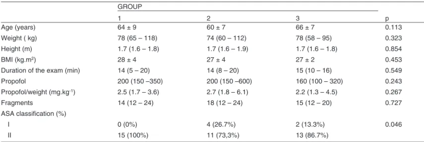
A significant difference was not observed among the three groups regarding anthropometric data (age, weight, height, body mass index), amount of propofol used, as well as exam-related variables: number of fragments and duration of the exam. Regarding the classification of physical status, patients in group 2 were more frequently classified as ASA I than in the other groups (Table I).
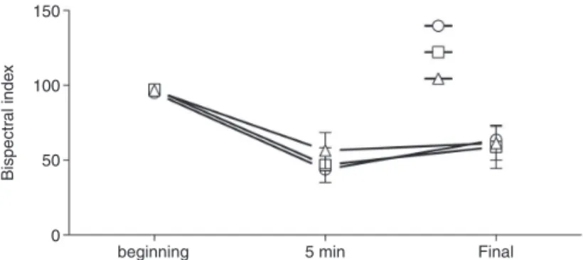
During the intraoperative period, the bispectral index had similar behavior in all three groups, with a fall 5 minu-tes after the beginning of the procedure and maintaining levels between 50 and 60 until the end of the observation period, as can be seen in Figure 1. Hemodynamic parame-ters, both in the intraoperative and postoperative periods, showed statistically similar behavior in all three groups. As shown in Figure 2, we observed that during the procedure a discrete fall in systolic, diastolic, and mean arterial pres-sures, but they recovered preoperative levels. A significant difference between the groups regarding heart rate and oxygen saturation was not observed (Figure 3).
Figure 1. Bispectral Index in Patients in Groups 1 (open circles), 2 (open squares), and 3 (open triangle) along the study.
150
50
Bispectr
al inde
x
0
beginning 5 min Final
Table I – Classification of Physical Status
GROUP
p
1 2 3
Age (years) 64 ± 9 60 ± 7 66 ± 7 0.113
Weight ( kg) 78 (65 – 118) 74 (60 – 112) 78 (58 – 95) 0.323
Height (m) 1.7 (1.6 – 1.8) 1.7 (1.6 – 1.9) 1.7 (1.6 – 1.8) 0.854
BMI (kg.m2) 28 ± 4 27 ± 4 27 ± 2 0.453
Duration of the exam (min) 14 (5 – 20) 14 (8 – 20) 15 (10 – 16) 0.549
Propofol 200 (150 –350) 200 (150 –600) 160 (100 – 320) 0.243
Propofol/weight (mg.kg-1) 2.5 (1.7 – 3.6) 2.7 (1.8 – 6.1) 2.2 (1.3 – 4.5) 0.267
Fragments 14 (12 – 24) 18 (12 – 24) 15 (12 – 20) 0.727
ASA classification (%)
I 0 (0%) 4 (26.7%) 2 (13.3%) 0.046
II 15 (100%) 11 (73,3%) 13 (86.7%)
BMI – Body Mass Index. Data presented as mean ± standard deviation, median (minimum-maximum), or absolute value (percentage).
160
140
120
100
80
60
60 50 40
30 20 final SO 0
intraop
SBP
(mmHg)
postop
G1 G2 G3
100
90
80
70
60
50
40
60 50 40 30 20 final SO
Hear
t r
ate (BPM)
0
intraop postop
G1 G2 G3
Figure 2. Systolic (upper panel), Diastolic (middle panel), and Mean (lower panel) Arterial Pressure in patients in groups 1 (open circles), 2 (open squares), and 3 (open triangles) along the study.
Figure 3. Heart Rate (upper panel) and Peripheral Oxygen Satu-ration (lower panel) in patients in groups 1 (open circles), 2 (open squares), and 3 (open triangles) along the study.
After the procedure, awakening from anesthesia was very fast in all three groups, although group 1 had a significan-tly higher number of patients with elevated pain score at 15 and 60 minutes when compared to groups 2 and 3, as can be observed in Figure 4. Te need of rescue medication for pain control was significantly higher in group 1 and progressi-ve lower in groups 2 and 3 (Figure 5). In group 3, two cases of intraoperative respiratory depression were observed, which were reverted with positive pressure ventilation, without pos-toperative consequences.
120
100
80
60
60 50 40
40
30 20 20
final SO 0
0
intraop
DBP
(mmHg)
postop
140
120
100
80
60
60 50 40
40
30 20 final SO
time (min)
0 intraop
MAP
(mmHg)
postop
100
98
96
94
92
90
60 50 40 30 20 final SO
time (min)
SpO
2
(%)
0
BARBOSA, SILVA, TORNIZIELLO ET AL.
460 Revista Brasileira de Anestesiologia
Vol. 60, No 5, September-October, 2010 DISCUSSION
Early diagnosis of prostate neoplasias is fundamental for a successful treatment. Biopsy is the reference in the diagno-sis of prostate tumors. The higher the number of samples, the higher is the success of the procedure and the pain and discomfort reported by patients. Thus, realization of the pro-cedure with the patient sedated allows a greater number of samples to be collected, which leads to a higher success rate and, consequently, the need of subsequent procedures.
However, a consensus in the literature regarding the best anesthetic technique does not exist. The present study compared three sedation techniques for ultrasound-guided prostate biopsy. Propofol was the hypnotic agent used in all three groups. The op-tion for isolated propofol has been described in the literature as a technique associated with a high patient satisfaction index. Pos-toperative pain, in this case, is considered acceptable 13. Parks et
al. reported that this technique is safe and that the concentration at the site of action of the drug is around 1.5 µg.L-1 14. Although
hemodynamic parameters had shown a stable temporal beha-vior without statistically significant differences among the three groups, in patients who received isolated propofol, a tendency for increase in blood pressure and heart rate was observed. Even though the bispectral index was similar in all groups throughout the study, the severity of postoperative pain was significantly gre-ater in the group of patients who received isolated propofol. Al-though the bispectral index was similar in all three groups along the study, the intensity of postoperative pain was significantly higher in patients who receive isolated propofol. Despite being considered a technique that promotes great patient satisfaction, according to the literature 13, by reducing the discomfort
seconda-ry to the positioning of the ultrasound transducer, the fear of the patient regarding the procedure, and promoting a calm operative environment, it does not prevent postoperative pain. When those findings are compared to those of groups 2 and 3, it is clear that the impact of the use of an analgesic technique, both systemic and regional, is very significant in the reduction of postoperative pain, representing a considerable advantage, since the patient will return home after the procedure. Several local anesthetics have been used in prostatic plexus block 4,11,12,15. The duration of
the local anesthetic could be related to postoperative discomfort. Lidocaine has been used in prostatic plexus block in our depart-ment. Despite the absence of significant differences in the seve-rity of pain with lidocaine, a discrete increase in the scores of the visual analogue scale, which might mean the end of the effect of the local anesthetic used was observed, while in the fentanyl group, patients did not complaint of postoperative pain. This might indicate a discretely superior effect of the association propofol-fentanyl in pain control when compared to group 2. However, this association is not devoid of risks, and two cases of intraoperative respiratory depression, possibly due to the interaction fentanyl-propofol, that were reverted with positive pressure ventilation, were observed 12. The anesthetic technique used did not alter the
time of discharge, since all patients remained at least two hours in the post-anesthetic recovery room for observation of adverse events related to the procedure, such as bleeding.
Intercurrences suggestive of increased morbidity associated with the techniques evaluated, such as rectal bleeding, hematuria, or hematospermia, non-scheduled hospitalization, or infectious complications, were not observed. Changes in blood oxygenation or postoperative respiratory depression were also not observed.
To conclude, due to the pain and discomfort reported by patients, anesthesia in patients undergoing ultrasound-guided prostate biopsy is fundamentally important to promote comfort and increase the number of fragments collected. However, besides hypnosis, analgesia, regional or systemic, is also ne-cessary for adequate control of postoperative pain.
p = 0.02
15
10
Group 1
P
atients (n)
Group 2 Group 3
5
0
group 1
VA
S
10
8
6
4
2
0
VA
S
10
8
6
4
2
0
VA
S
group 2
*
*
*
*
group 315 min 30 min 45 min 60 min
10
8
6
4
2
0
Figure 4. Box Diagram of Pain Scores, According to the VAS, in Patients in Groups 1 (upper panel), 2 (middle panel), and 3 (lower panel). *means different from group 1.
REFERÊNCIAS / REFERENCES
01. Horinaga M, Nakashima J, Nakanoma T – Efficacy compared be-1. Horinaga M, Nakashima J, Nakanoma T – Efficacy compared be-tween caudal block and periprostatic local anesthesia for transrec-tal ultrasound-guided prostate needle biopsy. Urology, 2006;68: 348-351.
02. Turgut AT, Ergun E, Kosar U et al. – Sedation as an alternative meth-od to lessen patient discomfort due to transrectal ultrasonography-guided prostate biopsy. Eur J Radiol, 2006;57:148-153.
03. Presti JC – Prostate biopsy: current status and limitations. Rev Urol, 2007;9:93-98.
04. Vanni AP, Schaal CH, Costa RP et al. – Is the periprostatic anes-thetic blockade advantageous in ultrasound-guided prostate biopsy? Int Braz J Urol, 2004;30:114-118.
05. Anatomia orientada para a clínica. 5 ed. Rio de Janeiro: Guanabara Koogan, 2007;249-250.
06. Visapaa H, Taari K – Combination of paracetamol, codeine and lido-6. Visapaa H, Taari K – Combination of paracetamol, codeine and lido-caine for pain relief during transrectal ultrasound guided biopsy of the prostate. Scand J Surg, 2009;98:55-57.
07. Obek C, Ozkan B, Tunc B et al. – Comparison of 3 different methods of anesthesia before transrectal prostate biopsy: a prospective ran-domized trial. J Urol, 2004;172:502-505.
08. Richman JM, Carter HB, Hanna MN et al. – Efficacy of periprostatic lo-cal anesthetic for prostate biopsy analgesia: a meta-analysis. Urology, 2006;67:1224-1228.
09. Montoliu Garcia A, Juan Escudero J, Ramos de Campos M et al. – Prospective randomized study on the use of lidocaine local anesthe-sia in prostate biopsy. Arch Esp Urol, 2009;62:339-347.
10. Bingqian L, Peihuan L, Yudong W et al. – Intraprostatic local anes-thesia with periprostatic nerve block for transrectal ultrasound guided prostate biopsy. J Urol, 2009;182:479-483; discussion 483-474. 11. Akpinar H, Tufek I, Atug F et al. – Doppler ultrasonography-guided
pelvic plexus block before systematic needle biopsy of the prostate: A prospective randomized study. Urology, 2009;74:267-271.
12. Yurdakul T, Taspinar B, Kilic O et al. – Topical and long-acting local anesthetic for prostate biopsy: a prospective randomized placebo-controlled study. Urol Int, 2009;83:151-154.
13. Awsare NS, Green JA, Aldwinckle B et al. – The use of propofol se-dation for transrectal ultrasonography-guided prostate biopsy is as-sociated with high patient satisfaction and acceptability. Eur J Radiol, 2007;63:94-95.
14. Park JY, Park SJ, Choi SU et al. – Target-controlled propofol infu-sion for sedation in patients undergoing transrectal ultrasound-guided prostate biopsy. J Int Med Res, 2007;35:773-780.
15. Kandirali E, Ulukaradag E, Uysal B et al. – Is only perianal anesthesia with lidocaine-prilocaine cream sufficient to decrease the pain during transrectal ultrasound-guided prostate biopsy? A prospective rando-A prospective rando-mized study. Urol Int, 2009;82:262-265.
Resumen: Barbosa RAG, Silva CD, Torniziello MYT, Cerri LMO, Carmona MJC, Malbouisson LMS – Estudio Comparativo entre Tres Técnicas de Anestesia General para Biopsia de Próstata Dirigida por Ultrasonido Transrectal.
Justificativa y objetivos: La biopsia de próstata dirigida por ultra-sonido transrectal constituye una referencia en el diagnóstico de las neoplasias de la próstata. Mientras mayor es el número de muestras escogidas, mayores son el dolor y la incomodidad relatados por el pa-ciente. El objetivo del estudio fue comparar tres técnicas anestésicas en ese grupo de pacientes.
Método: Fueron estudiados 45 pacientes divididos en tres grupos: 1– Propofol; 2– Propofol + Bloqueo de Plexo Prostático; 3– Propofol + Fentanil. Los pacientes fueron monitorizados con presión arterial
no invasiva, electrocardioscopia continua, oximetría de pulso (SpO2)
e Índice Bispectral. No recibieron medicación preanestésica. Se eva-luaron los parámetros hemodinámicos en el intra y postoperatorio, índice bispectral en el intraoperatorio, el dolor por la escala numéri-ca verbal (ENV) en el postoperatorio inmediato y el uso de dipirona como tratamiento del dolor postoperatorio.
Resultados: No hubo diferencia significativa entre los tres grupos: en las variables antropométricas, cantidad de propofol, número de fragmentos y tiempo del examen. Los parámetros hemodinámicos y
la SpO2 presentaron un comportamiento semejante en los tres
gru-pos durante el estudio. En el grupo 1, el dolor evaluado por la ENV fue más elevado y hubo una mayor necesidad de aplicar la dipirona que en los otros grupos.