Low Na, High K Diet and the Role of
Aldosterone in BK-Mediated K Excretion
Ryan J. Cornelius, Donghai Wen, Huaqing Li, Yang Yuan, Jun Wang-France, Paige C. Warner, Steven C. Sansom*
Department of Cellular and Integrative Physiology, University of Nebraska Medical Center, Omaha, Nebraska, United States of America
Abstract
A low Na, high K diet (LNaHK) is associated with a low rate of cardiovascular (CV) disease in many societies. Part of the benefit of LNaHK relies on its diuretic effects; however, the role of aldosterone (aldo) in the diuresis is not understood. LNaHK mice exhibit an increase in renal K secretion that is dependent on the large, Ca-activated K channel, (BK-αwith ac-cessory BK-β4; BK-α/β4). We hypothesized that aldo causes an osmotic diuresis by in-creasing BK-α/β4-mediated K secretion in LNaHK mice. We found that the plasma aldo concentration (P[aldo]) was elevated by 10-fold in LNaHK mice compared with control diet (Con) mice. We subjected LNaHK mice to either sham surgery (sham), adrenalectomy (ADX) with low aldo replacement (ADX-LA), or ADX with high aldo replacement (ADX-HA). Compared to sham, the urinary flow, K excretion rate, transtubular K gradient (TTKG), and BK-αand BK-β4 expressions, were decreased in ADX-LA, but not different in ADX-HA.
BK-β4 knockout (β4KO) and WT mice exhibited similar K clearance and TTKG in the ADX-LA groups; however, in sham and ADX-HA, the K clearance and TTKG ofβ4KO were less than WT. In response to amiloride treatment, the osmolar clearance was increased in WT Con, decreased in WT LNaHK, and unchanged inβ4KO LNaHK. These data show that the high P[aldo] of LNaHK mice is necessary to generate a high rate of BK-α/β4-mediated K secre-tion, which creates an osmotic diuresis that may contribute to a reduction in CV disease.
Introduction
Cardiovascular disease and stroke are more prevalent in societies subjected to a“Western”diet, as opposed to the“ancient”or“Mediterranean”diet, which comprises a preponderance of fruits and vegetables and is lower in Na, higher in K and more alkaline [1–3]. However, the low Na, high K, alkaline diet is associated with a high plasma aldosterone (aldo) concentration (P [aldo]), which is known as the end product of the renin-angiotensin system and is often associ-ated with hypertension [4,5]. It is therefore interesting that the low Na, high K diet is associated with low cardiovascular disease [3,6,7].
This laboratory has recently studied the handling of a low Na, high K, alkaline diet (LNaHK) in mice [8,9]. The LNaHK was designed to mimic that of the South American
OPEN ACCESS
Citation:Cornelius RJ, Wen D, Li H, Yuan Y,
Wang-France J, Warner PC, et al. (2015) Low Na, High K Diet and the Role of Aldosterone in BK-Mediated K Excretion. PLoS ONE 10(1): e0115515. doi:10.1371/ journal.pone.0115515
Academic Editor:Utpal Sen, University of Louisville,
UNITED STATES
Received:August 12, 2014
Accepted:November 25, 2014
Published:January 21, 2015
Copyright:© 2015 Cornelius et al. This is an open
access article distributed under the terms of the Creative Commons Attribution License, which permits unrestricted use, distribution, and reproduction in any medium, provided the original author and source are credited.
Data Availability Statement:All relevant data are
within the paper.
Funding:This project was funded by National
Institute of Diabetes and Digestive and Kidney Diseases Grants RO1 DK071014 and RO1 DK092474 (SCS), and a fellowship (#11PRE7530018) from the American Heart Association Midwest Affiliate (RJC). The funders had no role in study design, data collection and analysis, decision to publish, or preparation of the manuscript.
Competing Interests:The authors have declared
Yanomami, who exhibited very low Na, high K and low Cl concentrations in urinary samples. Little is understood about maintaining electrolyte homeostasis and the role of aldo in animals on LNaHK. Because LNaHK contains a 5% K content, compared with 0.6% K of a control diet (Con), the K must be eliminated at a very rapid rate. However, mice maintain K balance with no change in blood volume and only a slightly elevated plasma [K] (P[K]) [9]. When consum-ing LNaHK, filtered Na bypasses the Na-Cl-cotransporter (NCC) in the early distal tubule and is delivered for reabsorption to the epithelial Na channel (ENaC) in the connecting tubule (CNT) and the cortical collecting duct (CCD) [10]. Therefore, thiazides, which are effective NCC inhibiting diuretics in Con mice, do not elicit a natriuretic diuresis in LNaHK mice [9].
The beneficial cardiovascular effects of a high K diet may be due to its strong diuretic effect [11,12]. High K intake decreases NaCl reabsorption in the thick ascending limb via the Na-K-Cl-cotransporter (NKCC2) [13] and in the early distal tubule via NCC [14,15]. However, an-other mechanism should be involved in LNaHK diet diuresis since Na excretion is minimal due to the low NaCl consumption. Moreover, high aldo should enhance Na reabsorption rather than oppose it.
The diuretic effect of LNaHK might rely on aldo-induced K secretion mediated by the renal large, Ca-activated K channels (BK). Activation of BK channels in the distal nephron and vas-cular smooth muscle has been associated with reduced blood pressure [16–21]. Although the renal outer medullary K channel (ROMK) of CCD principal cells (PC) mediates K secretion in Con animals [22–25], K excretion during high K, alkaline conditions is mediated by BK of in-tercalated cells (IC), comprised of the BK-αpore and the BK-β4 subunit (BK-α/β4), along with an aldo-induced increase in PC-mediated K excretion [8,9]. Despite its localization in IC, BK-α/β4-mediated K secretion is driven by ENaC-mediated Na reabsorption in PC and may en-hance the K secreted to Na reabsorption ratio in the CNT/CCD to a value greater than one [9].
In animals on a regular Na, high K diet (HK) aldo increases the expression of ENaC [26–28] and Na-K-ATPase [29,30] in the CNT and CCD to enhance the Na reabsorption that drives K secretion. The combination of low Na and high K consumption results in both P[K] and angiotensin II stimulating very high levels of aldo production from the adrenal glomerulosa. However, we do not know whether the very high P[aldo] is necessary to increase BK expres-sion, BK-α/β4-mediated K excretion, or urinary flow.
In this study, we determined the role of P[aldo] to maintain BK-α/β4-dependent K homeo-stasis in LNaHK mice by employing adrenalectomy (ADX) for P[aldo] reduction and osmotic pump infusion for aldo repletion. The P[aldo], which is elevated to greater than 10-fold in mice on LNaHK, maintains K homeostasis, independent of P[K], by increasing BK-α/β 4-medi-ated K secretion, which creates a kaliuretic osmotic diuresis in the aldo-sensitive distal neph-ron. In contrast to a high Na diet that tends to expand blood volume, the high aldo-induced osmotic diuresis would counteract volume expansion and reduce the probability of developing cardiovascular disease and stroke.
Results
Role of aldo in K homeostasis in LNaHK mice
We examined the role of aldo, as opposed to high P[K], in maintaining K homeostasis in LNaHK mice. We first compared P[aldo] levels of sham mice that received Con, HK, or LNaHK diet. As shown by the bar plot ofFig. 1A, the P[aldo] of HK had a value of 753.2 ± 54.4 pg/ml, twice the value of Con. The P[aldo] of LNaHK was 15-fold greater than Con levels and more than 4-fold that of HK.
exhibited a significant increase in P[K], and significant decreases in transtubular K gradient (TTKG) and K excretion rate (UKV). ADX Con mice exhibited minimal but significant reduc-tions in the ability to secrete and eliminate K.
We determined the effects of ADX in LNaHK mice and whether aldo replacement could re-store the capacity of the mice to maintain K homeostasis. We could not obtain urinary or plas-ma values for ADX mice on LNaHK because these mice expired within 24 h after being placed on LNaHK. However, ADX mice on LNaHK thrived and maintained K homeostasis when re-ceiving a replacement dose of aldo that resulted in a P[aldo] similar to that of the sham on LNaHK (Fig. 1B). ADX mice receiving a low aldo replacement dose that resulted in a P[aldo] of 606.7 pg/ml (Fig. 1B), a value not different from that of sham Con mice (Fig. 1A), survived long enough for analysis of plasma and urinary electrolyte values after four days on LNaHK. Consequently, we compared (Table 2) sham LNaHK mice along with these two groups of aldo replacement mice: ADX LNaHK with partial, low aldo replacement (ADX-LA), and ADX LNaHK with complete, high aldo replacement (ADX-HA).
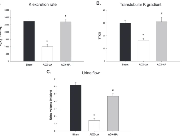
The complete replacement dose of aldo, ADX-HA, restored K homeostasis in mice on LNaHK; however, LNaHK mice receiving ADX-LA, demonstrated significant disruption of K homeostasis. Both the UKV (Fig. 2A) as well as the TTKG (Fig. 2B) decreased significantly in ADX-LA (1004.7 ± 154.5μmol/day and 16.5 ± 1.5,n= 6) compared with sham (2731.6 ±
155.0μmol/day and 29.9 ± 2.0,n= 7) and ADX-HA (2693.4 ± 186.5μmol/day and 31.0 ± 3.3,
n= 7). The UKV and TTKG of ADX-HA were not different from sham. The urinary flow of
ADX-LA mice decreased compared with sham and ADX-HA, to a value of 1.4 ml/day (Fig. 2C), which was similar to Con mice (Table 1). There was no difference in plasma [creatinine] between sham (0.087 ± 0.005 mg/dl,n= 4), ADX-LA (0.118 ± 0.014 mg/dl,n= 5), and ADX-HA (0.118 ±
0.008 mg/dl,n= 8) mice, a result consistent with the notion that glomerular filtration was not
compromised in the ADX-LA group.
Figure 1. The effect of high K diets, adrenalectomy (ADX), and aldo replacement on plasma aldosterone (aldo) concentration (P[aldo]). (A)Bar plot of P[aldo] in sham mice on control (Con;n= 5), high K (HK;n= 7), and low Na, high K (LNaHK;n= 6) diets.*P<0.001 vs. Con; #P<0.001 vs. HK.(B)Bar
plot of P[aldo] in sham (n= 4) and ADX mice receiving a low dose replacement of aldo (ADX-LA;n= 6), and ADX mice receiving high dose aldo (ADX-HA;n= 7).*P<0.001 vs. sham; #P<0.001 vs. ADX-LA.
Role of aldo in BK-
α
/
β
4 expression
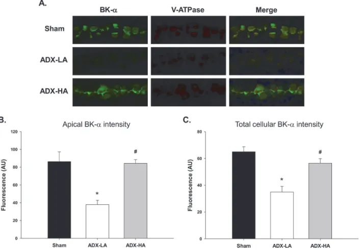
We showed previously that BK-αexpression is increased in HK mice [31].Fig. 3illustrates the staining intensities of sham, ADX-LA and ADX-HA. As shown by the representative staining (Fig. 3A) and the summary bar plots (Fig. 3B and 3C), both apical and total cellular BK-α ex-pression of ADX-LA were decreased compared to sham. BK-αexpression of ADX-HA was not different from sham, and was increased compared to ADX-LA.
Fig. 4illustrates the BK-β4 protein expression as measured by Western blot. The representa-tive immunoblots (Fig. 4A) and densitometry analysis (Fig. 4B) demonstrated that BK-β4 ex-pression decreased in ADX-LA mice compared to sham. BK-β4 expression of ADX-HA mice was increased compared to ADX-LA, but was not different from sham.
Role of aldo in BK-
α
/
β
4-mediated K secretion
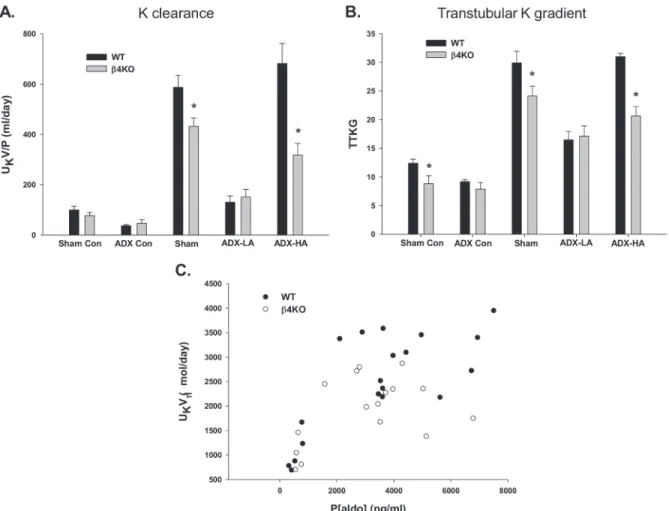
We showed previously that the apical BK-αexpression is increased by the presence of the BK-β4 subunit in IC of the mouse distal nephron and that BK-α/β4-mediated K secretion is attenu-ated in the absence of BK-β4 [31]. Here we determined aldo regulation on BK-α/β4-mediated K excretion by comparing K handling of WT with BK-β4 knockout mice (β4KO), which re-ceived the same experimental treatment as the WT mice described above. The various plasma and urinary values for WT andβ4KO are reported inTable 2andTable 3, respectively. The K excretion of LNaHK sham, ADX-LA, and ADX-HA WT andβ4KO mice are shown inFig. 5. The K clearance (Fig. 5A) and TTKG (Fig. 5B) were considerably reduced in LNaHK sham β4KO (431.9 ± 33.8 ml/day,n = 7and 24.1 ± 1.7,n= 7) compared with WT (587.4 ± 47.2 ml/
day,n= 10 and 29.9 ± 2.0,n= 7). Similarly, in the ADX-HA groups, the K clearance (317.9 ±
Table 1. Characteristics of WT sham and ADX mice on control diet.
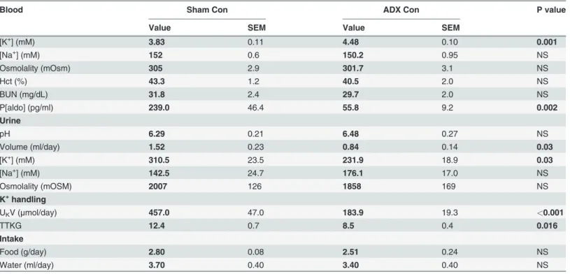
Blood Sham Con ADX Con P value
Value SEM Value SEM
[K+] (mM) 3.83 0.11 4.48 0.10 0.001
[Na+] (mM) 152 0.6 150.2 0.95 NS
Osmolality (mOsm) 305 2.9 301.7 3.1 NS
Hct (%) 43.3 1.2 40.5 2.0 NS
BUN (mg/dL) 31.8 2.4 29.7 2.0 NS
P[aldo] (pg/ml) 239.0 46.4 55.8 9.2 0.002
Urine
pH 6.29 0.21 6.48 0.27 NS
Volume (ml/day) 1.52 0.23 0.84 0.14 0.03
[K+] (mM) 310.5 23.5 231.9 18.9 0.03
[Na+] (mM) 142.5 24.7 176.1 17.0 NS
Osmolality (mOSM) 2007 126 1858 169 NS
K+handling
UKV (μmol/day) 457.0 47.0 183.9 19.3 <0.001
TTKG 12.4 0.7 8.5 0.4 0.016
Intake
Food (g/day) 2.80 0.08 2.51 0.24 NS
Water (ml/day) 3.70 0.40 3.40 0.40 NS
Abbreviations: Con, control; ADX, adrenalectomy; SEM, standard error of mean; NS, not significant; Hct, hematocrit; BUN, blood urea nitrogen; P[aldo]; plasma [aldosterone]; UKV, rate of K excretion; TTKG, transtubular K gradient. Sham Con,n= 5; ADX Con,n= 6.
46.2 ml/day,n= 5) and TTKG (20.7 ± 1.6,n= 5) ofβ4KO were significantly less compared with WT (681.8 ± 79.6 ml/day,n= 7 and 31.0 ± 0.6,n= 11). However, we observed no
differ-ence in K clearance and TTKG between WT (130.4 ± 25.1 ml/day and 16.5 ± 1.5,n= 4) and β4KO (151.6 ± 30.1 ml/day and 17.1 ± 1.8,n= 5) in the ADX-LA groups. These results indicate
that BK-α/β4 mediates K secretion in mice given LNaHK only when P[aldo] is increased to the high levels shown inFig. 1.
Fig. 5Cshows the relation between UKV and P[aldo] of WT andβ4KO mice receiving vari-ous doses of aldo replacement. As shown, WT andβ4KO UKV increased steeply at a P[aldo] of ~500 pg/ml to 3,000 pg/ml; however, the UKV leveled and did not increase additionally be-tween the values of 3,000 to 8,000 pg/ml. The UKV ofβ4KO (2022.4 ± 270.6μmol/day,n= 5)
was lower (P= 0.035) than WT (2731.6 ± 155.0μmol/day,n= 7) mice on LNaHK.
Amiloride-sensitive osmotic diuresis
Mice on LNaHK [9], or HK [8,32,33] exhibit a urinary flow that is 5-fold that of Con mice. A high urine volume is necessary to dilute the K in the CNT/CCD and excrete K at a high rate be-cause the concentration of K that can be secreted in the CNT/CCD is limited to approximately 120 mM [9].
The present study attempted to answer whether high urinary flow was the result of aldo—
induced high rate of K secretion in the CNT/CCD, thereby creating a high K osmotic gradient that would oppose the extraction of water via aquaporin 2 stimulated by ADH. If the secreted distal [K] is responsible for a high osmotic diuresis in the LNaHK mice then the osmolar clear-ance (COsm) should be decreased by amiloride, which inhibits the Na reabsorption for K se-cretion exchange in the CNT/CCD.
Fig. 6shows the COsm for WT Con, WT LNaHK, andβ4KO LNaHK mice with vehicle or amiloride treatment. Compared to vehicle, amiloride treatment caused COsm to increase in
Table 2. Characteristics of WT LNaHK sham, ADX-LA, and ADX-HA mice.
Blood Sham ADX-LA ADX-HA
Value SEM Value SEM Value SEM
[K+] (mM) 4.25 0.15 7.40 0.50 4.23 0.52
[Na+] (mM) 147.5 1.9 141.2 0.4 147.6 0.6
Osmolality (mOsm) 296 3 292 4 304 2
Hct (%) 43.4 1.0 51.5 2.0 48.1 1.5
BUN (mg/dL) 23.3 1.7 38.2 5.0 22.3 1.1
P[aldo] (pg/ml) 3629.7 20.5 606.7 89.4 4292.8 530.0
Urine
pH 8.43 0.24 8.61 0.15 7.83 0.20
Volume (ml/day) 6.19 0.36 1.43 0.27 4.69 0.33
[K+] (mM) 423.9 26.5 719.6 42.6 579.5 29.1
[Na+] (mM) 6.5 0.6 12.7 1.6 2.9 0.3
Osmolality (mOsm) 1005 69 1681 78 1454 60
Intake
Food (g/day) 4.32 0.55 1.98 0.22 3.21 0.22
Water (ml/day) 13.90 1.72 3.73 0.27 9.32 0.50
Abbreviations: LNaHK, low Na, high K; ADX-LA, adrenalectomy with low dose aldosterone replacement; ADX-HA, adrenalectomy with high dose aldosterone; SEM, standard error of mean; Hct, hematocrit; BUN, blood urea nitrogen; P[aldo]; plasma [aldosterone]. Sham,n= 7; ADX-LA,n= 6; ADX-HA,n= 7.
WT Con (vehicle = 8.0 ± 0.8 ml/day,n= 6; amiloride = 11.6 ± 1.3 ml/day,n= 5) and decrease
in WT LNaHK (vehicle = 13.3 ± 1.2 ml/day,n= 9; amiloride = 7.8 ± 1.1 ml/day,n= 9), while β4KO LNaHK mice remained unchanged (vehicle = 9.5 ± 0.8 ml/day,n= 9; amiloride = 7.0 ±
1.1 ml/day,n= 5). These results are consistent with an aldo-enhanced osmotic diuresis as part
of the mechanism for the increased urine flow.
Discussion
A diet comprised of low Na and high K has been touted by cardiovascular researchers and health experts for many years. The“Paleo”diet, as well as vegetarian or vegan diets, is a varia-tion of the LNaHK. A dietary plan was developed by the The Navaria-tional Heart, Lung, and Blood Institute and approved by the USDA and called Dietary Approaches to Stop Hypertension (DASH). The DASH diet, shown to decrease blood pressure and risk of cardiovascular disease, emphasizes eating K rich foods (4,700 mg) and limiting Na consumption to 1,500 mg per day.
The LNaHK diet is an extreme model of the“Paleo”diet [1–3], but is probably similar to the diet of the Yanomami [6,7]. Although the mechanism is still uncertain, high K diets are known as natural diuretics [11,12] and therefore would tend to lower the long-term mean
Figure 2. The effects of ADX and aldo replacement on K balance of LNaHK mice. (A)The rate of urinary K excretion (UKV;*P<0.001 vs. sham;
#P<0.001 vs. ADX-LA),(B)Transtubular K gradient (TTKG;*P= 0.002 vs. sham;#
P= 0.002 vs. ADX-LA), and(C)Urinary flow (*P<0.001 vs. sham;
#P<0.001 vs. ADX-LA) in sham (n= 7), ADX-LA (n= 6), and ADX-HA (n= 7) mice.
arterial pressure. This study determined the mechanism by which high aldo levels of LNaHK mice induced a diuresis, despite the normal role of aldo to retain Na.
The P[aldo] is elevated in mice on LNaHK to levels 15 times greater than Con mice (Fig. 1A). ADX Con mice exhibited a mild deficiency in K handling (Table 1). However, on LNaHK, ADX mice did not survive without aldo repletion and survived only six days with par-tial aldo repletion (ADX-LA). The rate of K excretion of LNaHK mice with ADX and complete aldo repletion (ADX-HA) was equal to sham (Fig. 2). The expressions of BK-αand BK-β4 were directly related to the P[aldo] achieved by repletion.β4KO mice with ADX-HA exhibited diminished K clearance, compared with WT (Fig. 5). Amiloride, which prevents Na reabsorp-tion dependent K secrereabsorp-tion in the CNT/CCD, increased COsm in Con mice but diminished COsm in WT LNaHK (Fig. 6).
Influence of diet on P[aldo]
We initially measured P[aldo] of mice on three different diets: Con, HK, and LNaHK using ELISA (Fig. 1A). The P[aldo] for Con mice of 239 pg/ml (Table 1) using ELISA was close to the values of 322 pg/ml [17] and 400 pg/ml [34] of previous studies using radioimmunoassay (RIA). For HK diet, our value of 753 pg/ml using ELISA was less than the value of 1797 ± 630
Figure 3. The effect of aldo on BK-αexpression in the distal nephron of LNaHK mice.Representative images(A)and bar plots summarizing intensity of fluorescent immunohistochemical staining of apical(B)and total(C)BK-αin cortical kidney sections of sham, ADX-LA, and ADX-HA mice. BK-α(green) was co-stained with V-ATPase (red), a marker of intercalated cells in the connecting tubule and collecting duct. Merged images also contain Hoechst nuclear stain (blue).*P<0.001 vs. sham; #P<0.001 vs. ADX-LA;n= 9.
Figure 4. The effect of aldo on BK-β4 expression of LNaHK mice.Western blot representative immunoblots(A)and summary bar plots(B)of BK-β4 expression in kidney cortex of sham, ADX-LA, and ADX-HA mice.β-actin was used as a loading control.*P= 0.004 vs. sham; #P= 0.019 vs. ADX-LA;n= 4. All bands shown were from the same blot.
doi:10.1371/journal.pone.0115515.g004
Table 3. Characteristics ofβ4KO LNaHK sham, ADX-LA, and ADX-HA mice.
Blood Sham ADX-LA ADX-HA
Value SEM Value SEM Value SEM
[K+] (mM) 5.17 0.34 7.02 0.68 6.46 0.27
[Na+] (mM) 147.4 1.2 147.2 0.7 149.4 0.8
Osmolality (mOsm) 290 7 300 0 304 3
Hct (%) 43.3 1.1 45.0 2.1 43.6 1.6
BUN (mg/dL) 21.4 1.0 40.3 4.4 26.6 0.9
P[aldo] (pg/ml) 2771.9 277.7 554.4 84.7 3656.0 352.0
Urine
pH 8.51 0.17 7.43 0.41 7.65 0.35
Volume (ml/day) 5.36 0.39 1.42 0.30 2.43 0.17
[K+] (mM) 431.8 24.0 743.4 63.4 826.0 89.8
[Na+] (mM) 5.0 0.9 14.8 2.3 8.6 1.1
Osmolality (mOsm) 1027 48 1932 126 1867 258
Intake
Food (g/day) 3.22 0.31 1.56 0.09 1.69 0.21
Water (g/day) 7.81 1.12 4.25 0.22 5.39 0.42
Abbreviations: LNaHK, low Na, high K; ADX-LA, adrenalectomy with low dose aldosterone replacement; ADX-HA, adrenalectomy with high dose aldosterone; SEM, standard error of mean; Hct, hematocrit; BUN, blood urea nitrogen; P[aldo]; plasma [aldosterone]. Sham,n= 7; ADX-LA,n= 5; ADX-HA,n= 5.
Figure 5. The effect of aldo onβ4KO LNaHK mice.The K clearance(A)and TTKG(B)of WT andβ4KO mice on Con given sham or ADX surgery or on LNaHK given sham, ADX-LA, or ADX-HA.*P<0.05 vs. WT.(C)The relation between P[aldo] and UKV determined in WT andβ4KO ADX mice receiving
various doses of aldo replacement.
doi:10.1371/journal.pone.0115515.g005
Figure 6. The effects of amiloride on osmolar clearance.The osmolar clearance was determined in WT Con (n5), WT LNaHK (n= 9), andβ4KO LNaHK (n5) mice given vehicle or amiloride.*P<0.05 vs.
vehicle; #P<0.05 vs. WT LNaHK.
using RIA [32]; however, the SEM of the RIA value indicates some readings in the range of our value.
Our study showed that mice consuming a diet of LNaHK exhibited a very high P[aldo] that was greater than 3,000 pg/ml (Fig. 1,Table 2). This value is much greater than the value of 1369 pg/ml detected in another study of mice on another low Na, high K diet [35]. However, the dietary K content in that study was 2%, compared with the 5% of our study.
Our results show that the P[aldo] levels of the ADX-HA group were necessary to achieve the high rate of K excretion in the LNaHK mice. When the P[aldo] of ADX mice on LNaHK were partially replete (ADX-LA), with a value of 606 pg/ml (Fig. 1B,Table 2), the mice were unable to achieve a substantial rate of K excretion and the P[K] increased to lethal levels within one week.
Aldo-dependent BK-
α
/
β
4-mediated K secretion
Aldo is the primary hormone regulating K secretion. The mechanism involves increasing Na reabsorption via ENaC [36] in exchange for K secretion via ROMK [37] and BK [38] channels in the CNT/CCD. An increase in P[K], independent of aldo, could regulate the transporters that mediate K secretion. In ADX HK mice, P[K], independent of P[aldo], increased Na-K-ATPase by 200% [39] and increased urine flow and K excretion [40]. K secretion was reported-ly independent of ENaC-mediated Na reabsorption in rats with a high K intake, suggesting an aldo-independent mechanism that increases K secretion [41]. However, our results here dem-onstrate that K secretion in LNaHK mice is completely aldo-dependent.
To establish aldo-independent K secretion it is important that the ADX is complete, because residual adrenal glomerulosa cells can hypertrophy in response to ACTH and secrete substan-tial aldo [42]. Using ELISA, Con ADX mice exhibited a P[aldo] of 56 pg/ml (Table 1), which is the same value for ADX mice using RIA in a previous study [34]. We were unable to determine the P[aldo] of ADX LNaHK mice because they did not survive for two days without restoring the P[aldo] to at least Con levels. Potassium excretion in mice on ADX-LA, which exhibits near Con levels of P[aldo], is probably mediated by ROMK [10]. Moreover, the ADX-LA mice had P[K]>7 at 4 days and survived for only 6 days. Therefore, the high rate of K elimination
of LNaHK mice is dependent on high levels of P[aldo].
If there was a mechanism to enhance K secretion, independent of aldo-induced Na reab-sorption, it would be manifested by LNaHK fed animals, which exhibit minimal distal Na de-livery and a very high demand for K secretion. However, the results of our study did not support an aldo-independent mechanism for K secretion in mice on LNaHK. Because aldo reg-ulates Na reabsorption via ENaC in the distal nephron, our result is consistent with a previous study that could not demonstrate Na-independent K secretion in mice on LNaHK [9]. It is pos-sible that there is aldo-independent K excretion in mice on LNaHK but this mechanism is in-adequate for survival.
The very high LNaHK P[aldo], which is 4-fold more than the P[aldo] of HK mice, may be necessary to ensure all Na delivery to ENaC. Thiazide diuretics, which inhibit the Na reabsorbing NCC, are ineffective in LNaHK mice because the NCC is inhibited and all Na is delivered to the CNT/CCD for ENaC-mediated Na for K exchange [9,10]. Although aldo enhances NCC-mediat-ed Na reabsorption [43,44], a very high P[aldo] in LNaHK mice may be required to inhibit Na re-absorption via the NCC and maximize Na delivery to ENaC. A paradoxical effect on the NCC for aldo at higher plasma concentrations has not been reported; however, it may involve a non-genomic effect that rapidly activates signaling pathways via an unidentified receptor [45].
the P[K], is enhancing BK-α/β4-mediated K excretion. An aldo-induced increase in BK activity is consistent with an early study showing that isolated CCDs from mineralocorticoid treated rabbits exhibited an elevated K conductance [46], and a more recent study showing that a HK diet caused increased BK-αexpression in IC [31].
The present study shows that the high levels of P[aldo], and not an elevation of P[K], causes the increase in BK-αexpression. In our previous study, the effects of spironolactone, an aldo antagonist, did not affect the expression of BK-β4 in HK mice [31]. However, the present study shows that BK-β4 expression is enhanced by aldo in LNaHK mice. Thus, the very high P[aldo] of the LNaHK diet is necessary for the enhanced expression of BK-β4. BK-β4 functions to pre-vent the lysosomal breakdown of BK-αthereby maintaining its presence in the apical mem-brane [31].
Aldo-induced increase in urine flow
At least two mechanisms are involved in the high urinary flow associated with high K diets. With a high K intake, high urinary flow is increased by medullary K recycling, which inhibits the thick ascending limb NaCl reabsorption and the concentrating mechanism [13]. In addi-tion, we have shown here that the high urine flow of LNaHK mice is associated with a high P [aldo], which increased an amiloride-sensitive COsm, indicating a K secreted per Na reabsorp-tion ratio greater than 1. The glomerular filtrareabsorp-tion rate may also be a factor in diuresis in LNaHK mice. An increased GFR due to either the LNaHK diet or the increased aldo could lead to increased urine flow. The GFR in LNaHK cannot be ruled out as a partial cause for an in-crease in urine flow.
ADX-LA mice had a food and water consumption (Table 2) similar to Control diet mice but reduced compared to sham LNaHK and ADX-HA. It is unlikely that ADX-LA mice had a re-duced urine flow due to an inability to drink water rather than an aldo-dependent decrease in K secretion as the primary defect. If ADX-LA mice drank less water as the primary defect of less P[aldo], then they would be volume depleted with an increased hematocrit. However, the hematocrit levels for ADX-LA were not different compared to sham and ADX-HA (Table 2), indicating similar blood volumes. Therefore, the reduced food and water intake of ADX-LA was probably an adaptation to the increasing P[K] resulting in impaired diuresis.
Our finding that increased flow in mice on LNaHK was aldo dependent is consistent with a recent study examining the handling of a high (5%) K diet in mice with a knockout of aldo synthase (AS-/-). In this study, flow increased by nearly three-fold in WT, but remained the same in AS-/- on a 5% K diet [47].
This relation between P[aldo] and high osmotic distal flow seemingly contradicts a previous study showing that acute aldo infusion to ADX rats does not increase K excretion because aldo stimulates Na reabsorption in the distal tubule, thereby decreasing urine flow [40]. However, these were acute aldo infusions whereas we treated with aldo for several days. It is also impor-tant to distinguish between treating with aldo alone and giving aldo with high K as we did in the present study. We recently learned that high K intake turns off the NCC [14,15]. Thus, giv-ing aldo without K reduces P[K] and favors reabsorption of Na via NCC while reducgiv-ing ENaC-mediated Na reabsorptive exchange for K. Consistent with our results, KCl infusion alone in-creased P[K], urine flow, and K excretion [40]. Increasing evidence indicates that the WNK4-SPAK signaling pathway can distinguish between high P[aldo] associated with high and low K intake [48].
Glucocorticoid hormone enhances water excretion and decreases urine osmolality by inhibit-ing urea transporters (UT-A1) in the inner medullary tip, thereby inhibitinhibit-ing the concentratinhibit-ing mechanism [49,50].
Limitations and assumptions
We determined that the K secretion in mice on LNaHK was creating an osmotic diuresis. This conclusion was based on the assumption that K, a non-reabsorbable ion, influences water reab-sorption in the collecting duct. However, when inhibiting vasopressin receptors (V2) with tolvaptan, water reabsorption is inhibited without affecting Na reabsorption [51]. In this case, Na is permeable and reabsorbed actively to a very low concentration, since the Na retention is regulated by aldo, without the influence of the diuretic effect of non-reabsorbing water. Inhibit-ing Na reabsorption with amiloride, in this case, would not influence water transport, which is already inhibited. However, water reabsorption in the collecting ducts depends on a combina-tion of the water permeability and the gradient of the non-permeable solute driving force. We assume the water channels are in the apical (AQP2) and basolateral (AQP3) membranes of the CNT and collecting ducts because the urine is concentrating to approximately 2,000 mOsm. In this case, the non-reabsorbable ions, such as Na or secreted K in the lumen (here due to amiloride inhibition) will create an osmotic force to prevent water transport from the lumen. This is the best explanation for the osmotic diuresis created by amiloride in mice on a normal diet and the decrease in osmotic diuresis when mice on LNaHK are treated with amiloride.
The accuracy of the TTKG may be questionable because there is probably K reabsorption in the collecting ducts. However, as shown in a previous study, the reabsorption of urea in this segment will offset the K flux thereby preserving the accuracy of the equation [9].
Conclusions
The P[aldo] levels of low Na diet mice result in antidiuresis and Na retention. However, the very high P[aldo] levels of LNaHK mice serve to enhance urinary volume increasing BK-α/β 4-mediated K secretion, which creates an osmotic diuresis. The high urinary flow created by BK-α/β4-mediated K secretion could activate more BK-α/β4 in a positive feedback manner or activate the BK-α/β1 of PC, which have flow detecting primary cilia.
Methods
Animal studies
The protocol was approved by and all animals were maintained in strict accordance with the Institutional Animal Care and Use Committee of the University of Nebraska Medical Center (protocol #’s: 09-050-07, 11-090-11-FC). All surgery was performed under anesthesia, and all efforts were made to minimize suffering. C57BL/6 (8–12 weeks, Charles River, Wilmington, MA) andβ4KO mice (12–20 weeks, generously provided by R. Brenner) were housed in the UNMC animal care facility and maintained on a 12-h day-night cycle, with access to food and water at all times. Mice underwent sham or ADX surgery and were given regular mouse chow (control; 0.3% Na, 0.6% K), high K (#TD.07278; 0.3% Na, 5.0% K) or a low Na, high K (#TD.08240; 0.01% Na, 5.0% K) pellet diet (Harlan Teklad, Madison, WI). The low Na, high K diet contained a mix of carbonate, citrate, and Cl as the counter anions to K. ADX mice received aldo replacement by osmotic pump, implanted subcutaneously, either at a low (25μg/kg/day) or high (500μg/kg/day) dose, and recovered for 3 days before being placed on
treated with an intraperitoneal bolus of vehicle or amiloride (5 mg/kg), at a concentration of 1 mg/ml, twelve hours before sacrifice. Aldo and amiloride were dissolved in polyethylene glycol. P [aldo] was measured using a competitive ELISA kit (DRG International, Springfield, NJ).
During the last two days of treatment, mice were placed in metabolic cages to record food and water consumption and urine volume in a 24 hr period. Urinary pH was measured and [Na] and [K] were determined using a flame photometer (Jenway Clinical PFP7) as previously described [16]. Urine osmolality was measured with a Model 3250 osmometer (Advanced In-struments). Mice were anesthetized by a ketamine/xylazine mixture and sacrificed by severing the carotid artery. The blood was extracted from the carotid artery and used for measurement of hematocrit and [K] and [Na] using an i-STAT 1 blood gas analyzer (Abbott Laboratories). Excess blood was centrifuged for measurement of plasma osmolality.
The TTKG is a calculation that estimates the K gradient across the final CCD before water is reabsorbed in the medullary collecting duct, concentrating K [9]. TTKG was calculated using the following formula: U[K]X POsm/ P[K]X UOsm. The COsm was calculated using the follow-ing formula:, UOsmX UV/ POsm.
Immunohistochemical staining
Kidneys were removed post-sacrifice and placed in Histochoice MB (Electron Microscopy Sci-ences, Hatfield, PA), embedded in paraffin and sectioned onto slides for immunofluorescent staining as previously performed [52]. Primary antibodies were applied overnight as follows: BK-α(mouse monoclonal, diluted 1:200, Neuromab), and V-ATPase (goat polyclonal, diluted 1:200, Santa Cruz). Fluorescent secondary antibodies (donkey anti-mouse IgG conjugated Alexa Fluor 488 and donkey anti-goat IgG conjugated Alexa Fluor 594, diluted 1:200,
Intitrogen) were then applied for 1 h. Hoechst stain was used to identify nuclei. Quantification of BK-αsignal intensity was determined in single-channel, gray scale images after background correction as performed previously [53]. Each group contained a kidney section from 3 different mice. Three different tubules per kidney section were used to quantify staining intensity.
Western blotting
Kidney cortex was separated from medulla tissue and used for Western blotting as performed following manufacturer’s protocol (Bio-Rad Laboratories, Hercules, CA) and as performed previously by our lab [53,54]. Primary antibodies used were anti-BK-β4 (rabbit polyclonal, di-luted 1:500, Alomone Laboratories), andβ-actin (mouse monoclonal, diluted 1:500, Santa Cruz Biotechnology). Secondary antibodies included goat anti-mouse IgG, and goat anti-rabbit IgG-conjugated horseradish peroxidase (diluted 1:30,000, Santa Cruz Biotechnology). Detection of protein was performed using enhanced chemiluminescence (Thermo Scientific, Rockford, IL). Densitometry analysis was performed using imageJ software.
Plasma [creatinine]
software. The reversed-phase separations were performed by means of a C18column (Waters Spherisorb 10.0 mm) as the stationary phase which was kept at 45°C. The mobile phase (fil-tered through a 22μm nylon filter) consisted of 5 mM Na acetate, with 40 ml/L of methanol,
and 10 ml/L of ACN:GAA and was pumped at a constant flow-rate of 1 ml/min. The retention time of creatinine was 1.1 min and was separated from the rest of the compounds present in the plasma in chromatographs obtained at 225 nm.
Statistical analysis
The data are presented as means ± SEM. Statistical analyses were performed using one-way ANOVA with Student-Newman-Keuls post hoc analysis (P<0.05 considered significant). We
performed data management and statistical analyses using SigmaPlot version 11.0.
Acknowledgments
This project was funded by National Institute of Diabetes and Digestive and Kidney Diseases Grants RO1 DK071014 and RO1 DK73070 (SCS), and a fellowship (#11PRE7530018) from the American Heart Association MWA Affiliate (RJC).
Author Contributions
Conceived and designed the experiments: RJC DW HL YY JWF PCW SCS. Performed the ex-periments: RJC DW HL YY JWF PCW SCS. Analyzed the data: RJC DW HL YY JWF PCW SCS. Contributed reagents/materials/analysis tools: RJC DW HL YY JWF PCW SCS. Wrote the paper: RJC DW HL YY JWF PCW SCS.
References
1. Eaton SB, Konner M (1985) Paleolithic nutrition. A consideration of its nature and current implications. N Engl J Med 312: 283–289. PMID:2981409
2. Frassetto L, Morris RC Jr., Sellmeyer DE, Todd K, Sebastian A (2001) Diet, evolution and aging—the pathophysiologic effects of the post-agricultural inversion of the potassium-to-sodium and base-to-chlo-ride ratios in the human diet. Eur J Nutr 40: 200–213. PMID:11842945
3. Cordain L, Eaton SB, Sebastian A, Mann N, Lindeberg S, et al. (2005) Origins and evolution of the Western diet: health implications for the 21st century. The American Journal of Clinical Nutrition 81: 341–354. PMID:15699220
4. Vasan RS, Evans JC, Larson MG, Wilson PW, Meigs JB, et al. (2004) Serum aldosterone and the inci-dence of hypertension in nonhypertensive persons. N Engl J Med 351: 33–41. PMID:15229305 5. Gordon RD, Stowasser M, Tunny TJ, Klemm SA, Rutherford JC (1994) High incidence of primary
aldo-steronism in 199 patients referred with hypertension. Clin Exp Pharmacol Physiol 21: 315–318. PMID:
7923898
6. Oliver WJ, Cohen EL, Neel JV (1975) Blood pressure, sodium intake, and sodium related hormones in the Yanomamo Indians, a“no-salt”culture. Circulation 52: 146–151. PMID:1132118
7. Mancilha-Carvalho JJ, Souza e Silva NA (2003) The Yanomami Indians in the INTERSALT Study. Arq Bras Cardiol 80: 289–300. PMID:12856272
8. Cornelius RJ, Wen D, Hatcher LI, Sansom SC (2012) Bicarbonate promotes BK-alpha/beta4-mediated K excretion in the renal distal nephron. Am J Physiol Renal Physiol 303: F1563–F1571. doi:10.1152/ ajprenal.00490.2012PMID:22993067
9. Wen D, Cornelius RJ, Rivero-Hernandez D, Yuan Y, Li H, et al. (2014) Relation between BK-alpha/ beta4-mediated potassium secretion and ENaC-mediated sodium reabsorption. Kidney Int 86: 139–
145. doi:10.1038/ki.2014.14PMID:24573316
10. Wen D, Cornelius RJ, Sansom SC (2014) Interacting influence of diuretics and diet on BK channel-reg-ulated K homeostasis. Curr Opin Pharmacol 15: 28–32. doi:10.1016/j.coph.2013.11.001PMID:
24721651
12. Treasure J, Ploth D (1983) Role of dietary potassium in the treatment of hypertension. Hypertension 5: 864–872. PMID:6360869
13. Stokes JB (1982) Consequences of potassium recycling in the renal medulla. Effects of ion transport by the medullary thick ascending limb of Henle’s loop. J Clin Invest 70: 219–229. doi:10.1172/JCI110609
PMID:6284797
14. Sorensen MV, Grossmann S, Roesinger M, Gresko N, Todkar AP, et al. (2013) Rapid dephosphoryla-tion of the renal sodium chloride cotransporter in response to oral potassium intake in mice. Kidney Int 83: 811–824. doi:10.1038/ki.2013.14PMID:23447069
15. Rengarajan S, Lee DH, Oh YT, Delpire E, Youn JH, et al. (2014) Increasing plasma [K+] by intravenous potassium infusion reduces NCC phosphorylation and drives kaliuresis and natriuresis. Am J Physiol Renal Physiol 306: F1059–F1068. doi:10.1152/ajprenal.00015.2014PMID:24598799
16. Grimm PR, Irsik DL, Settles DC, Holtzclaw JD, Sansom S (2009) Hypertension of Kcnmb1-/- is linked to deficient K secretion and aldosteronism. Proceedings of the National Academy of Sciences 106: 11800–11805. doi:10.1073/pnas.0904635106PMID:19556540
17. Grimm PR, Sansom SC (2010) BK channels and a new form of hypertension. Kidney Int 78: 956–962. doi:10.1038/ki.2010.272PMID:20720523
18. Brenner R, Perez GJ, Bonev AD, Eckman DM, Kosek JC, et al. (2000) Vasoregulation by the beta1 subunit of the calcium-activated potassium channel. Nature 407: 870–876. PMID:11057658 19. Amberg GC, Bonev AD, Rossow CF, Nelson MT, Santana LF (2003) Modulation of the molecular
com-position of large conductance, Ca(2+) activated K(+) channels in vascular smooth muscle during hyper-tension. J Clin Invest 112: 717–724. doi:10.1172/JCI18684PMID:12952920
20. Fernandez-Fernandez JM, Tomas M, Vazquez E, Orio P, Latorre R, et al. (2004) Gain-of-function muta-tion in the KCNMB1 potassium channel subunit is associated with low prevalence of diastolic hyperten-sion. J Clin Invest 113: 1032–1039. doi:10.1172/JCI20347PMID:15057310
21. Pluger S, Faulhaber J, Furstenau M, Lohn M, Waldschutz R, et al. (2000) Mice with disrupted BK chan-nel beta1 subunit gene feature abnormal Ca(2+) spark/STOC coupling and elevated blood pressure. Circ Res 87: E53–E60. PMID:11090555
22. Sansom SC, Welling PA (2007) Two channels for one job. Kidney Int 72: 529–530. PMID:17713560 23. Yoo D, Kim BY, Campo C, Nance L, King A, et al. (2003) Cell surface expression of the ROMK (Kir 1.1)
channel is regulated by the aldosterone-induced kinase, SGK-1, and protein kinase A. J Biol Chem 278: 23066–23075. PMID:12684516
24. Wade JB, Fang L, Coleman RA, Liu J, Grimm PR, et al. (2011) Differential regulation of ROMK (Kir1.1) in distal nephron segments by dietary potassium. Am J Physiol Renal Physiol 300: F1385–F1393. doi:
10.1152/ajprenal.00592.2010PMID:21454252
25. Welling PA (2013) Regulation of renal potassium secretion: molecular mechanisms. Semin Nephrol 33: 215–228. PMID:23953799
26. Huang DY, Wulff P, Volkl H, Loffing J, Richter K, et al. (2004) Impaired regulation of renal K+ elimination in the sgk1-knockout mouse. J Am Soc Nephrol 15: 885–891. PMID:15034090
27. Dijkink L, Hartog A, Deen PM, van Os CH, Bindels RJ (1999) Time-dependent regulation by aldoste-rone of the amiloride-sensitive Na+ channel in rabbit kidney. Pflugers Arch 438: 354–360. PMID:
10398866
28. Nesterov V, Dahlmann A, Krueger B, Bertog M, Loffing J, et al. (2012) Aldosterone-dependent and— in-dependent regulation of the epithelial sodium channel (ENaC) in mouse distal nephron. Am J Physiol Renal Physiol 303: F1289–F1299. doi:10.1152/ajprenal.00247.2012PMID:22933298
29. Garg LC, Knepper MA, Burg MB (1981) Mineralocorticoid effects on Na-K-ATPase in individual neph-ron segments. Am J Physiol 240: F536–F544. PMID:6264796
30. Tsuchiya K, Giebisch G, Welling PA (1996) Aldosterone-dependent regulation of Na-K-ATPase subunit mRNA in the rat CCD: competitive PCR analysis. Am J Physiol 271: F7–15. PMID:8760237
31. Wen D, Cornelius RJ, Yuan Y, Sansom SC (2013) Regulation of BK-alpha expression in the distal nephron by aldosterone and urine pH. Am J Physiol Renal Physiol 305: F463–F476. doi:10.1152/ ajprenal.00171.2013PMID:23761673
32. Kobayashi M, Yasuoka Y, Sato Y, Zhou M, Abe H, et al. (2011) Upregulation of calbindin D28k in the late distal tubules in the potassium-loaded adrenalectomized mouse kidney. Clin Exp Nephrol 15: 355–362. doi:10.1007/s10157-011-0414-4PMID:21347582
33. Wang XF, Zhou CX, Shi QX, Yuan YY, Yu MK, et al. (2003) Involvement of CFTR in uterine bicarbonate secretion and the fertilizing capacity of sperm. Nat Cell Biol 5: 902–906. PMID:14515130
mineralocorticoid receptor blocker. Eur J Gastroenterol Hepatol 25: 1086–1092. doi:10.1097/MEG. 0b013e328360554aPMID:23524523
35. Manolopoulou J, Bielohuby M, Caton SJ, Gomez-Sanchez CE, Renner-Mueller I, et al. (2008) A highly sensitive immunofluorometric assay for the measurement of aldosterone in small sample volumes: vali-dation in mouse serum. J Endocrinol 196: 215–224. doi:10.1677/JOE-07-0134PMID:18252945 36. Masilamani S, Kim GH, Mitchell C, Wade JB, Knepper MA (1999) Aldosterone-mediated regulation of
ENaC alpha, beta, and gamma subunit proteins in rat kidney. J Clin Invest 104: R19–R23. doi:10. 1172/JCI7840PMID:10510339
37. Hebert SC, Desir G, Giebisch G, Wang W (2005) Molecular diversity and regulation of renal potassium channels. Physiol Rev 85: 319–371. doi:10.1152/physrev.00051.2003PMID:15618483
38. Bailey MA, Cantone A, Yan Q, MacGregor GG, Leng Q, et al. (2006) Maxi-K channels contribute to uri-nary potassium excretion in the ROMK-deficient mouse model of Type II Bartter’s syndrome and in ad-aptation to a high-K diet. Kidney Int 70: 51–59. PMID:16710355
39. Garg LC, Narang N (1987) Effects of potassium bicarbonate on distal nephron Na-K-ATPase in adrenalectomized rabbits. Pflugers Arch 409: 126–131. PMID:3039448
40. Field MJ, Stanton BA, Giebisch GH (1984) Differential acute effects of aldosterone, dexamethasone, and hyperkalemia on distal tubular potassium secretion in the rat kidney. J Clin Invest 74: 1792–1802. doi:10.1172/JCI111598PMID:6501571
41. Frindt G, Palmer LG (2009) K+ secretion in the rat kidney: Na+ channel-dependent and—independent mechanisms. Am J Physiol Renal Physiol 297: F389–F396. doi:10.1152/ajprenal.90528.2008PMID:
19474187
42. Nickerson PA, Brownie AC, Skelton FR (1969) An electron microscopic study of the regenerating adre-nal gland during the development of adreadre-nal regeneration hypertension. Am J Pathol 57: 335–364. PMID:4311762
43. Velazquez H, Bartiss A, Bernstein P, Ellison DH (1996) Adrenal steroids stimulate thiazide-sensitive NaCl transport by rat renal distal tubules. Am J Physiol 270: F211–F219. PMID:8769842
44. Kim GH, Masilamani S, Turner R, Mitchell C, Wade JB, et al. (1998) The thiazide-sensitive Na-Cl cotransporter is an aldosterone-induced protein. Proc Natl Acad Sci U S A 95: 14552–14557. PMID:
9826738
45. Dooley R, Harvey BJ, Thomas W (2012) Non-genomic actions of aldosterone: from receptors and sig-nals to membrane targets. Mol Cell Endocrinol 350: 223–234. doi:10.1016/j.mce.2011.07.019PMID:
21801805
46. Sansom SC, O’Neil RG (1985) Mineralocorticoid regulation of apical cell membrane Na+ and K+ trans-port of the cortical collecting duct. Am J Physiol 248: F858–F868. PMID:4003557
47. Todkar A, Picard N, Loffing-Cueni D, Sorensen MV, Mihailova M, et al. (2014) Mechanisms of Renal Control of Potassium Homeostasis in Complete Aldosterone Deficiency. J Am Soc Nephrol.
48. Arroyo JP, Ronzaud C, Lagnaz D, Staub O, Gamba G (2011) Aldosterone paradox: differential regula-tion of ion transport in distal nephron. Physiology (Bethesda) 26: 115–123. doi:10.1152/physiol.00049. 2010PMID:21487030
49. Gertner RA, Klein JD, Bailey JL, Kim DU, Luo XH, et al. (2004) Aldosterone decreases UT-A1 urea transporter expression via the mineralocorticoid receptor. J Am Soc Nephrol 15: 558–565. PMID:
14978157
50. Green HH, Harrington AR, Valtin H (1970) On the role of antidiuretic hormone in the inhibition of acute water diuresis in adrenal insufficiency and the effects of gluco—and mineralocorticoids in reversing the inhibition. J Clin Invest 49: 1724–1736. doi:10.1172/JCI106390PMID:5449709
51. Onogawa T, Sakamoto Y, Nakamura S, Nakayama S, Fujiki H, et al. (2011) Effects of tolvaptan on sys-temic and renal hemodynamic function in dogs with congestive heart failure. Cardiovasc Drugs Ther 25 Suppl 1: S67–S76. PMID:22120095
52. Grimm PR, Irsik DL, Liu L, Holtzclaw JD, Sansom SC (2009) Role of BK{beta}1 in Na+ reabsorption by cortical collecting ducts of Na+-deprived mice. Am J Physiol Renal Physiol 297: F420–F428. doi:10. 1152/ajprenal.00191.2009PMID:19458125
53. Holtzclaw JD, Cornelius RJ, Hatcher LI, Sansom SC (2011) Coupled ATP and potassium efflux from in-tercalated cells. Am J Physiol Renal Physiol 300: F1319–F1326. doi:10.1152/ajprenal.00112.2011
PMID:21454249
54. Holtzclaw JD, Grimm PR, Sansom SC (2010) Intercalated cell BK-α/β4 channels modulate sodium and potassium handling during potassium adaptation. J Am Soc Nephrol 21: 634–645. doi:10.1681/ASN. 2009080817PMID:20299355
![Figure 1. The effect of high K diets, adrenalectomy (ADX), and aldo replacement on plasma aldosterone (aldo) concentration (P[aldo])](https://thumb-eu.123doks.com/thumbv2/123dok_br/18330943.350861/3.918.63.764.116.418/figure-effect-diets-adrenalectomy-replacement-plasma-aldosterone-concentration.webp)
![Fig. 5C shows the relation between U K V and P[aldo] of WT and β 4KO mice receiving vari- vari-ous doses of aldo replacement](https://thumb-eu.123doks.com/thumbv2/123dok_br/18330943.350861/5.918.54.871.133.467/shows-relation-mice-receiving-vari-doses-aldo-replacement.webp)