J of Evolution of Med and Dent Sci/ eISSN- 2278-4802, pISSN- 2278-4748/ Vol. 4/ Issue 35/ Apr 30, 2015 Page 6081
CLINICAL AND HISTOPATHO
LOGICAL CORRELATION IN HANSEN’S
DISEASE
M. Suryanarayana Murthy1, Gitu Duara2, K. Kasi Viswanath3, Kiran Kanth4
HOW TO CITE THIS ARTICLE:
M. Suryanarayana Murthy, Gitu Duara, K. Kasi Viswanath, Kiran Kanth. Clinical and Histopathological Correlation in Hansen’s Disease. Journal of Evolution of Medical and Dental Sciences 2015; Vol. 4, Issue 35, April 30; Page: 6081-6085, DOI: 10.14260/jemds/2015/886
ABSTRACT:Hansen’s disease or leprosy, caused by Mycobacterium leprae, is a spectral disease with
a variety of clinical manifestations depending on the host immune response. The classification of patients within this spectrum using Ridley and Jopling’s criteria is based on clinical diagnosis aided by bacteriological, histological and immunological features. Many a time, the clinical diagnoses do not match the histological features, causing difficulty in proper diagnosis and treatment. Hence in this study an attempt has been made to correlate the clinical finding with the histological features.
KEYWORDS:Hansen’s disease, clinico histopathological correlation.
INTRODUCTION: Leprosy is a chronic infectious granulomatous disease caused by M. leprae, that primarily affects the skin and the peripheral nerves. It is a global health problem. Although India has achieved elimination of leprosy, with a prevalence rate of 0.69/10, 000 in March 2013 (NLEP), many new cases are detected annually.(1) The principle of reducing the load of infection is the cornerstone
of leprosy control by early diagnosis and early adequate drug treatment. So confirmation of diagnosis in doubtful cases is an important indication for histopathological examination.
MATERIAL AND METHODS: One hundred patients of untreated leprosy of both sexes and all ages
attending the Dermatology Department of Konaseema Institute of Medical Sciences, Amalapuram, Andhra Pradesh, were selected for this study. The study extended for a period of two years from January 2013- December 2014. The patients were typed according to Ridley Jopling scale into TT, BT, BB, BL and LL. Indeterminate type was also included.(2) Polyneuritic types were excluded for the want
of nerve biopsy. Wherever possible, the classification was more specific like BB downgrading to BL, or BT upgrading to TT, etc. Bacteriological examination was performed by slit and scrape method. After Ziehl-Neilsen staining, the slides were examined under oil immersion for detection of bacilli. Biopsy specimens were taken by punch biopsy and sent to pathology department and the reports obtained.
OBSERVATION: The study was undertaken on 100 clinically diagnosed, untreated patients of
Hansen’s disease of both sexes and all ages and examined as per the schedule laid down on the protocol. The ages ranged between 5-75 years. Most of the cases belong to the 2nd and 3rd decade of
life, making a 52% of the total number. 17 patients were below 12 years of age and one patient was below 6 years. There were 12 cases in the 4th decade, 9 patients in the 5th and 6th decades and only
one patient in the 7th decade of life. The total number of male patients was 60 and female patients
were 42. Similar observations relating to age and sex have been seen in other studies.( 3)
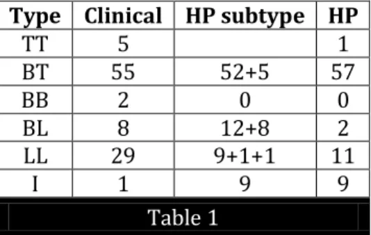
J of Evolution of Med and Dent Sci/ eISSN- 2278-4802, pISSN- 2278-4748/ Vol. 4/ Issue 35/ Apr 30, 2015 Page 6082 presence of bacilli only for 4 cases of BL and 28 cases of LL. Similar predominance of borderline group was also observed by others.(4)
Out of 100 patients, only 25 showed the presence of AFB in histological sections stained by Fite-Faraco and H&E staining. In 3 patients of BT where the skin smears were negative, tissue sections showed the presence of bacilli. This can be explained in the basis that organisms in borderline tuberculoid leprosy will be very few and may be missed on direct smear examination. 9 of the multibacillary type (LL and BL) which were smear positive did not show any bacilli on tissue section.
Type Clinical HP subtype HP
TT 5 1
BT 55 52+5 57
BB 2 0 0
BL 8 12+8 2
LL 29 9+1+1 11
I 1 9 9
Table 1
From the above table, it can be seen that the largest group was that of BT clinically (55) but there were 57cases histologically. Out of 29 cases of clinical LL, only 9 were histologically LL. Similarly there were 20 cases of BL histologically against 8 patient clinically. For one case of Indeterminate leprosy clinically, there were 9 cases of Indeterminate leprosy histologically. 5 patients of tuberculoid type showed only histological picture of BT. There was no concordance in the two patients of borderline leprosy clinically and histopathologically.
DISCUSSION: In this study there was a good deal of discrepancy between the clinical diagnosis and
histopathological findings as shown in the table below:
Clinical type
No. of cases
Histopathological disagreement
Type wise
Histopathological disagreement
total
Correlation
TT 5 BT- (5) 5 0
BT 55
TT-1 BB-1 BT BL -3
BL-4 BL LL-1
I -4
14 41
BB 2 BT-2 2 0
BL 8 BT-3
LL-1 4 4
LL 29
BL-10 BT-4 BB-1 I-5
20 9
I 1 BT-1 1 0
Total 100 46 54
J of Evolution of Med and Dent Sci/ eISSN- 2278-4802, pISSN- 2278-4748/ Vol. 4/ Issue 35/ Apr 30, 2015 Page 6083 All the 5 TT cases diagnosed clinically turned out to be BT histologically, this is always possible because of minor difference between TT and BT clinically and this variation is a minor disaggrement. Though there is 100% disagreement in all 5cases, it is of not much significance except that from the practical point of view it is safer to err on the side of BT than TT.
Out of 55 cases of BT diagnosed clinically, one case turned out to be TT histologically showing typical localised granuloma with Langhan’s giant cell. Clinically, it was BT in reversal. Four cases turned out to be BL histologically. Three of them diagnosed clinically as BT & one was BT in reversal. Three cases were showing features of both BT & BL and so diagnosed as BT downgrading to BL. Surprisingly one case which showed no bacilli in smear as well as in tissue section showed feature of BL downgrading to LL. Clinically the patient was a young adult male with a single erythematous plaque with ill-defined margins in the elbow. This is surprising & difficult to explain, but the possibility of the patient developing multiple lesions with lepromatous feature may be forewarned histologically in a given case. The variation histologically in TT & BT cases was observed earlier by other authors.(5) All these incidences showed that wherever possible histopathological examination at
different times during the evolution of the lesions will help in correct typing.(6)
Out of the eight cases diagnosed as BL, 3 cases showed features of BT. On tissue section, they were negative for AFB and weakly positive on direct smear. Possibly they were clinically also BT but because of numerous lesions and bilateral distribution they were diagnosed as BL. One case was LL histologically and this is understandable. But more number of cases have to be studied before a proper conclusion could be drawn.
Out of 29 LL cases clinically, 10 cases turned out to be BL. This is possible because of two reasons. The patient could have downgraded from Bl to LL and the lesion biopsied was BL.It is sometimes very difficult Top differentiate Bl and LL cases clinically specially in dark skinned individuals. What is unexplainable is the finding that 4 of LL cases were histologically BT and 5 indeterminate. In the 3 cases diagnosed as histoid HPE did not support the diagnosis. Understandably, 1 indeterminate case clinically in our study showed histopathology of BT which is not a serious disagreement.
On the whole, out of 100 cases, 46 cases showed disagreement i. e 46% and serious disagreement was in 21% and minor disagreement in 25%. In other words there is total, clinical and histopathological correlation in about 54%. Our study correlates very well with the study of Ridley & Jopling (1966) where there was complete agreement in 56 out of 82 cases studied and minor disagreement in 21 patients.(7)
The table III below shows a comparative study of clinicopathological correlation of different workers with our study.
Type of Leprosy
Bhatia A.S et al 1993
Moorthy et al 2001
Sharma A et al 2008
S Thakkar et al 2014
Present study
TT 50% 46.15% 47.37% 71.4% 0%
BT 77% 66.34% 53.01% 50% 74.54%
BB 26% 50% 37.35% 33.3% 0%
BL 43% 70% 58.82% 60% 50%
LL 91% 80% 75.86% 80% 31%
IL 36% 20% 100% 100% 0%
J of Evolution of Med and Dent Sci/ eISSN- 2278-4802, pISSN- 2278-4748/ Vol. 4/ Issue 35/ Apr 30, 2015 Page 6084 In our study there was no correlation between clinical and histological findings in TT, BB and Indeterminate in comparison to others this is probably due to the very few number of patients of this type in our study.
SUMMARY AND CONCLUSIONS: One hundred untreated cases comprising all varieties of leprosy
were classified clinically using Ridley-Jopling’s criteria. Skin smear examination was done for all cases; all the cases were also subjected to histopathological study, including acid fast staining of tissue sections for bacilli. The decree of correlation of clinical and histological diagnosis was carried out. The results were evaluated and compared with those of others who have undertaken similar study before.
1. The correlation without immunological assessment can never be 100%. But properly applied, with the 3 parameters alone namely, clinical, bacteriological and histological about 50-80% correlations can be achieved.
2. Histopathological study in this series has certainly raised awareness, in that some of the clinically tuberculoid cases showed definite LL change therby giving a warning to undertake careful follow up.
3. All clinically diagnosed Hansen’s disease should be submitted to histopathology whenever possible, so that it will help in determing the type of the disease and duration of treatment.
BIBLIOGRAPHY:
1. National leprosy eradication programme. (March 2013)
2. Ridley, D. S and Jopling W H (1966): A classification of leprosy according to immunity. A five group system. Ind. Leprosy. 34: 255.
3. Bhatia AS, Katoch K, Narayanan RB. Ramu G, Mukherjee A, Lavania RK: Clinical and histopathological correlation in the classification of leprosy. Int J Lepr Other Mycobact Dis 1993 Sept 61 (3) 433-8.
4. Moorthy BN, Kumar P, Chatura KR, Chandrasekhar HR, Basavaraja P. K: Histopathological correlation of skin biopsies in Leprosy Ind. J. Derma, Ven, Leprol 2001; 67; 299-301 [Pub Med]. 5. Sharma A, Sharma RK, Goswami KC, Bardwaj S: Clinico histopathological correlation in Leprosy
JK Science 2008: 10: 120-123: 3.
6. Sejal Thakkar, Sangeta V Patel. A Clinical profile of leprosy patients- A prospective study. Indian Journal & Dermatology: 2014 Mar-Apr; 59 (2): 158-162[Pub Med]. Med know publication. 7. Jopling WH, Mc Dougall AC. Handbook of leprosy 5th Edition, Delhi, CBS Publication and
J of Evolution of Med and Dent Sci/ eISSN- 2278-4802, pISSN- 2278-4748/ Vol. 4/ Issue 35/ Apr 30, 2015 Page 6085
AUTHORS:
1. M. Suryanarayana Murthy 2. Gitu Duara
3. K. Kasi Viswanath 4. Kiran Kanth
PARTICULARS OF CONTRIBUTORS:
1. Assistant Professor, Department of DVL, Dr. NTRUHS, Vijayawada.
2. Associate Professor, Department of DVL, Dibrugarh University, Assam.
3. Post Graduate, Department of DVL, Dr. NTRUHS, Vijayawada.
FINANCIAL OR OTHER
COMPETING INTERESTS: None
4. Post Graduate, Department of DVL, Konaseema Institute of Medical Sciences & Research Foundation, Amalapuram.
NAME ADDRESS EMAIL ID OF THE CORRESPONDING AUTHOR:
Dr. M. Suryanarayana Murthy, Assistant Professor,
Department of DVL, Vijayawada. E-mail: [email protected]