Oroantral fistula and genian
mucosal flap : a review of 25
cases
Summary
Roberto Campos Meirelles 1,Roberto Machado Neves-Pinto 2
1 Master’s degree - UFRJ; Doctoral degree - USP; Associate Professor - UERJ and UNIRIO; Adjunct Professor, Rio de Janeiro State University. 2 Associate Professor, Rio Grande do Sul Federal University. Professor colaborador, Rio de Janeiro State University.
Rio de Janeiro State University (Universidade do Estado do Rio de Janeiro).
Address for correspondence: Roberto Campos Meirelles - Rua Sorocaba 706 Botafogo Rio de Janeiro RJ 22271-110 Brasil. Tel. (0xx21) 2103-1500 Fax: (0xx21) 2579-3713 - E-mail: [email protected]
Paper submitted to the ABORL-CCF SGP (Management Publications System) on December 20th, 2005 and accepted for publication on December 21th, 2007. cod. 1653.
T
he oroantral fistula is a pathological connection between the maxillary sinus and with the oral cavity. The condition mostly follows dental extraction. Aim: To present the experience of 25 cases. Material and methods: Retrospective cases between 1996-2000. The ORL examination included nasal or sinusal endoscopy, a CT scan and histopathological analysis. Results: Twenty-five cases were found: ten 2nd molar cases, eight 1st molar cases, six 2nd premolar cases, and one canine case. All patients underwent a Caldwell-Luc operation plus excision of the epithelium lining the fistula, that was then completely covered by a flap of mucosa rotated from the genian region. Discussion: In cases of major fistulae a bone autograft taken from the anterior sinus wall was used. Bacterial cultures (n=19) revealed streptococus pneumoniae (13), haemophillus influenza (6), Moraxella catharralis (2) and staphylococus aureus (2). Aspergillus niger was found in one case presenting as a “fungic ball”. Conclusions: The only case of surgical failure, after 30 days postoperatively, was reoperated, using a bone graft. After a 6-month follow up all of the patients progressed satisfactorily, including the reoperated patient.Keywords: oroantral fistula, surgical flaps, sinusitis. ORIGINAL ARTICLE
INTRODUCTION
There are three types of maxillary sinus floor fis-tulae: the oronasal, the oroantral and the oroantronasal
fistula.1 The oroantral fistula (OAF) is a pathological
communication between the maxillary sinus and the oral
cavity,1,2,3 mostly of the alveolar type, resulting from
trau-ma from endodontic treatment or tooth extraction1,4-7 that
leaves a gap in the bone on the maxillary sinus floor. This gap becomes contaminated by food and saliva, leading to bacterial infection, impaired healing and chronic sinusitis. The OAF occurs mostly in the second and first molars,
and the second premolar teeth.4,5 It is more frequent in
males compared to females.1,8 Usual radiologic findings in
bone include sinus floor discontinuity, a communication between the oral cavity and the sinus, opacification of the sinus, focal alveolar atrophy, and associated periodontal
disease.9 Antibiotics are of no use for closing the fistulae.
Small fistulae tend to heal spontaneously, whereas larger fistulae rarely heal.8 Surgery is indicated if a fistula does
not heal within three weeks.1,4,8
Surgery aims to promote ventilation and aeration of the maxillary sinus, to remove diseased bone and to resect the thickened epithelium along the borders of the fistula. Surgical success depends on the technique, the size and site of the fistula and the presence or absence of sinus
disease.4,8 It is rare for the sinus to be infection-free in this
condition. Sinus disease is commonly treated surgically by a maxillary sinusectomy, according to the Caldwell-Luc technique, followed by middle meatotomy.
Fistula may be closed by using alveolar, palatal or jugal mucosa flaps, after having removed the diseased
mucosa and bone.1,5
No surgical flap is superior to another;2,4 each one
has its advantages and disadvantages. The palatal flap has better blood perfusion, but the technique is difficult and time-consuming. These flaps are preferred and indicated for wide, high output fistulae. In such cases, the palatal bone structure is exposed, leading to an increased healing time, meaning that the postoperative period is prolonged
and troublesome.6 The gingivolabial sulcus is narrowed in
the oral mucosa flap, which might lead to a second
pro-cedure or definitive sequelae.1-3,5 An alveolar flap may be
used for the treatment of small fistulae.2 The jugal mucosa
flap is well irrigated and has a higher possibility of covering
the fistula and the bone graft.8 Special attention must be
given to avoid injuring Stenon’s duct. The disadvantage of this flap is that it crosses and partially obliterates the gingivolabial sulcus, making it difficult to use prostheses; the flap is also under stress from continuous lip and cheek movements. Furthermore, this operation requires a second procedure to release the sulcus. Palatal rotation may be
indicated if the buccal flap is unsuccessful.3,7 This may be
problematic in the presence of a third molar fistula, as flap
rotation may affect negatively the vascular pedicle.6 This flap may result in arterial injury and hemorrhage; it is also more technically challenging and may expose more of the palatal bone if the fistula is larger, requiring a longer
procedure and a second operation.10
A variety of graft materials have been used in place of bone, including tantalum and gold plates, fascia lata,
dura mater and freeze-dried collagen.1 In general, allografts
are preferred to heterografts.4 Although much used in facial
bone reconstruction, albeit costly, porous block hydroxya-patite does not yield good results when in contact with the oral mucosa, and is not recommended for the treatment of
fistulae.11 An original technique is that which transplants
a closed apex third molar to the dental extraction site to
occlude the gap and close the fistula.12
Given the lack of standard protocols for choosing the best technique, the authors present the results of the genian mucosal flap rotation surgery for the treatment of alveolar type oroantral fistulae.
SERIES AND METHOD
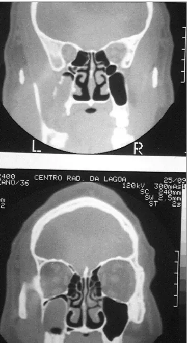
A retrospective study was made based on com-piled data from the charts and medical files of patients diagnosed with alveolar type OAF and treated surgically between 1996 and 2000 at the University Hospital Otorhi-nolaryngology Unit and at a private clinic. The routine diagnostic sequence included an otorhinolaryngological exam, rigid telescope (30º/4mm) nasal endoscopy, and computed tomography showing coronal, axial and in some cases sagittal sections of the paranasal cavities (Figure 1). Sinus telescope (30º/2.7mm) endoscopy was done if fistulae were equal to or larger than 0.5 mm. Additionally, resected tissue was tested for bacteria and fungi, and sent for pathology.
Figure 1. Opacification and erosion of the lower maxillary sinus bone wall.
followed by intranasal middle meatotomy and rotation of a contiguous genian mucosal flap of variable width and adequate blood supply. In high-output fistulae (equal to or > 0.5 cm) (Figure 2), a bone graft removed from the anterior wall of the maxillary sinus was placed on the site. Postoperative care included antibiotics during 14 days, instructions to avoid tooth brushing or touching the site with the tongue, to avoid blowing and exercising the cheek or using dental prostheses for seven days. Patients were seen postoperatively on the 7th, 14th, 30th and 180th day. After 30 days a second surgical procedure was undertaken under local anesthesia to release the pedicle of the flap that was linked to the jugal region, followed by hemostasis and suturing.
already undergone surgery at another institution.
All of the cases were operated by the Caldwell-Luc type maxillary sinusectomy technique, including stripping of mucosal and bony edges of the fistula to healthy tissue, intranasal middle meatotomy and rotation of a genian mucous flap measuring 1 to 3 cm in width (Figure 3). A bone graft was place on the site in high-output fistula or those that measured 0.5 cm or more (14 cases); bone was removed for grafting from the anterior maxillary sinus wall. All of the cases progressed favorably, except for one. A good result was considered as clinically verified absence of a fistula and of sinusitis.
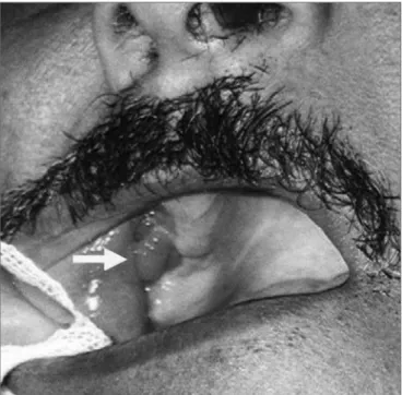
Figure 2. Ample fistula measuring over 1 cm following tooth extrac-tion.
RESULTS
There were 25 cases of alveolar fistulae, as follows: 10 second molar fistulae, 8 first molar fistulae, 6 second premolar fistulae and 1 canine fistula. There were 14 cases of high-output secretion fistulae equal to or larger than 0.5 cm. In 23 cases, patients reported previous attempts to correct the fistula, such as allowing second intention healing (10 cases), suturing with simple interrupted sti-tches to bring together the edges of the fistula (8 cases) and cauterization with a variety of chemical substances (5 cases). Only two cases were referred to an otorhinolaryn-gologist as the first therapeutic measure. The time needed for diagnosis varied from 21 to 137 days. Three cases had
Figure 3. Final aspect of the flap after 30 days.
Bacteriological cultures were done in 19 cases that presented secretion with pus, revealing Streptococcus pneumoniae (13 cases), Haemophilus influenzae (6 cases), Moraxella catarrhalis (2 cases), and Staphylococcus aureus (2 cases). Anaerobes were not investigated. Investigation for fungi was done in one case that had a radiologic ima-ge of a mycetoma and a suspect dark green tissue within the sinus; this was found to be Aspergilus niger (Figure 4). Pathology of 25 cases revealed non-specific chronic inflammation of variable degree.
A second surgical procedure was done in 19 patients. Six patients chose not to undergo the second procedure, considering themselves pleased with the first operation.
whi-ch was successful. After 60 days this patient had progressed favorably. Six months following the treatment, 23 patients were examined and considered as cured. Two patients could not be contacted by telephone or telegrams.
DISCUSSION
It seems that there is a delay in the diagnosis of oro-antral fistulae caused by manipulation of teeth; frequently dentists chose inefficient treatments, such as chemical cau-terization, second intention healing and simple interrupted sutures. No study has been conducted on how many of these patients achieved success with the abovementioned
methods; such a study would require a comprehensive approach to dental offices and outpatient clinics. More than 30 days with no resolution of the fistula seems to us an excessive time period, particularly considering that these patients end up developing maxillary sinus infection originating from direct contamination from food, and thus require both medical and surgical treatment.
Antibiotics can resolve sinusitis, but have no
in-fluence on fistula closure.8 Small fistulae tend to heal
spontaneously, as opposed to fistulae measuring over 5
mm.8 Von Wowern13 investigated 90 cases and concluded
that spontaneous closure of an OAF of any size was rare, and that surgery was required for closure. Abuabara et
al.14 found that fistula smaller than 2 mm tended to close,
contrary to fistula larger than 3 mm. Sinus disease or fo-reign bodies impair closure. We found no sinusal fofo-reign bodies. Surgical treatment is usually indicated after three
weeks from the diagnosis.1,4,8 All of the patients had been
treated previously by dental surgeons, and as we have no experience with their approaches, we could not evaluate whether the foregoing methods resulted in closure or not; we dealt only with those cases that were referred to us. As surgical treatment involves accessing the maxillary sinus, we believe that otorhinolaryngologists should undertake this procedure, since these professionals are able to deal with sequelae or complications of sinusectomies.
The surgical principles of this operation seek to attain solid and final closure of the fistula and to cure sinus disease. Success depends on the technique, the size and the location of the fistula, as well as the presence or
not of sinus disease.4,8
Although rare, it is worth remembering a case that was described in the literature in which the patient deve-loped a subdural empyema, left hemiplegia, and eventual
death as complications of a fistula.15 We believe that this
is another reason for referring the surgical treatment of these cases to physicians.
One of our cases presented a mycetoma, seen on radiology (Figure 3); it appears that the incidence of sinusal aspergillosis is increasing, usually resulting from inhalatory contamination, which has been observed even
in non-immunodeficient patients.16 A fistula
communica-ting the oral cavity to the maxillary sinus creates an access route for fungal penetration into the sinus, although the origin of the infection could not be established in the case above. Surgical treatment is mandatory and solves the problem; systemic antifungal medication is used only in specific cases, if needed. These are usually severe cases of
this disease in immunodeficient patients.17 In our patient,
abundant rinsing of the sinus with saline and topical an-tifungal medication solved the problem, and flap rotation closed the fistula successfully.
Although there was one canine OAF case that
resulted from traumatic tooth extraction, fistulae of the canine tooth are infrequent. We found cases reported in veterinary journals, which describe this condition as relatively common in small carnivorous animals following tooth extraction. It is interesting to note that repair
tech-niques are similar to those we use.18 Normally, the canine
relation with the maxillary sinus is not maintained; our one case was an anatomical variant. We opted to place a bone graft in this case, for safety, although it was a low-output fistula.
Many techniques, including flaps, have been
pro-posed for the closure of OAFs.8
Our unsuccessful case was a small fistula; later on the patient, who was a nurse, told us that she cleaned the site many time a day with cotton swabs imbibed in oxygenated water. Possibly constant trauma on the loose flap might have increased the diameter of the fistula.
It is rare not to have sinusitis when there is a fistula. Therefore, it is essential to establish the extension of sinus disease, which is usually restricted to the maxillary sinus. Sinusitis was present in all of our cases, which is why we chose the Caldwell-Luc technique for providing the best access to the floor of the sinus for complete cleaning.
The choice of the procedure is controversial. Some prefer simple interrupted sutures (60%), followed by a
variety of flaps (39%)14. Most of the authors prefer flaps,
with no preference for any specific type.2,4 All of them have
their advantages and disadvantages.1,2,3,5 Güven8 treated 90
patients (out of a 98-case series) by Rehrmann’s technique, using an advancement buccal flap similar to the one we used. In his series, complications were loss of sensitivity of the infraorbital nerve (2 cases), a reduced gingivolabial sulcus (6 cases), the need for resuturing the flap (3 cases) and granulation tissue along the suture (2 cases).
Similarly, we chose the jugal mucosa flap for all of the cases; this flap is wider, well irrigated and offers a better chance for covering the whole fistula, including the bone graft. The flap should be rotated and fixed free from any stretching force. All of the diseased bone should be removed, including any bone spiculae that might exert pressure on the flap. Special efforts were made to avoid injuring Stenon’s duct, so that we did not have this complication. The disadvantage of this flap is that it crosses and partially obliterates the gingivolabial sulcus; thus, prostheses are difficult to use and the flap is under stress from continuous lip and cheek movements. It also requires a second procedure to release the sulcus.
There are various other techniques that might be
used in such cases, such as three-layer closure19 and
third-molar transplantation for closure of the fistula12; both
techniques have been investigated in small series. In general, any communication between the ma-xillary sinus and the oral cavity lasting for more than three
weeks should be surgically repaired. It is also essential to cure the sinus disease; otherwise closure of the fistula will not take place.
CONCLUSION
Our results with a simple rotation of a jugal muco-sal flap were good in 96% of 25 cases during the first 30 days, and good in 100% after 60 days, although a larger number of cases would be necessary to critically assess the efficacy of this technique.
REFERENCES
1. Yilmaz T, Suslu AE, Gursel B. Treatment of oroantral fistula: experience with 27 cases. Am J Otolaryngol 2003;24(4):221-3. 2. Bluestone CD, The management of oroantral fistulas. Otolaryngol
Clin North Am 1971;4:179-91.
3. Amaratunga NA. Oro-antral fistulae. A study of clinical, radiologi-cal and treatment aspects. Br. J Oral Maxillofac Surg 1986;24:433-7.
4. Haas R, Watzak G, Baron M, Tepper G, Mailath G, Watzek G. A preliminary study of monocortical bone grafts for oroantral fis-tula closure. Oral Surg Oral Med Oral Pathol Oral Radiol Endod 2003;96(3):263-6.
5. Killey HC, Kay LW Observations based on surgical closure of 362 oroantral fistulas. Int Surg 1972;57:545-9.
6. Lee JJ, Kok SH, Chang HH, Yang PJ, Hahn LJ, Kuo YS. Repair of oroantral communications in the third molar region by random palatal flap. Int J Oral Maxillofac Surg 2002;31(6):677-80. 7. Anavi Y, Gal G, Silfen R, Calderon S. Palatal
rotation-advance-ment flap for delayed repair of oroantral fistula. A retrospective evaluation of 63 cases. Oral Med Oral Pathol Oral Radiol Endod 2003;96(5):527-34.
8. Güven O. A clinical study on oroantral fistulae. J Craniomaxillofac Surg 1998;26(4):267-71.
9. Abraham JJ, Berger SB. Oral-maxillary sinus fistula (oroantral fistula): clinical features and findings on multiplanar CT. Am J Roentgenol 1995;165(5):1273-6.
10. Car M, Juretic M. Treatment of oroantral communications after tooth extraction. Is drainage into the nose necessary or not? Acta Otolaryngol 1998;118(6):844-6.
11. Cottrell DA, Wolford LM. Long-term evaluation of the use of co-ralline hydroxyapatite in orthognathic surgery. J Oral Maxillofac Surg 1998;56(8):935-41.
12. Kitagawa Y, Sano K, Nakamura M, Ogasawara T. Use of third molar transplantation for closure of the oroantral communication after tooth extraction: a report of 2 cases. Oral Surg Oral Med Oral Pathol Oral Radiol Endod 2003;95(4):409-15.
13. Von Wovern. Closure of oroantral fistula with buccal flap: Rehr-mann versus Môczár. Int J Oral Surg 1982;11(3):156-65. 14. Abuabara A, Cortez ALV, Passeri LA, de Moraes M, Moreira RWF.
Evaluation of different treatments for oroantral communications: experience of 112 cases. Int J Oral Maxillofac Surg 2006;35:155-8.
16. Shams MG, Motamedi MH. Aspergilloma of the maxillary sinus complicating an oroantral fistula. Oral Surg Oral Med Oral Pathol Oral Radiol Endod 2003;96(1):3-5.
17. Schulte B; Beyer D. Radiologic diagnosis of maxillary sinus as-pergillosis. Radiologe 1992;32(11):553-7.
18. Smith MM. Oronasal Fistula repair. Clin Tech Small Anim Pract 2000;15(4):243-50.