Program in
Staphylococcus aureus
Ahmed S. Attia1,2, Meredith A. Benson3, Devin L. Stauff1, Victor J. Torres3, Eric P. Skaar1*
1Department of Microbiology and Immunology, Vanderbilt University Medical Center, Nashville, Tennessee, United States of America,2Department of Microbiology and Immunology, Faculty of Pharmacy, Cairo University, Cairo, Egypt,3Department of Microbiology, New York University School of Medicine, New York, New York, United States of America
Abstract
The Staphylococcus aureus HrtAB system is a hemin-regulated ABC transporter composed of an ATPase (HrtA) and a
permease (HrtB) that protect S. aureusagainst hemin toxicity. S. aureusstrains lacking hrtA exhibit liver-specific hyper-virulence and upon hemin exposure over-express and secrete immunomodulatory factors that interfere with neutrophil recruitment to the site of infection. It has been proposed that heme accumulation in strains lackinghrtABis the signal which triggers S. aureusto elaborate this anti-neutrophil response. However, we report here that S. aureusstrains expressing catalytically inactive HrtA do not elaborate the same secreted protein profile. This result indicates that the physical absence of HrtA is responsible for the increased expression of immunomodulatory factors, whereas deficiencies in the ATPase activity of HrtA do not contribute to this process. Furthermore, HrtB expression in strains lackinghrtAdecreases membrane integrity consistent with dysregulated permease function. Based on these findings, we propose a model whereby hemin-mediated over-expression of HrtB in the absence of HrtA damages the staphylococcal membrane through pore formation. In turn,S. aureussenses this membrane damage, triggering the increased expression of immunomodulatory factors. In support of this model, wildtypeS. aureustreated with anti-staphylococcal channel-forming peptides produce a secreted protein profile that mimics the effect of treatingDhrtAwith hemin. These results suggest thatS. aureussenses membrane damage and elaborates a gene expression program that protects the organism from the innate immune response of the host.
Citation:Attia AS, Benson MA, Stauff DL, Torres VJ, Skaar EP (2010) Membrane Damage Elicits an Immunomodulatory Program inStaphylococcus aureus. PLoS Pathog 6(3): e1000802. doi:10.1371/journal.ppat.1000802
Editor:Michael S. Gilmore, Harvard Medical School, United States of America ReceivedSeptember 10, 2009;AcceptedFebruary 3, 2010;PublishedMarch 12, 2010
Copyright:ß2010 Attia et al. This is an open-access article distributed under the terms of the Creative Commons Attribution License, which permits unrestricted use, distribution, and reproduction in any medium, provided the original author and source are credited.
Funding:This work was supported by United States Public Health Service Grants AI69233 and AI073843 from the National Institute of Allergy and Infectious Diseases (E.P.S). E.P.S. holds an Investigator in Pathogenesis of Infectious Disease Award from the Burroughs Wellcome Fund and is a Searle Scholar. V.J.T. was supported by a Ruth L. Kirschstein NRSA postdoctoral fellowship from the National Institute of Allergy and Infectious Diseases (AI071487). D.L.S. was supported by T32 HL069765 from the National Institute of Allergy and Infectious Diseases. The funders had no role in study design, data collection and analysis, decision to publish, or preparation of the manuscript.
Competing Interests:The authors have declared that no competing interests exist. * E-mail: [email protected]
Introduction
Staphylococcus aureus is a Gram-positive commensal bacterium that colonizes the skin and anterior nares of approximately 25 % of the population [1]. Upon breaching these initial sites of colonization, S. aureusis capable of causing a range of infections [2]. Staphylococcal infections affect almost every organ in the human body ranging from minor skin and soft tissue infections to more serious diseases such as endocarditis, septicemia, pneumonia and toxic shock syndrome [3,4]. In order to cause such a diverse array of pathologies, S. aureus employs an arsenal of virulence factors including proteins that contribute to immune evasion and alter immune system function [5].
During infection,S. aureusfaces several barriers that interfere with its ability to replicate and colonize its host. One of these barriers is the paucity of free iron, which is a critical component of several reactions within the bacterial cell [6]. To circumvent this barrier,S. aureuscan satisfy its iron needs through acquisition of the metalloporphyrin heme, which is a cofactor of host hemoglobin and myoglobin.S. aureus binds, transports, and releases heme into the cytoplasm through the combined action of the iron regulated surface determinant (Isd)
system and the heme transport system (Hts) [7,8,9,10,11]. Although heme is a valuable nutrient iron source at low concentrations, high concentrations of heme are toxic and therefore heme acquisition necessitates the presence of heme detoxification systems. In this regard, S. aureus senses heme exposure through the HssRS two-component system [12,13] resulting in the up-regulation of the heme regulated transporter (HrtAB). HrtAB is an ABC-type transporter that consists of an ATPase (HrtA) and a permease (HrtB) which work together to alleviate heme toxicity and protect the cell from the adverse effects of heme accumulation [13,14].
S. aureusstrains lacking HrtA (DhrtA) exhibit liver-specific hyper-virulence in an animal model of infection suggesting that heme toxicity is linked to staphylococcal virulence. Consistent with this supposition, hemin-exposed DhrtA exhibits increased expression and secretion of several immunomodulatory proteins that are modeled to interfere with immune cell migration to the site of infection. Taken together, these findings have led to a model wherebyS. aureusstrains unable to alleviate heme toxicity through HrtAB activate an immunomodulatory program resulting in decreased killing by immune cells and increased virulence [13].
In this manuscript, we propose an alternative model to explain the hypervirulence of DhrtA.We report that the stimulus which leads to the increased expression of immunomodulatory proteins in hemin-exposedDhrtAis membrane damage caused by permease over-expression rather than intracellular hemin accumulation. Consistent with this, we present a series of data suggesting that permease expression in the absence of a cognate ATPase produces dysregulated pores in the membrane. This membrane damage is somehow sensed byS. aureusleading to increased expression and secretion of immunomodulatory factors. This model is further supported by the observation that exposing wildtype S. aureusto the channel forming antimicrobial peptide (AMP) gramicidin results in a secreted protein profile that mimics that of DhrtA exposed to hemin. The pathological relevance of this response is demonstrated by results reported here which show that up-regulation of secreted proteins is responsible for the hyperviurlence of strains lackinghrtA. Taken together, these data suggest a model wherebyS. aureussenses pore-formation in the cell membrane to elicit an immunomodulatory program that interferes with neutrophil recruitment to the site of infection.
Results
S. aureusDhrtAincreases expression of immunomodulatory proteins
In an effort to elucidate the mechanism responsible for the hypervirulence ofS. aureusDhrtA,we analyzed the secreted protein
profiles of staphylococcal strains with mutations in the Hss and Hrt systems. Upon exposure of DhrtA to hemin in concentrations of either 0.5 or 2mM, up-regulation of the production and secretion
of at least five proteins was observed (Fig. 1A). These proteins have been previously identified through mass spectrometry to be immunomodulatory proteins that interfere with neutrophil recruitment to the site of infection [13,19,20,21]. This finding suggests that strains unable to alleviate hemin toxicity through
Author Summary
Staphylococcus aureus infects almost every tissue within
the human body utilizing a range of virulence factors to combat host defenses. The expression of these virulence factors is a tightly regulated process; however, the signals sensed byS. aureusduring infection remain elusive. It has been hypothesized that heme toxicity is a signal sensed by S. aureusduring infection. This hypothesis is based on the observation thatS. aureusmutants which are incapable of relieving heme-toxicity due to inactivation of the ATPase HrtA elicit an immunomodulatory program that interferes with neutrophil recruitment to the site of infection. In keeping with this, S. aureus hrtA mutants exhibit liver-specific hypervirulence. Herein, we provide evidence for an alternative model to explain the hypervirulent phenotype
of S. aureus DhrtA. We demonstrate that instead of
accumulation of heme toxicity being the trigger for the observed immunomodulatory program, dysregulated pore formation caused by the HrtB permease triggers the anti-neutrophil response. In support of this model, over-expression of HrtB in wildtype S. aureus or exposing S.
aureusto channel-forming antimicrobial peptides induces
a similar immunomodulatory program. Our work provides evidence that S. aureus senses membrane damage and induces an immunomodulatory circuit that helps the pathogen evade immune-mediated clearance.
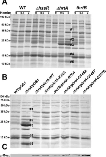
Figure 1. Up-regulation of immunomodulatory proteins is
caused by loss of expression of HrtA.(A) Bacterial strains were
grown in (0, 0.5 and 2mM) hemin in RPMI/CAS for 18 hours and proteins in culture supernatants were precipitated using 10% TCA, separated using 15% SDS-PAGE, and stained with Coomassie blue. (B) Exoprotein profiles of wildtype transformed with control plasmid pOS1 andDhrtA transformed with control plasmid or plasmids expressing WT HrtA, catalytically inactive HrtA variants (K45A, G145A, G145T, and E167Q) or partially catalytically active HrtA (R76A) [14]. All strains were grown in the presence of 1mM hemin. Proteins up-regulated under the indicated condition that have been identified previously using mass spectrometry are marked with a#. The predicted identities of the proteins in these bands are as follows; #1 (Exotoxin 8, SACOL0472), #2 (Exotoxin, SACOL0478), #3 (Exotoxin 3 and 5, SACOL0468/0473), #4 (Efb, SACOL1168), and#5 (FLIPr, SACOL1166). Positions of protein molecular mass markers in kilodaltons (kDa) are indicated on the left side of panels A and B. (C) Immunoblot analysis of protoplast lysates of strains analyzed in (B). Proteins were resolved using 15% SDS-PAGE, transferred to nitrocellulose membrane, probed with 9E10 anti-C-Myc monoclonal primary and AlexaFluor-680-conjugated anti-mouse secondary antibod-ies. Membranes were then dried and scanned using an Odyssey Infrared Imaging System.
HrtAB increase the expression of immunomodulatory proteins. However, a hemin-dependent increase in protein expression was not observed in either wildtype S. aureus or a strain lacking functional HssRS (DhssR), the regulatory system which controls hemin-dependenthrtABexpression (Fig. 1A) [12]. In addition, the secreted protein profile of a hemin-exposed S. aureus strain containing a transposon insertion that inactivates the permease HrtB (thrtB) closely resembled that of wildtype and did not show changes similar to those observed in DhrtA exposed to hemin (Fig. 1A). These data indicate that hemin-exposedS. aureusDhrtA increases the secretion of immunomodulatory proteins, but this immunomodulatory response does not occur upon loss of HrtB.
The presence of a catalytically inactive HrtA prevents the hemin-dependent immunomodulatory response
One potential explanation for the discrepancy in secreted protein profiles observed betweenDhrtAandthrtBis that theDhrtA strain has accumulated secondary mutations that are responsible for altering the secretome. This possibility was eliminated by the demonstration that the secreted protein profile of hemin-exposed
DhrtAcan be complemented by providing a full-length copy ofhrtA in trans (Fig. 1B). HrtA is an ATPase which provides the energy required for hemin-detoxification by its cognate permease HrtB [13,14]. Based on the phenotype of theDhrtAmutant, we reasoned that strains producing a catalytically inactive HrtA would elaborate a similar secreted protein profile upon hemin exposure. To test this hypothesis, we transformed DhrtA with plasmids encoding HrtA proteins that are mutated in key residues in the conserved nucleotide-binding and hydrolysis motifs (K45A, G145A, G145T, E167Q) or partially catalytically active (R76A) [14]. Surprisingly, expression of catalytically inactive versions of HrtA complemented the secreted protein profile of hemin-exposed
DhrtA(Fig. 1B). These strains expressed comparable levels of HrtA, eliminating expression level as a possible factor in this analysis (Fig. 1C). These data indicate that it is not the catalytic activity of
the HrtAB system that is required to prevent up-regulation of the immunomodulatory proteins upon hemin exposure, but instead it is the physical presence of HrtA that is required to prevent this phenotype.
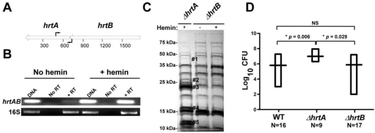
ThehrtBand thehrtAgenes are transcriptionally linked Based on the genomic organization of the hrtAB locus, it is expected that hemin exposure should increase HrtB expression in strains lackinghrtA. One potential explanation for the phenom-enon described above is that HrtB acts as an unregulated pore in the absence of HrtA, sending a stress signal to the cell that leads to the up-regulation of immunomodulatory proteins. This model assumes thathrtBandhrtAare co-transcribed and over-expressed when S. aureus is exposed to hemin. To address this, RT-PCR experiments were performed using two primers that bind to the 39 -end of thehrtBgene and the 59-end of thehrtAgene, respectively (Fig. 2A). The results of this experiment showed thathrtAandhrtB are indeed co-transcribed and transcription of the hrt operon increases upon hemin exposure (Fig. 2B). These results demon-strate that hrtA and hrtB comprise a heme-regulated bicistronic operon.
Loss of HrtB alone does not affect protein secretion or virulence in mice
To further confirm that it is not inactivation of the HrtAB system that is responsible for the up-regulation of the immuno-modulatory proteins, we constructed a strain containing a clean knock-out of hrtB(DhrtB). Upon examining the secreted protein profile ofDhrtB, no notable changes were observed upon hemin exposure (Fig. 2C). We next sought to determine if the secreted protein profiles ofDhrtAandDhrtBcorrelate with virulence levels in a murine model of systemic staphylococcal infection. Consistent with what has been reported previously, inactivation ofhrtAleads to a significant increase in liver-specific virulence in this model [13]. However, when mice were infected withDhrtB,no significant
Figure 2. Loss of HrtB alone does not affect protein secretion or virulence in mice.(A) Diagrammatic representation of thehrtABlocus; the relative positions of the primers used for RT-PCR are indicated by black arrows. (B) Agarose gels showing the results of an RT-PCR experiment involving thehrtABlocus and the ribosomal 16S RNA as the loading control. Bacteria were grown61mM hemin and RT-PCRs were done using DNA as the template (DNA), RNA with no reverse transcriptase (No RT) and RNA with reverse transcriptase (+RT). (C) Exoprotein profile ofDhrtA+1mM hemin andDhrtB61mM hemin. The#indicates the positions of proteins up-regulated under the indicated condition and the predicted identity of these proteins is as described in Figure 1. Positions of protein molecular mass markers in kilodaltons (kDa) are shown on the left side of the gel. (D) Bacterial multiplication in infected BALB/c mouse livers as measured by tissue homogenization, dilution, and colony formation on agar media 96 hours post infection. The horizontal bars represent the mean of log10CFU, and the boxes cover the range of log10CFU obtained in each group. * indicates statistically significant differences fromDhrtAas determined by Student’sttest with the indicatedpvalues. NS indicates a non-statistically significant difference. N indicates the number of mice included in each group.
difference was observed in liver colonization as compared to wildtype (Fig. 2D). These data link the secreted protein profile of hemin-exposed DhrtA to increased virulence and suggest that inactivation of HrtAB activity is not responsible for this hypervirulence.
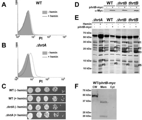
Hemin-exposedDhrtAhas decreased membrane integrity
A potential explanation for the results obtained above is that hemin-exposedDhrtA over-expresses HrtB which localizes to the membrane as a dysregulated permease and causes membrane damage. In turn, this membrane damage is sensed by S. aureus leading to changes in the secreted protein profile. As an initial test of this hypothesis, both wildtype andDhrtAwere grown in61mM hemin and membrane integrity was determined by propidium iodide (PI) staining. PI is a membrane impermeant cationic stain that produces strong red fluorescence when bound to nucleic acids, and hence can be detected using FACS [22]. When wildtype S. aureuswas exposed to hemin there was a small shift in PI staining indicating minor changes in membrane integrity (Fig. 3A). In contrast,DhrtAexposed to hemin exhibited a pronounced shift in PI staining indicative of substantial changes in membrane integrity (Fig. 3B). Cell death was excluded as a possible cause of the
increased permeability of hemin exposedDhrtA, as hemin exposure at this concentration did not affect viability of the tested strains (Fig. 3C). These results indicate that exposure ofDhrtA to 1mM
hemin compromises cell membrane integrity without affecting cellular viability.
Hemin-independent over-expression of HrtB elicits a secreted protein profile that mimics that of
hemin-exposedDhrtA
The data presented above are consistent with a model whereby HrtB expression in the absence of its cognate ATPase is responsible for increased secretion of immunomodulatory proteins. This phenotype can be complemented by catalytically inactive versions of HrtA suggesting that it is not HrtA ATPase activity but the physical presence of HrtA that prevents alterations in protein secretion. In keeping with this, it is predicted that discordant regulation of HrtB and HrtA would lead to a phenotype that mimics DhrtA exposed to hemin. To test this hypothesis, myc-tagged HrtB was constitutively expressed in wildtype,DhrtBand thrtBbackgrounds. The three strains expressed comparable levels of HrtB as assessed by immunoblots using anti-c-Myc monoclonal antibody (Fig. 3D). Upon comparing the secreted protein profiles of these three strains in the absence of hemin exposure to matched
Figure 3. Over-expression of HrtB without hemin exposure mimics the effect of exposingDhrtAto hemin.(A and B) FACS analysis of
strains harboring empty vector, we observed alterations in protein abundance that closely resemble those produced by hemin-exposed DhrtA (Fig. 3E). This result indicates that expressing disproportional ratios of HrtB:HrtA triggers a response similar to that obtained when HrtB is expressed in the absence of HrtA. To confirm that over-expressed HrtB localizes to the cell membrane, cell fractionation was performed on wildtype cells harboring the phrtB-myc plasmid grown in the absence of hemin. When equivalent protein amounts of cellular fractions were immuno-blotted using monoclonal antibody against c-Myc, a reactive band with the predicted molecular mass (,37 kDa) was detected exclusively in the membrane fraction (Fig. 3F). A second band migrating at approximately 20 kDa was detected in the membrane fraction, however the identity of this band in unknown at this point. These observations support a model whereby HrtB expression in the absence of HrtA acts as an unregulated pore resulting in the production of membrane stress and the up-regulation of immunomodulatory proteins.
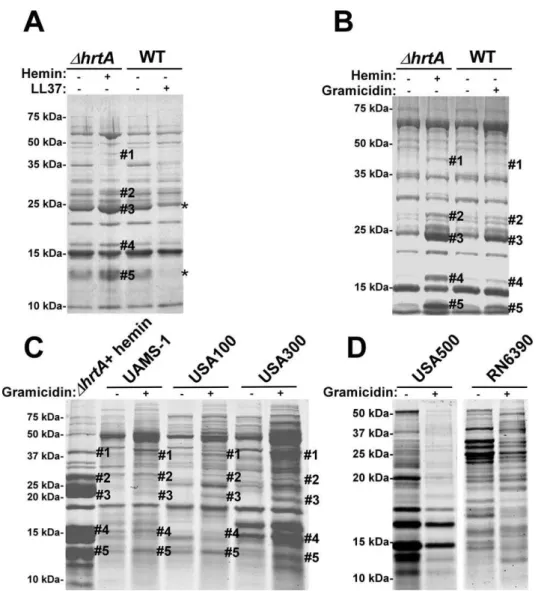
Membrane pore formation rather than generalized membrane damage in wildtypeS. aureusmimics the effect of hemin onDhrtA
To test if the secreted protein profile of hemin-exposedDhrtAis due to non-specific membrane damage,S. aureuswas treated with sub-inhibitory concentrations of the anti-microbial peptide (AMP) LL37. LL37 elicits antimicrobial activity through membrane disruption caused by widespread random intercalation into the bacterial membrane [23]. We chose to use 40mg/ml LL37 for these experiments as this concentration slightly inhibitedS. aureus growth but permitted growth to cell densities that were similar to untreated cultures (data not shown). When the secreted protein profiles ofS. aureusgrown in the absence and presence of LL37 were compared to those of DhrtA grown in the absence and presence of 1mM hemin, similar effects regarding the secreted proteins were not observed (Fig. 4A). None of the five bands that were visibly up-regulated in hemin-exposed DhrtA were
up-Figure 4. Membrane pore formation but not generalized membrane damage mimics the effect of hemin onDhrtA.(A) Exoprotein
profile ofDhrtAtreated61mM hemin and WT640mg/ml of the antimicrobial peptide LL37. (B) Exoprotein profile ofDhrtAtreated61mM hemin and WT616mg/ml of the antimicrobial peptide gramicidin. (C) Exoprotein profile ofDhrtAtreated with 1mM hemin and different staphylococcal strains632mg/ml gramicidin. (D) Exoprotein profile of wildtype strains USA500 and RN6390632mg/ml gramicidin. Samples were prepared and analyzed as in Figure 1. The#indicates the positions of proteins up-regulated under the indicated condition and the predicted identity of these proteins is as described in Figure 1. The * indicates the positions of proteins down-regulated under the indicated condition. Positions of protein molecular mass markers in kilodaltons (kDa) are indicated on the left side of each panel.
regulated in wildtype exposed to LL37. In fact, some of the bands that were up-regulated inDhrtAexposed to hemin seemed to be down-regulated when wildtype was exposed to LL37 (Fig. 4A). This result indicates that membrane damage caused by LL37 treatment does not induce the same response withinS. aureus as over-expression of the HrtB permease without its cognate ATPase. Therefore, non-specific membrane damage is not responsible for alterations in the secretome observed upon HrtA and HrtB dysregulation.
We next sought to test the impact of a pore-forming AMP on the staphylococcal secretome to more closely mimic the hypoth-esized membrane damage elicited upon HrtB over-expression. To this end, wildtype S. aureus was treated with sub-inhibitory concentrations of the pore-forming AMP gramicidin. The growth of S. aureus in the presence of 16mg/ml gramicidin was slightly
inhibited, but the treated cultures were able to reach similar optical densities to those of untreated cells (data not shown). Upon comparing the secreted protein profile of wildtype treated with gramicidin to that of DhrtA treated with hemin a remarkable conservation in the expression patterns was observed (Fig. 4B). Notably, all of the five bands that were up-regulated in hemin-exposed DhrtA were also up-regulated in gramicidin-treated wildtype, but to varying extents (Fig. 4B). This result supports the contention that dysregulated pore formation through the staphylococcal membrane leads to a specific alteration in protein secretion. Gramicidin exposure induces significant alterations in protein expression across a panel of S. aureus isolates (UAMS-1, USA100, USA300, USA500, and RN6390) (Fig. 4C & D). Notably, a subset of these strains (UAMS-1, USA100, and USA300) exhibit altered expression of proteins migrating to positions similar to those of the immunomodulatory proteins affected in the Newman strain (Fig. 4C). It is interesting to point out that not all strains analyzed were affected by gramicidin treatment equally. More specifically, USA500 and RN6390 do not seem to increase expression of the immunomodulatory proteins (based on migration pattern) upon gramicidin exposure despite exhibiting significant changes in protein expression (Fig. 4D). Taken together, these experiments suggest that treatment of distinctS. aureusstrains with pore forming toxins produces changes in protein expression.
The S. aureus Aps/Gra system is a two-component system responsible for resistance to antimicrobial peptides suggesting a potential involvement in the response to gramicidin reported here [24,25]. To evaluate the involvement of Aps/Gra in this response to gramicidin, we created aS. aureusstrain inactivated for Aps/Gra (DapsR) and measured the impact of HrtB over-expression and gramicidin exposure on this strain. The secreted protein profile of
DapsRdid not differ noticeably from that of wildtype (Fig. S1A & B). These experiments revealed that the Aps/Gra system is not involved in the response to gramicidin or HrtB overexpression.
The secretome of wildtypeS. aureustreated with gramicidin resembles that ofDhrtAexposed to hemin
Gel based comparisons of protein secretion between gramicidin-treated wildtype and hemin-exposed DhrtA suggest that these distinct but similar stressors lead to analogous alterations in the staphylococcal secretome (Figs. 3E and 4B). In an effort to increase the sensitivity and resolution of this comparison beyond Coomas-sie blue-stained SDS-PAGE analysis, a mass spectrometry-based approach known as shotgun proteomics (see Materials and Methods) was employed. Using shotgun proteomics we deter-mined the proteomes of the culture supernatants ofDhrtAgrown in the presence and absence of hemin and wildtypeS. aureusgrown in the presence and absence of gramicidin. The analysis was
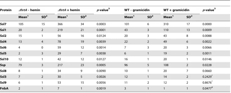
performed on three independent samples from each condition. Quantitation of the proteins in each sample was performed using spectral counting of tandem spectra acquired for each protein normalized to the total number of spectra detected in the same sample. Analysis of the culture supernatants ofDhrtA with and without hemin revealed the presence of at least 137 proteins exhibiting an average of at least 2 spectra in the three samples analyzed. Among these, 21 proteins were significantly up-regulated and 50 proteins were down-up-regulated in the presence of hemin (Table S2). Analysis of known secreted proteins demonstrated that there were 12 proteins significantly up-regulated and 18 proteins significantly down-up-regulated between these conditions (Tables 1 & 2, Figs. 5A & B). In the case of wildtype grown in the presence and absence of gramicidin, 101 proteins were detected among which 32 proteins were significantly up-regulated and 29 were down-regulated (Table S3). Among the 12 secreted proteins that were up-regulated inDhrtA exposed to hemin, 8 proteins (i.e. 75%) were also up-regulated in WT exposed to gramicidin (Table 1 and Fig. 5A). All 8 of these proteins have potential immunomodulatory functions including seven Staphylo-coccal Superantigen-Like proteins (Ssl1, Ssl2, Ssl4, Ssl5, Ssl6, Ssl7, and Ssl10) and the extracellular matrix and plasma binding protein (Ssp, also known as Emp). In addition three other Ssls (Ssl9, Ssl8 and Ssl3) were up-regulated under both conditions but their up-regulation did not reach the level of significance in wildtype treated with gramicidin (Table 1).
Among the 18 secreted proteins that were down-regulated in
DhrtA exposed to hemin, 16 proteins (i.e. 89%) were also significantly down-regulated in wildtype treated with gramicidin (Table 2 and Fig. 5B). This group of proteins included several hemolysins, leukotoxins, and other known virulence factors. Two additional proteins (SspB and CHIPS) that were significantly down-regulated in DhrtA exposed to hemin were also regulated in wildtype exposed to gramicidin but their down-regulation did not reach the level of significance (Table 2). Taken together, these results demonstrate that pore formation through gramicidin treatment or permease dysregulation results in alterations in the staphylococcal exoprotein profile highlighted by an increased abundance of immunomodulatory proteins with known anti-neutrophil functions [19,26,27].
Alterations in protein secretion upon pore formation occur at the transcriptional level
Table 1.Secreted proteins up-regulated in (DhrtA+hemin) and (WT+gramicidin).
Protein DhrtA- hemin DhrtA+hemin p-value3 WT - gramicidin WT+gramicidin p-value3
Mean1 SD2 Mean1 SD2 Mean1 SD2 Mean1 SD2
Ssl7 105 15 366 34 0.0003 101 6 310 17 0.0000
Ssl1 20 2 219 21 0.0001 43 3 110 13 0.0009
Ssl2 15 1 56 16 0.0124 20 3 43 8 0.0088
Ssl4 13 4 78 19 0.0039 22 2 49 6 0.0022
Ssl6 4 0 59 12 0.0014 7 3 20 3 0.0066
Ssl5 2 3 29 7 0.0038 6 1 19 2 0.0011
Ssl10 12 1 42 12 0.0127 16 1 20 1 0.0146
Ssp 79 3 217 23 0.0005 96 5 108 2 0.0228
Ssl8 8 1 34 9 0.0090 10 1 20 7 0.0660
Ssl3 7 2 30 5 0.0026 12 1 14 2 0.2429*
Ssl9 6 1 53 15 0.0056 11 2 12 2 0.8676*
FnbA 2 1 7 1 0.0019 3 1 1 1 0.0477¥
1Mean of the number of spectra detected in the three samples analyzed for this condition.
2Standard deviation among the number of the spectra detected in the three samples analyzed for this condition. 3
p-value as obtained by applying Student’st-test.
*The up-regulation of these proteins in the presence of gramicidin was not statistically significant. ¥This protein was significantly down-regulated in the presence of gramicidin.
doi:10.1371/journal.ppat.1000802.t001
Table 2.Secreted proteins down-regulated in (DhrtA+hemin) and (WT+gramicidin).
Protein DhrtA- hemin DhrtA+hemin p-value3 WT - gramicidin WT+gramicidin p-value3
Mean1 SD2 Mean1 SD2 Mean1 SD2 Mean1 SD2
LukS 212 14 87 2 0.0001 243 21 103 12 0.0005
HlgC 141 4 46 19 0.0011 120 5 62 8 0.0004
HlgA 132 6 78 18 0.0076 104 6 62 9 0.0028
LukF 100 14 33 3 0.0013 132 4 45 4 0.0000
Truncated MAP 80 16 19 6 0.0035 53 3 14 0 0.0000
Autolysin 58 4 15 1 0.0000 88 8 61 4 0.0057
LukD 56 4 17 5 0.0005 82 3 30 2 0.0000
LukE 53 3 21 4 0.0003 68 4 35 10 0.0060
Hla 46 4 28 5 0.0091 41 3 3 3 0.0001
Truncated lipase 45 4 36 2 0.0280 54 9 33 6 0.0265
Sbi 38 4 24 5 0.0237 52 1 23 2 0.0000
Coa 26 4 8 2 0.0028 29 5 19 2 0.0343
Lip 25 1 13 1 0.0004 36 4 26 1 0.0159
SsaA-like 12 2 5 1 0.0080 15 2 8 1 0.0151
NAMLA amidasey¥ 18 5 4 1 0.0126 8 2 1 1 0.0105
59-nucleotidase¥ 150 14 93 2 0.0024 148 13 119 11 0.0395
SspB 8 1 1 1 0.0009 6 1 3 2 0.0764*
CHIPS 169 20 108 9 0.0088 164 11 151 33 0.5285*
1Mean of the number of spectra detected in the three samples analyzed for this condition.
2Standard deviation among the number of the spectra detected in the three samples analyzed for this condition. 3
p-value as obtained by applying Student’st-test.
*The down-regulation of these proteins in the presence of gramicidin was not statistically significant. y
The hypervirulence of strains lackinghrtAis due to the increased expression and secretion of Ssl1-11 and Ssp
Strains lacking hrtA exhibit liver-specific hypervirulence and increased secretion of immunomodulatory proteins such as Ssls and Ssp. To determine if the increased secretion of these immunomodulatory proteins is responsible for the hypervirulence ofhrtAmutants, we created strains lackingssl1-11orsspin athrtA
background (thrtADssls, thrtADssp,thrtADsslsDssp).Next we assessed the liver-specific virulence of S. aureuswildtype, thrtA, thrtADssls, thrtADssp, andthrtADsslsDsspin the systemic model of staphylococ-cal infection described above. As expected, thrtA exhibited increased virulence in these studies as compared to wildtype. Inactivation of eitherssl1-11orsspreduced the virulence ofthrtAto levels approximately equivalent to wildtype despite the fact that
Figure 5. Treatment ofS. aureuswildtype with gramicidin andDhrtAwith hemin elicit similar secretomes.(A) Average number of
spectra per protein of secreted proteins that were significantly up-regulated inDhrtA+1mM hemin and WT+32mg/ml gramicidin. (B) Average number of spectra per protein of secreted proteins that were significantly down-regulated under both conditions. The data presented are means of three independent samples and the error bars represent the standard deviation. The differences betweenDhrtA+hemin (black bars) andDhrtA -hemin (white bars) and WT+gramicidin (diagonally streaked bars) and WT - gramicidin (grey bars) are statistically significant in all the presented proteins withpvalues less than 0.05 as indicated by Student’sttest. * T stands for truncated. (C-F) Results of quantitative real-time RT-PCR using RNA samples isolated from samples ofDhrtA6hemin and WT6gramicidin. The levels of the transcripts were normalized using the levels of the ribosomal RNA 16S and the levels of the normalized transcripts from samples without treatment (either hemin or gramicidin) were used as calibrators. The data presented are the means of two independent experiments with each done in triplicate and the error bars represent the standard deviation. The differences betweenDhrtA+hemin (black bars) andDhrtA- hemin (white bars) and WT+gramicidin (diagonally streaked bars) and WT -gramicidin (grey bars) are statistically significant in all the presented transcripts withpvalues less than 0.05 as indicated by Student’sttest except the one data point marked with NS.
these mutations had no adverse effect on growthin vitro (Fig. 6). Inactivation of ssp had a more pronounced effect on the hypervirulence of thrtAas compared to ssl1-11 underscoring the significant contribution of Ssp to the hypervirulence ofthrtA. When the mutations of thesslswere combined with that of thesspin the hrtA mutant background, the hypervirulence of the hrtA mutant was significantly reduced almost to the level of wildtype (Fig. 6). Taken together, these data demonstrate that the hypervirulence of hrtAmutants is due to the increased expression of Ssl1-11 and Ssp in response to dysregulated pore formation.
Discussion
S. aureusHrtAB is an ABC-type transport system that is essential for alleviating hemin toxicity [13,14]. Upon hemin exposure HrtAB is up-regulated approximately 45-fold [28]. Inactivation of hrtAresults in liver specific hyper-virulence that is associated with a hemin-induced over expression of immunomodulatory proteins
[13]. Here we propose a revised model to explain the hyper-virulence ofS. aureusDhrtAas summarized in Fig. 7A; whenDhrtA is exposed to hemin, the HssRS system is activated leading to the over-expression of HrtB in the cell membrane without its cognate ATPase. In addition to an inability to relieve heme-toxicity [13], this mutant experiences membrane stress caused by the over-expressed HrtB permease acting as an unregulated pore. This membrane stress is sensed by S. aureus through an as-yet-unidentified mechanism, leading to over-expression and secretion of several immunomodulatory molecules. The combined action of these immunomodulatory proteins inhibits neutrophil migration to the site of infection enabling DhrtA to exhibit hypervirulence during liver colonization. This model is supported by the observation thatS. aureusexposed to a pore forming AMP exhibits a similar secreted protein profile to hemin-exposed DhrtA, potentially providing a physiologically relevant explanation for the existence of this response (Fig. 7B). When S. aureus senses membrane damage in the form of pore formation, the organism appears to over-express and secrete immunomodulatory molecules that interfere with the recruitment of phagocytic cells to the site of infection. The observation that strains lacking hssR also exhibit hypervirulence suggest that disruption of HssRS signaling impacts HrtAB expression in a way that leads to a similar form of membrane damage [13].
The precise mechanism by which HrtB over-expression leads to membrane stress is not completely understood, but lack of expression of the HrtA protein may lead to misfolding of its cognate permease (HrtB), or lock the HrtB pore in a conformation that no longer allows the passage of its substrate, and/or allows the passage of ions involved in maintaining membrane potential. These possibilities are in accordance with the observation that over-expression of membrane proteins usually leads to reduced growth rates that are generally assumed to be due to negative effects on membrane integrity [29]. An indication that DhrtA exposed to hemin experiences membrane damage is provided by the observation that this strain showed increased permeability to propidium iodide (Fig. 3B) [22].
During infection, S. aureus faces an array of membrane damaging peptides and small proteins that perturb the cell membrane leading to cell leakage and eventually death [30,31]. In fact, mammalian cells increase the production of several AMPs upon sensingS. aureusand its components [31,32,33]. In general, AMPs exert their antimicrobial activity through pore formation or membrane barrier disruption, however some AMPs mediate their antibacterial activity through altering septum formation or inhibiting cell-wall, nucleic-acid, or protein synthesis [34,35,36]. Few AMPs have been described that are considered channel-forming peptides that act through what is known as the ‘‘barrel-stave’’ mechanism [35,37]. On the other hand, several AMPs, including the widely expressed human LL37, have been described to perturb membranes through what is known as the ‘‘carpet’’ or ‘‘detergent like’’ mechanism [23,37].
To counter the bactericidal effects of AMPs, bacteria express regulatory systems designed to sense these innate immune threats to generate an anti-AMP response. Among these systems, the Gram negative PhoPQ system is the best characterized [38]. Recently, the staphylococcal counterpart of the PhoPQ system was identified [25,39,40]. The Aps/Gra system is a three component bacterial sensing system that senses AMPs to induce a resistance response that includes modification of cell surface components and increased expression of putative AMP transporters [25,39,40]. The Aps/Gra system is not involved in the phenotypes reported here, implying the existence of an additional system responsible for sensing membrane damage inS. aureus[40,41,42]. The S. aureus
Figure 6. Ssls1-11 and Ssp are responsible for the liver-specific
hyper-virulence of hrtA mutants. (A) Growth curve analysis of
wildtype, thrtA, thrtADssls, thrtADssp, and thrtADsslsDssp. Overnight cultures were diluted 1:20 in TSB and the absorbance at 600 nm was recorded at the indicated time points. A representative growth curve is presented. (B) Bacterial multiplication in infected BALB/c mice livers as measured by tissue homogenization, dilution, and colony formation on agar media 96 hours post infection. The horizontal bars represent the mean of log10CFU, and the boxes cover the range of log10CFU obtained in each group. * indicates statistically significant differences fromthrtA as determined by Student’s t test with the indicated p values. NS indicates a non-statistically significant difference. N indicates the number of mice included in each group.
immunomodulatory factors described here were found to be up-regulated upon exposure to the pore-forming peptide gramicidin which is produced byBacillus brevis. In contrast, this response is not elicited upon exposure to the membrane-disintegrating mamma-lian peptide LL37. Considering the apparent specificity of this secreted protein response to inhibit neutrophil migration, it is likely that as-yet-unidentified channel-forming AMPs elicit this response during mammalian infection.
S. aureusregulation of virulence factors in response to phagocytes and phagocytosis-related signals have been previously reported and the response varies according to the setting of the experiment and the strain tested. For instance, theS. aureusvirulence regulator Sae is highly activated upon exposure toa-defensins and H2O2but
not LL37 [43]. When S. aureus strain SG511 was exposed to human b-defensin 3, the only pathogenic factors that were significantly up-regulated were fmtA and sdrE [44]. In a more comprehensive study [45], different S. aureus strains were phagocytosed by human PMNs and transcriptome analyses of these bacteria showed up-regulation of genes encoding multiple virulence factors such ashlgA,hlgB,hlgC,extracellular matrix and plasma binding protein (ssp), staphylocoagulase, and clumping factor. Interestingly, genes encoding several toxins, such as ssl7, ssl11,ssl10(set14),lukD, andlukEwere up-regulated only in strains causing community acquired (CA) infections. When the CA-MRSA strain MW2 was exposed to neutrophil azurophilic granule proteins [46], there was an up-regulation of genes encoding numerous virulence factors including hemolysins (hla, hld, hlgA, hlgB, andhlgC) and leukotoxins (lukSandlukF) in addition to several immunomodulatory toxins belonging to the Ssl group (ssl7, ssl1 and ssl10). In this manuscript we report that theS. aureus strain Newman response to either over-expression of the HrtB permease or the pore-forming AMP gramicidin is different from previous reports. We have observed up-regulation of the immunomodula-tory Ssl exoproteins (Ssl7, 1, 2, 4, 6, 5, and 10) and extracellular matrix and plasma binding protein (Ssp) while simultaneously observing a down-regulation of several hemolysins, cytotoxins and other secreted virulence proteins. As observed withS. aureusstrain Newman, we found that UAMS-1, USA100, USA300, USA500 and RN6390 undergo profound changes in protein expression
upon exposure to gramicidin (Fig. 4C and D). A subset of these strains (UAMS-1, USA100, and USA300) exhibit altered expres-sion of proteins migrating at the predicted size for the immunomodulatory proteins affected in the Newman strain. It is interesting to point out that not all strains analyzed were affected by gramicidin treatment equally. More specifically, USA500 and RN6390 do not seem to increase expression of the immunomod-ulatory proteins upon gramicidin exposure despite exhibiting significant changes in protein expression (Fig. 4D). From these data, we conclude that S. aureus alters exoprotein expression in response to pore-forming toxins.
The Ssl proteins have sequence and structural homology with superantigens; however they lack the superantigenic activity [19]. Functions have yet to be ascribed to each of the Ssl proteins, but those with known functions have been shown to have an immunomodulatory effect. For instance Ssl7 binds IgA and complement C5, blocking IgA-FcR interactions and complement activation [26]. Both Ssl5 and Ssl11 inhibit neutrophil rolling while Ssl5 was recently shown to inhibit leukocyte activation by chemokines and anaphylatoxins [27,47]. In addition, Ssl10 inhibits CXCL12-induced human tumor cell migration [48]. However, very little is known about the regulation of these exotoxins [19]. It was shown that ssl4 (set9) is up-regulated in a SarA-dependent manner while ssl3 (set8) is up-regulated in an agr-dependent manner [49]. Combining these observations with the fact that many of the virulence factors that are down-regulated in our experiments such as Hla, HlgABC, Lip, and Map are also regulated through these two majorS. aureus virulence regulators (agrand SarA) [49], it is tempting to speculate that one or both of these regulators may play a role in orchestrating the immuno-modulatory program elicited by membrane damage caused by pore formation.
In conclusion, the data presented here shed light on a potentially new virulence regulatory circuit inS. aureus that can modulate the immune response in the host and the components of such a circuit represent potential therapeutic targets. The significance of this circuit stems from the fact that an inducer of this program, the antimicrobial agent gramicidin, is a component in preparations that have shown efficacy against S. aureus
Figure 7. Membrane damage triggers immunomodulatory proteins secretion.(A) WhenDhrtAis exposed to hemin(1)the HssS/R system is
activated(2)leading to over-expression of thehrtBgene(3)causing membrane damage by the over-expressed HrtB proteins acting as unregulated pores(4). This in turn activates an internal signal(5)that turns on the expression(6)and secretion(7)of the immunomodulatory proteins. (B) When WT S. aureusis attacked by antimicrobial peptides(1)that form pores in the membrane(2), this in turn activates an internal signal(3)that turns on the expression(4)and secretion(5)of immunomodulatory proteins.
colonization and infections [50,51]. The identification of the factors involved in transducing the signal from the membrane which activates the expression and secretion of these immuno-modulatory factors is critical for the full understanding of this program and is the focus of future research.
Materials and Methods
Ethics statement
All procedures involving animals were approved by Vanderbilt University’s Institutional Animal Care and Use Committee (IACUC). All animal experiments were performed in accordance to NIH guidelines, the Animal Welfare Act, and US federal law.
Bacterial strains, plasmids and growth conditions Staphylococcus aureusclinical isolate Newman [52] was used in all experiments as the wildtype strain (unless explicitly stated) and mutants were generated in its background. Isogenic mutants lacking thehrtAandhssR gene were previously described [13]. A strain containing a transposon insertion into hrtB (thrtB) was obtained from the Phoenix (N) library clone PhiNE 05560 (SAV2360) [53]. Plasmids expressing WT and mutatedhrtAgenes were described previously [14].S. aureuswere grown on tryptic soy broth (TSB) solidified with 1.5 % agar at 37uC or in TSB with shaking at 180 rpm, unless otherwise indicated. When appropriate TSB was supplemented with chloramphenicol at a final concen-tration of 10mg/ml.Escherichia coliwere grown in Luria broth (LB)
and when needed, the media were supplemented with ampicillin at a final concentration of 100mg/ml.
Construction of mutants
Construction of the hrtB deletion mutant strain. The
DhrtB mutant lacking the hrtB gene was constructed using established methods [54]. Briefly, a PCR amplicon containing the first 12 bp of the hrtB ORF together with approximately 600 bp upstream of the hrtB ORF was amplified using primers AA431 (59 -GGGGACCACTTTGTACAAGAAAGCTGGGTT-AACGTCATCGTTTTA-39) and AA407 (59 -GCACCTCCA-ATTGCTTCGATCGCTAATTTCATATCGATTCACTT-39). Another amplicon containing the last 24 bp of the hrtB ORF together with approximately 750 bp downstream of thehrtBORF was amplified using primers AA404 (59 -ATCGAAGCAAT-TGGAGGTG-39) and AA428 (59 -GGGGACAAGTTTGTAC-AAAAAAGCAGGCTGCCTAAGAACTTAATG-39). The first amplicon contained in its 39-end 20 bp that are complementary to the first 20 bp in the 59-end of the second amplicon. Both amplicons were used together as a template in a second cycle of PCR using primers AA431 and AA428. The resultant PCR product was recombined into pKOR1 [54] and used for allelic replacement into Newman as described [54].
Construction of the ssl deletion mutant strain. The S. aureus mutant strain lacking ssls 1-11 (Dssl1-11) was constructed using the pKOR-1 plasmid as described by [54]. Briefly, sequences flanking the ssllocus were PCR amplified with primers VJT160 (59 -GGGGACAAGTTTGTACAAAAAAGCAGGCTAATAGT-CCTCTTGCTCCTGC-39) and VJT172 (59 -TCCCCCCGGG-TTCAGACACAAAACAGACATC-39) for the upstream frag-ment and primers VJT124 (59 -GGGGACCACTTTGTAC-AAGAAAGCTGGGTTAAAACCATACCTTCATCATCC- 39) and VJT173 (59 -TCCCCCCGGGGAGTTAATCACTGACT-TCTAC-39) for a downstream fragment. The PCR amplicons were cloned into pCR2.1 (Invitrogen), and then digested with XmaI and assembled into pCR2.1. A PCR amplicon of the joined DNA fragment was recombined into pKOR1 resulting in the
pKOR-1Dssl1-11plasmid. Deletion of thessllocus in Newman was achieved by allelic replacement with pKOR-1Dssl1-11.
Construction of the hrtA::bursa ssl double mutant strain. An erythromycin cassette insertion mutant ofhrtA was obtained from the Phoenix (N) library (thrtA) [13,53]. ThehrtA::erm mutation was transduced into Newman lackingssls(Dssl1-11) with phageW85 [10].
Construction of thesspdeletion mutant strain. Primers AA547 (59- GGGGACAAGTTTGTACAAAAAAGCAGGCT-GAAAACACCTGTAGTGTCATTAGA-39) and AA543 (59 -AACCCGGGGCTCATAGTTAAAACTAATAA-39, XmaI site underlined) were used to amplify the upstream region of thessp ORF (NWMN_0758) and primers AA545 (59- CACCCG-GGGTATAAAAATTGGCACTAAGT-39, XmaI site under-lined) and AA548 (59- GGGGACCACTTTGTACAA-GAAAGCTGGGTGGTTTGCTTAATGTGTTAACTTTT-39) were used to amplify the region downstream of thesspORF. Both PCR amplicons were digested with XmaI then ligated, PCR-amplified and ligated into pCR2.1 vector. The resulting plasmid was digested with XmaI and ligated to the non-polar spectinomycin cassette from plasmid pSL60-1[55]. The PCR fragment containing the flanking regions and the spectinomycin resistance cassette was then amplified and recombined into pKOR1. The Dssp::spc mutant was constructed in strain Newman as described above then transduced into the thrtA mutant andthrtA.Dsslsmutant background.
Construction of the apsR deletion mutant strain. The
DapsRmutant lacking theapsRgene (NWMN_0628) encoding the response regulator of the Aps/Gra system was constructed using a similar approach to that described above for the construction of theDhrtBandDssl1-11mutants.
Construction of a plasmid over-expressing HrtB
For the construction of a plasmid constitutively expressing Myc-tagged HrtB, a PCR amplicon was made containing thehrtBORF into which the sequence encoding a c-Myc-epitope (EQKLI-SEEDL) was inserted just prior to the stop codon using the primers DS218 (59- GGGGCATATGAAATTAGCGATAAAAGAG-39, NdeI site underlined) for the 59-end and AA494 (59- TA- GATCTTCTTCAGATATCAGTTTCTGTTCGCCGCCTT-CTGCACCTCCAATTGCTTCGATAGGATCCACTTT-39) together with AA495 (59 -CCTCGAGTTATAGATCTTCTTCA-GATATCAGTTT-39, XhoI site underlined) for the 39-end. The PCR product was then digested with both NdeI and XhoI and ligated into the E. coli/S. aureus shuttle vector pOS1-plgt [56] which had been digested with the same restriction enzymes. Ligation mixtures were then transformed intoE. coliDH5a and the resultant plasmid was designated phrtB-myc. After DNA sequence verification the plasmid was transformed into the restriction-deficient modification-positive S. aureus RN4220 [57] followed by transformation into the respective electrocompetentS. aureusstrain using the protocol described [58].
Cell fractionation
S. aureuscells were grown to mid-log phase and sedimented by centrifugation at 3,2006g for 10 min. The supernatants were
removed and the pellet was suspended in 500ml TSM (100 mM Tris-HCl (pH 7.0), 500 mM sucrose, 10 mM MgCl2) supplemented
with 100mg lysostaphin and incubated for 1 hr at 37uC. The
resulting protoplasts were sedimented by centrifugation at 16,0006g for 15 min; the supernatant was collected [cell wall (CW) fraction], and the pellet was suspended in 500ml membrane buffer (50 mM
Tris-HCl (pH 7.0), 10 mM MgCl2, 60 mM KCl) and subjected to
100,0006gfor 45 min. The supernatant was collected [cytoplasm
(Cyt) fraction] and the pellet [membrane (Mem) fraction] was suspended in 200ml membrane buffer. Equivalent amounts of protein from each fraction were separated by SDS-PAGE and analyzed by immunoblotting as described below.
Measuring membrane integrity using propidium iodide staining
In order to monitor staphylococcal membrane integrity, bacterial cells were grown in Roswell Park Memorial Institute (RPMI) medium supplemented with 1% (wt/vol) Casamino Acids (CAS) 61mM hemin until the cultures reached mid-log phase
(OD600,0.4), washed and suspended in phosphate buffered saline (PBS) supplemented with 1% wt/vol bovine serum albumen (BSA). All samples were adjusted to the same OD600and 50ml
aliquots corresponding to approximately 107cells were mixed with 2mg of propidium iodide (PI) (Sigma) in 1 ml PBS/1%BSA and analyzed by fluorescence-activated cell sorting (FACS). Analysis was performed on a FACSCalibur system, (BD, Franklin Lakes, NJ) using Cell Quest Pro software (BD). Equal aliquots of each sample were serially diluted and plated on TSA for viability counting.
Antimicrobial peptides and MIC determination
The AMP LL37 was obtained from Phoenix Pharmaceuticals, Inc (Burlingame, CA) and it was dissolved directly in RPMI/CAS. The AMP gramicidin was obtained from Sigma (St. Louis, MO) and dissolved in absolute ethanol to a final concentration of 10 mg/ml. MIC determination was done by incubating 105CFU/ml S. aureus with 2 fold serial dilutions of the tested peptide in RPMI/CAS at 37uC. The MIC was considered as the lowest concentration that prevented growth as indicated by lack of visible turbidity following incubation at 37uC for 24 hr.
Analyses of secreted proteins
S. aureus cultures from a fresh streak on TSA plates were inoculated in 5 ml of RPMI/CAS overnight at 37uC. The overnight cultures were then used to inoculate a new 5 ml culture in a 15 ml concical tube to which hemin was added at final concentrations of 0.5, 1, or 2mM. The AMP LL37 was added to a final concentration of 40mg/ml and gramicidin was added to a final concentration of 16 or 32mg/ml. As a negative control, cells
were grown in RPMI/CAS with equivalent volume of ethanol (WT – gramicidin). All the cultures were allowed to grow overnight (,18 hrs) then the samples were normalized to the same OD600. Bacterial cells were sedimented by centrifugation
and proteins in the culture supernatants were precipitated using 10% (vol/vol) TCA at 4uC overnight (,16 hrs). The precipitated proteins were sedimented by centrifugation, washed with absolute ethanol, dried, resuspended in 1X SDS-loading buffer, and boiled for 10 min. Proteins in the samples were resolved using 15% wt/ vol SDS-PAGE, and stained with Coomassie blue. At times, we observed slight variations in protein expression from identical strains used in distinct experiments as shown in Figures 1, 2, 3, 4, and S1. This variation correlated with changes in the source of medium, suggesting that batch-to-batch variation in media preparations affects this phenotype. Despite these minor varia-tions, similar patterns of protein expression were observed across all experiments.
Shotgun proteomics analysis ofS. aureusexoproteins TCA precipitated proteins from filtered culture supernatants of
DhrtA 61mM hemin and WT 632mg/ml gramicidin were
prepared as described above. Higher concentration of gramicidin than what was used above was aimed to increase the sensitivity of the assay. Proteins were resuspended in 1X SDS-digestion buffer and loaded into 12% SDS-PAGE gel (without a stacking gel) and electrophoresed 2 cm into the gel. The gel was stained with Colloidal Blue (Invitrogen, Carlsbad, CA) then destained with distilled water over night. Proteins were then subjected to in-gel trypsin digestion and peptide extraction and the resulting peptides were then analyzed using a Thermo Finnigan LTQ ion trap instrument and separated as described [59]. Tandem spectra were acquired using a data dependent scanning mode with a one full MS scan (m/z 400–2000) followed by 9 MS-MS scans. The SEQUEST algorithm was then used to search the tandem spectra against the Newman strain of theS. aureussubset of the UniRef100 database. To determine false positive rates, the database was concatenated with the reverse sequences of all proteins in the database. The SEQUEST outputs were filtered through the ID Picker suite with a false positive ID threshold of 5% and proteins were required to be identified by 2 or more unique peptides. Protein reassembly from identified peptide sequences was done as previously described [60]. The number of spectra identified for each protein under a given condition were normalized to the number of total spectra detected in the same injection.
RNA isolation and manipulation
S. aureus cultures where grown in RPMI/CAS with the appropriate additive to an OD600 ,1.0. Cultures were then mixed with equal volume of 1:1 ethanol: acetone mixture and frozen at -80uC. For RNA extraction, frozen samples were allowed to thaw on ice; cells were then sedimented and washed two times with TE buffer. Cells were broken mechanically using fastprep bead beater (MP Biomedicals, Solon, OH) then RNA was isolated using the RNeasy mini kit (Qiagen, Valencia, CA) according to the manufacturer’s recommendation. On-column DNase digestion was performed and after RNA elution, the samples were cleaned from any residual DNA contamination using the MessageClean kit (GenHunter, Nashville, TN).
RT-PCR and real time RT-PCR
RT-PCR was performed to determine if thehrtBandhrtAgenes were transcriptionally linked. 2mg of RNA isolated as described
Immunoblots
S. aureus cultures were grown in RPMI/CAS overnight, cells were then treated with lysostaphin to digest the cell wall and the resulting protoplasts were pelleted and resuspended in BugBuster Protein Extraction Reagent (Novagen, Gibbstown, NJ) with proteinase inhibitor and sonicated for 10 s. Proteins in protoplasts were then resolved in 15% SDS-PAGE, transferred to nitrocel-lulose membranes, immunoblotted with 9E10 anti-C-Myc mono-clonal antibody as a primary antibody and AlexaFluor-680-conjugated anti-mouse as secondary antibody. Membranes were then scanned using an Odyssey Infrared Imaging System (LI-Cor Biosciences, Lincoln, NE).
Murine model of infection
Six- to eight-week-old BALB/c female mice (Jackson Labora-tories, Bar Harbor, Maine) were infected retro-orbitally with approximately 16107CFU ofS. aureus strains. Ninety-six hours
post-infection, mice were euthanized with CO2, livers were
removed, homogenized in sterile PBS, serially diluted and plated on TSA for colony forming unit (CFU) counts. At least seven mice were infected with each strain and statistical analyses were performed using the Student’sttest, where pvalues,0.05 were considered statistically significant.
Supporting Information
Figure S1 The Aps/Gra system is not involved in the observed changes in the secreted proteins profile phenotype. (A) Exoprotein profile of wildtype strain andDapsRcontaining plasmids pOS1-plg
and phrtB-myc. (B) Exoprotein profile of wildtype strain andDapsR
632mg/ml gramicidin. The#indicates the positions of proteins up-regulated under the indicated condition and the predicted identity of these proteins is as described in Figure 1. Positions of protein molecular mass markers in kilodaltons (kDa) are indicated on the left side of each panel.
Found at: doi:10.1371/journal.ppat.1000802.s001 (0.66 MB TIF)
Table S1 Primers used for real time RT-PCR analysis. Found at: doi:10.1371/journal.ppat.1000802.s002 (0.03 MB XLS)
Table S2 Proteins significantly changed in the culture superna-tants of (DhrtA+hemin)compared to (DhrtA- hemin).
Found at: doi:10.1371/journal.ppat.1000802.s003 (0.05 MB XLS)
Table S3 Proteins significantly changed in the culture superna-tants of (WT+gramicidin) compared to (WT - gramicidin). Found at: doi:10.1371/journal.ppat.1000802.s004 (0.05 MB XLS)
Acknowledgments
We thank members of the Skaar lab for critical reading of this manuscript, and Dr. W. Hayes McDonald from The Mass Spectrometry Research Center, Vanderbilt University Medical Center for performing the shotgun proteomics analyses.
Author Contributions
Conceived and designed the experiments: ASA DLS VJT EPS. Performed the experiments: ASA MAB VJT. Analyzed the data: ASA MAB VJT EPS. Contributed reagents/materials/analysis tools: ASA MAB DLS EPS. Wrote the paper: ASA EPS.
References
1. Wertheim HF, Vos MC, Ott A, van Belkum A, Voss A, et al. (2004) Risk and outcome of nosocomialStaphylococcus aureusbacteraemia in nasal carriers versus non-carriers. Lancet 364: 703–705.
2. Diekema DJ, Pfaller MA, Schmitz FJ, Smayevsky J, Bell J, et al. (2001) Survey of infections due toStaphylococcusspecies: frequency of occurrence and antimicrobial susceptibility of isolates collected in the United States, Canada, Latin America, Europe, and the Western Pacific region for the SENTRY Antimicrobial Surveillance Program, 1997-1999. Clin Infect Dis 32 Suppl 2: S114–132. 3. Klevens RM, Morrison MA, Nadle J, Petit S, Gershman K, et al. (2007) Invasive
methicillin-resistantStaphylococcus aureusinfections in the United States. JAMA 298: 1763–1771.
4. Lowy FD (1998)Staphylococcus aureusinfections. N Engl J Med 339: 520–532. 5. DeLeo FR, Diep BA, Otto M (2009) Host defense and pathogenesis in
Staphylococcus aureusinfections. Infect Dis Clin North Am 23: 17–34. 6. Bullen DJ, Griffiths E, eds (1999) Iron and Infection: Molecular, Physiological
and Clinical Aspects, 2nd Edition ed. New York, NY: John Wiley and Sons. 7. Skaar EP, Gaspar AH, Schneewind O (2004) IsdG and IsdI, heme-degrading
enzymes in the cytoplasm ofStaphylococcus aureus. J Biol Chem 279: 436–443. 8. Reniere ML, Torres VJ, Skaar EP (2007) Intracellular metalloporphyrin
metabolism inStaphylococcus aureus. Biometals 20: 333–345.
9. Pishchany G, Dickey SE, Skaar EP (2009) Subcellular localization of the Staphylococcus aureus heme-iron transport components IsdA and IsdB. Infect Immun.
10. Mazmanian SK, Skaar EP, Gaspar AH, Humayun M, Gornicki P, et al. (2003) Passage of heme-iron across the envelope ofStaphylococcus aureus. Science 299: 906–909.
11. Torres VJ, Pishchany G, Humayun M, Schneewind O, Skaar EP (2006) Staphylococcus aureus IsdB is a hemoglobin receptor required for heme iron utilization. J Bacteriol 188: 8421–8429.
12. Stauff DL, Torres VJ, Skaar EP (2007) Signaling and DNA-binding activities of theStaphylococcus aureusHssR-HssS two-component system required for heme sensing. J Biol Chem 282: 26111–26121.
13. Torres VJ, Stauff DL, Pishchany G, Bezbradica JS, Gordy LE, et al. (2007) A Staphylococcus aureusregulatory system that responds to host heme and modulates virulence. Cell Host Microbe 1: 109–119.
14. Stauff DL, Bagaley D, Torres VJ, Joyce R, Anderson KL, et al. (2008) Staphylococcus aureusHrtA is an ATPase required for protection against heme toxicity and prevention of a transcriptional heme stress response. J Bacteriol 190: 3588–3596.
15. Davidson AL, Chen J (2004) ATP-binding cassette transporters in bacteria. Annu Rev Biochem 73: 241–268.
16. Higgins CF, Linton KJ (2004) The ATP switch model for ABC transporters. Nat Struct Mol Biol 11: 918–926.
17. Holland IB, Blight MA (1999) ABC-ATPases, adaptable energy generators fuelling transmembrane movement of a variety of molecules in organisms from bacteria to humans. J Mol Biol 293: 381–399.
18. Davidson AL, Dassa E, Orelle C, Chen J (2008) Structure, function, and evolution of bacterial ATP-binding cassette systems. Microbiol Mol Biol Rev 72: 317–364.
19. Fraser JD, Proft T (2008) The bacterial superantigen and superantigen-like proteins. Immunol Rev 225: 226–243.
20. Williams RJ, Ward JM, Henderson B, Poole S, O’Hara BP, et al. (2000) Identification of a novel gene cluster encoding staphylococcal exotoxin-like proteins: characterization of the prototypic gene and its protein product, SET1. Infect Immun 68: 4407–4415.
21. Prat C, Bestebroer J, de Haas CJ, van Strijp JA, van Kessel KP (2006) A new staphylococcal anti-inflammatory protein that antagonizes the formyl peptide receptor-like 1. J Immunol 177: 8017–8026.
22. Weller K, Lauber S, Lerch M, Renaud A, Merkle HP, et al. (2005) Biophysical and biological studies of end-group-modified derivatives of Pep-1. Biochemistry 44: 15799–15811.
23. Porcelli F, Verardi R, Shi L, Henzler-Wildman KA, Ramamoorthy A, et al. (2008) NMR structure of the cathelicidin-derived human antimicrobial peptide LL-37 in dodecylphosphocholine micelles. Biochemistry 47: 5565–5572. 24. Kraus D, Herbert S, Kristian SA, Khosravi A, Nizet V, et al. (2008) The GraRS
regulatory system controlsStaphylococcus aureussusceptibility to antimicrobial host defenses. BMC Microbiol 8: 85.
25. Lai Y, Villaruz AE, Li M, Cha DJ, Sturdevant DE, et al. (2007) The human anionic antimicrobial peptide dermcidin induces proteolytic defence mecha-nisms in staphylococci. Mol Microbiol 63: 497–506.
26. Langley R, Wines B, Willoughby N, Basu I, Proft T, et al. (2005) The staphylococcal superantigen-like protein 7 binds IgA and complement C5 and inhibits IgA-Fc alpha RI binding and serum killing of bacteria. J Immunol 174: 2926–2933.
27. Bestebroer J, Poppelier MJ, Ulfman LH, Lenting PJ, Denis CV, et al. (2007) Staphylococcal superantigen-like 5 binds PSGL-1 and inhibits P-selectin-mediated neutrophil rolling. Blood 109: 2936–2943.
28. Friedman DB, Stauff DL, Pishchany G, Whitwell CW, Torres VJ, et al. (2006) Staphylococcus aureus redirects central metabolism to increase iron availability. PLoS Pathog 2: e87. doi:10.1371/journal.ppat.0020087.
29. Wagner S, Bader ML, Drew D, de Gier JW (2006) Rationalizing membrane protein overexpression. Trends Biotechnol 24: 364–371.
31. Komatsuzawa H, Ouhara K, Yamada S, Fujiwara T, Sayama K, et al. (2006) Innate defences against methicillin-resistant Staphylococcus aureus (MRSA) infection. J Pathol 208: 249–260.
32. Harder J, Bartels J, Christophers E, Schroder JM (2001) Isolation and characterization of human b-defensin-3, a novel human inducible peptide antibiotic. J Biol Chem 276: 5707–5713.
33. Dinulos JG, Mentele L, Fredericks LP, Dale BA, Darmstadt GL (2003) Keratinocyte expression of humanb2defensin 2 following bacterial infection: role in cutaneous host defense. Clin Diagn Lab Immunol 10: 161–166. 34. Koo SP, Bayer AS, Yeaman MR (2001) Diversity in antistaphylococcal
mechanisms among membrane-targeting antimicrobial peptides. Infect Immun 69: 4916–4922.
35. Brogden KA (2005) Antimicrobial peptides: pore formers or metabolic inhibitors in bacteria? Nat Rev Microbiol 3: 238–250.
36. Hale JD, Hancock RE (2007) Alternative mechanisms of action of cationic antimicrobial peptides on bacteria. Expert Rev Anti Infect Ther 5: 951–959. 37. Shai Y (2002) Mode of action of membrane active antimicrobial peptides.
Biopolymers 66: 236–248.
38. Bader MW, Sanowar S, Daley ME, Schneider AR, Cho U, et al. (2005) Recognition of antimicrobial peptides by a bacterial sensor kinase. Cell 122: 461–472.
39. Li M, Lai Y, Villaruz AE, Cha DJ, Sturdevant DE, et al. (2007) Gram-positive three-component antimicrobial peptide-sensing system. Proc Natl Acad Sci U S A 104: 9469–9474.
40. Herbert S, Bera A, Nerz C, Kraus D, Peschel A, et al. (2007) Molecular basis of resistance to muramidase and cationic antimicrobial peptide activity of lysozyme in staphylococci. PLoS Pathog 3: e102. doi:10.1371/journal.ppat.0030102. 41. Novick RP (2003) Autoinduction and signal transduction in the regulation of
staphylococcal virulence. Mol Microbiol 48: 1429–1449.
42. Cheung AL, Bayer AS, Zhang G, Gresham H, Xiong YQ (2004) Regulation of virulence determinants in vitro and in vivo in Staphylococcus aureus. FEMS Immunol Med Microbiol 40: 1–9.
43. Geiger T, Goerke C, Mainiero M, Kraus D, Wolz C (2008) The virulence regulator Sae of Staphylococcus aureus: promoter activities and response to phagocytosis-related signals. J Bacteriol 190: 3419–3428.
44. Sass V, Pag U, Tossi A, Bierbaum G, Sahl HG (2008) Mode of action of human b2defensin 3 against Staphylococcus aureus and transcriptional analysis of responses to defensin challenge. Int J Med Microbiol 298: 619–633. 45. Voyich JM, Braughton KR, Sturdevant DE, Whitney AR, Said-Salim B, et al.
(2005) Insights into mechanisms used byStaphylococcus aureusto avoid destruction by human neutrophils. J Immunol 175: 3907–3919.
46. Palazzolo-Ballance AM, Reniere ML, Braughton KR, Sturdevant DE, Otto M, et al. (2008) Neutrophil microbicides induce a pathogen survival response in community-associated methicillin-resistantStaphylococcus aureus. J Immunol 180: 500–509.
47. Bestebroer J, van Kessel KP, Azouagh H, Walenkamp AM, Boer IG, et al. (2009) Staphylococcal SSL5 inhibits leukocyte activation by chemokines and anaphylatoxins. Blood 113: 328–337.
48. Walenkamp AM, Boer IG, Bestebroer J, Rozeveld D, Timmer-Bosscha H, et al. (2009) Staphylococcal superantigen-like 10 inhibits CXCL12-induced human tumor cell migration. Neoplasia 11: 333–344.
49. Dunman PM, Murphy E, Haney S, Palacios D, Tucker-Kellogg G, et al. (2001) Transcription profiling-based identification ofStaphylococcus aureusgenes regulat-ed by theagrand/orsarAloci. J Bacteriol 183: 7341–7353.
50. Fung S, O’Grady S, Kennedy C, Dedier H, Campbell I, et al. (2000) The utility of polysporin ointment in the eradication of methicillin-resistantStaphylococcus aureuscolonization: a pilot study. Infect Control Hosp Epidemiol 21: 653–655. 51. Schubert C, Moosa MR (2007) Infective endocarditis in a hemodialysis patient:
a dreaded complication. Hemodial Int 11: 379–384.
52. Duthie ES, Lorenz LL (1952) Staphylococcal coagulase; mode of action and antigenicity. J Gen Microbiol 6: 95–107.
53. Bae T, Banger AK, Wallace A, Glass EM, Aslund F, et al. (2004)Staphylococcus aureusvirulence genes identified by bursa aurealis mutagenesis and nematode killing. Proc Natl Acad Sci U S A 101: 12312–12317.
54. Bae T, Schneewind O (2006) Allelic replacement inStaphylococcus aureuswith inducible counter-selection. Plasmid 55: 58–63.
55. Lukomski S, Hoe NP, Abdi I, Rurangirwa J, Kordari P, et al. (2000) Nonpolar inactivation of the hypervariable streptococcal inhibitor of complement gene (sic) in serotype M1 Streptococcus pyogenes significantly decreases mouse mucosal colonization. Infect Immun 68: 535–542.
56. Bubeck Wardenburg J, Williams WA, Missiakas D (2006) Host defenses against Staphylococcus aureusinfection require recognition of bacterial lipoproteins. Proc Natl Acad Sci U S A 103: 13831–13836.
57. Novick RP (1991) Genetic systems in staphylococci. Methods Enzymol 204: 587–636.
58. Schenk S, Laddaga RA (1992) Improved method for electroporation of Staphylococcus aureus. FEMS Microbiol Lett 73: 133–138.
59. Tabb DL, Fernando CG, Chambers MC (2007) MyriMatch: highly accurate tandem mass spectral peptide identification by multivariate hypergeometric analysis. J Proteome Res 6: 654–661.